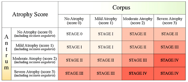
[OLGA staging system]
위축성위염, 화생성위염의 중증도 분류는 매우 다양하지만 저는 보통 내시경 소견에 의한 Kimura 분류를 좋아합니다. 편하기 때문이지요. 병리과 의사의 도움을 받아서 분류하는 방법도 많습니다. 최근 OLGA라는 분류가 제안되었고 몇몇 연구자들이 이용하고 있습니다.
2026년 대한헬리코박터학회지에 유럽 병리학자들의 리뷰가 실렸습니다.
[Abstract] Gastric mucosa atrophy is the cancerization field for gastric cancer development. Atrophic changes in the gastric mucosa can be assessed endoscopically and histologically. Gastric serology (e.g., pepsinogens and their ratio, and gastrin) may provide information on the functional status of the mucosa. However, outside large-scale epidemiological studies, the clinical reliability of serology at the individual patient level remains controversial. Histology-based estimates of atrophy-associated neoplastic risk require mucosal specimens from both the antral/mucus-secreting and corpus/fundus oxyntic compartments. Inspired by the clinical/biological framework of cancer staging, the Operative Link on Gastritis Assessment (OLGA) staging system classifies the severity and topography of mucosal atrophy into five stages, ranging from 0 to IV. International trials have validated the stepwise increasing risk of cancer with disease progression. Based on this evidence, OLGA stages are classified into low-risk (0-I) and high-risk (III-IV), with stage II remaining clinically controversial. The prevalence of high-risk stages varies according to patient- and population-specific risk factors. Among these, the most significant is the prevalence of Helicobacter pylori, a recognized oncogenic agent. Bacterial infection at the time of the initial endoscopy requires eradication therapy regardless of the stage of gastritis. Histological stage reliably identifies the endoscopic population at a high risk of cancer progression and shapes the schedule of endoscopy/histology-based surveillance. Serology at the index endoscopy may circumvent the inherent inconsistencies of population-based studies. This patient-specific serology could serve as an interval for non-invasive monitoring within the endoscopy/histology follow-up framework.
2026년 Rugge의 리뷰에 혈청검사에 대한 부분이 있어 옮깁니다. 병리학자라서 그런지 혈청검사를 거의 부정하고 있습니다. 혈청검사에 작은 의미라도 부여하려면 조직검사와 함께 하라는 것입니다. 말도 안되는 헛소리라고 생각합니다. 고위험 환자면 내시경을 해야지 혈청검사로 추적관찰 한다는 것이 말이나 되는 이야기입니까?
THE SEROLOGICAL COUNTERPART OF GASTRIC MUCOSA ATROPHY
For at least three decades, “functional” gastric serology (i.e., Pg-1, Pg-2, Pg-1:Pg-2, and gastrin) has been proposed as a non-invasive and inexpensive method to identify subjects at increased risk for GC. Large-scale epidemiological studies have shown a strong correlation between mucosal atrophy and the pepsinogen ratio. Based on this evidence, Pg-serology has been proposed as a screening tool for patients who require further invasive diagnostic procedures. However, beyond epidemiology, the clinical reliability of serology for ranking the severity of atrophy in individual patients remains controversial.
The limited use of serology in diagnostic practice is affected by significant biases, including demographic and lifestyle variables, local H. pylori epidemiology, current or prior H. pylori status, and ongoing therapies. To minimize the confounding effect of these variables, the patient’s specific serology could be tested along with histology at index endoscopy. This serology could serve as a reference for a multimodal approach to atrophy monitoring that may replace non-invasive and invasive (more expensive EGD with biopsy) surveillance.
[Home]