![]() [Description exercise 1]
[Description exercise 1]
![]() Case 1
Case 1
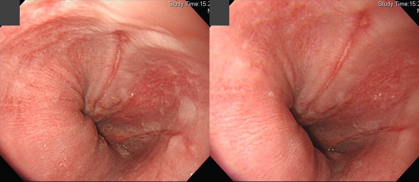
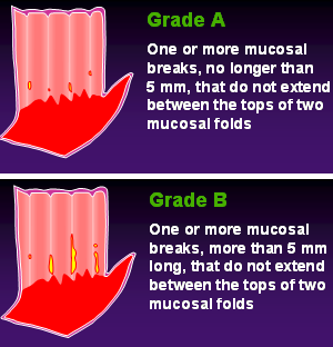
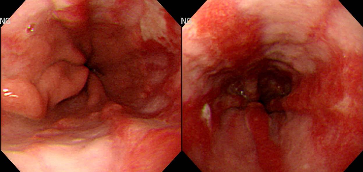
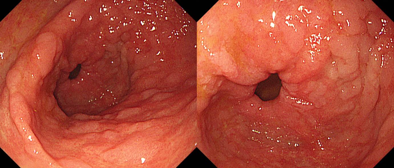
Findings: At far distal esophagus, a linear mucosal break longer than 5 mm is located at 12 o'clock direction. Another short mucosal break less than 5 mm is seen at 3 o'clock direction. Surrounding mucosa shows dirty-white mucosal hypertrophy.)
Impression: Reflux esophagitis, LA-B
[Comment]
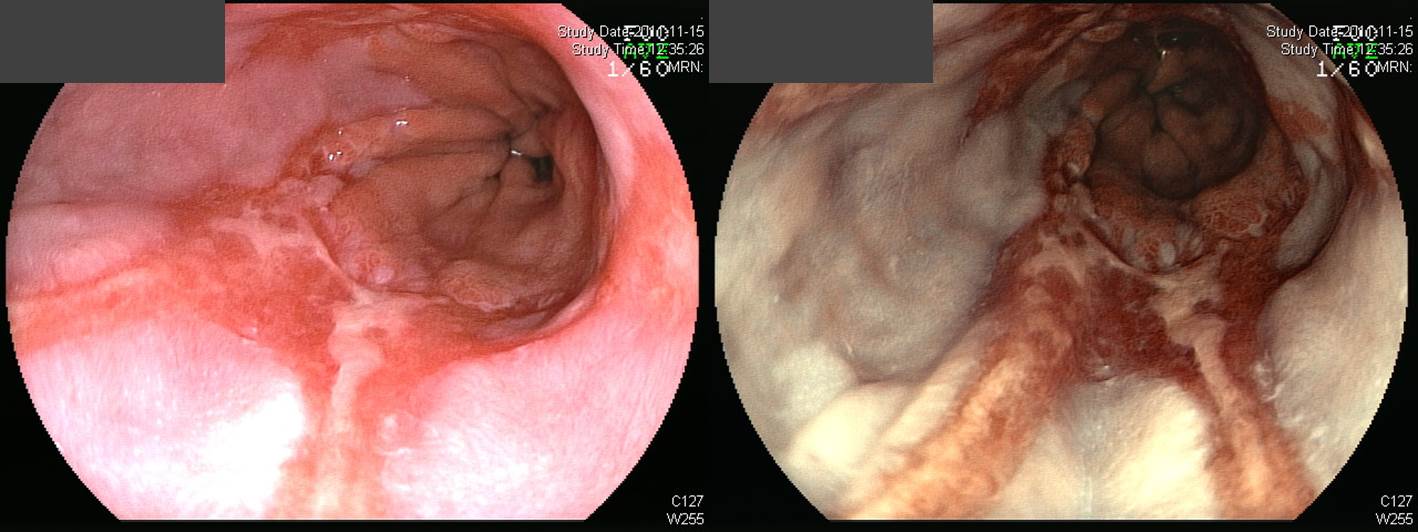
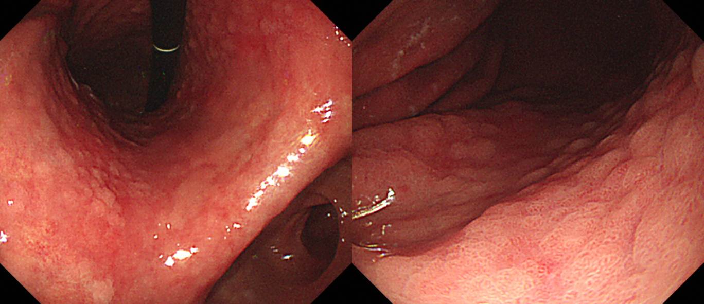
There is no confluent erosions in this case. Each mucosal breaks are separated. Followings are examples of confluent mucosal breaks.
![]() Case 2. Mid-esophagus
Case 2. Mid-esophagus
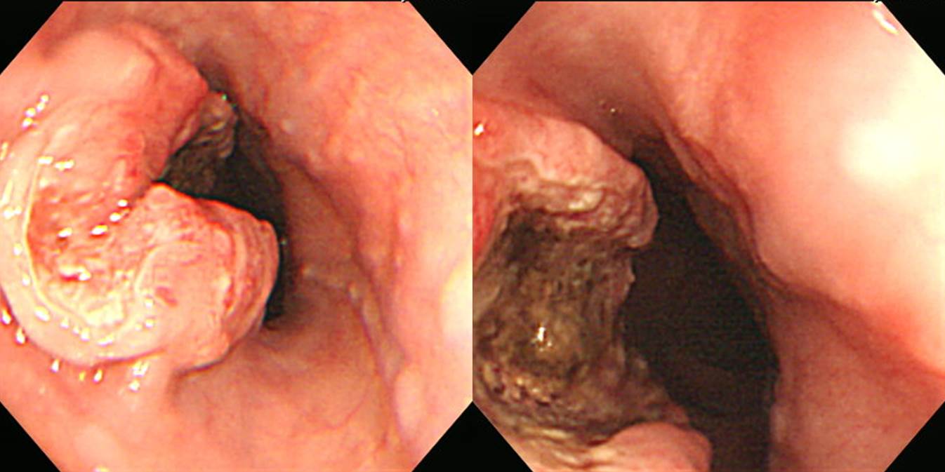
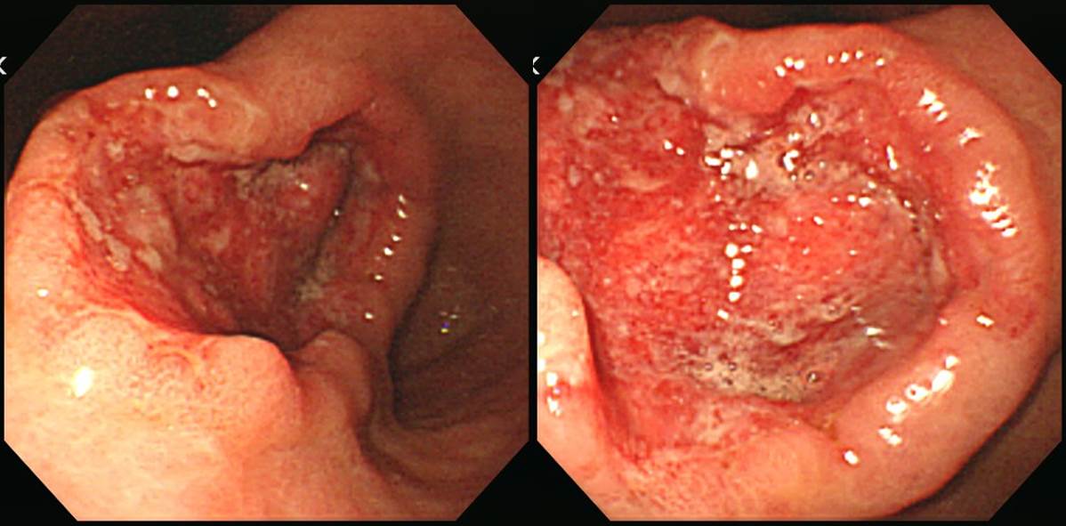
Findings: At mid-esophagus, a 4x2 cm sized luminal-narrowing mass with deep central ulceration was seen. The ulcer base was covered with dirty necrotic exudate. Protruded portion showed irregular surface, erythema, and mucosal friability.
Impression: Advanced esophageal cancer, type 3
[Comment]
Longitudinal diameter is bigger than the horizontal diameter.
Final pathologic size was 4 cm.
Invasive squamous cell carcinoma, moderately differentiated, esophagus:
1) tumor size: 4x2 cm
2) extension to perimuscular adventitia
3) endolymphatic tumor emboli: not identified
4) perineural invasion: present
5) involvement of radial margin
6) negative resection margins (proximal, 3 cm ; distal, 10 cm)
7) metastasis to 3 out of 62 regional lymph nodes (3/62: "LC omentum", 0/1; "RRLN", 0/0; "LRLN", 0/9; "5", 0/6; "7", 2/15; "8u", 0/1; "R9", 0/2; "L10", 0/1; "G1", 0/2; "G2", 0/8; "G3", 1/17)
![]() Case 3
Case 3
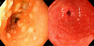
Findings: At antrum, multiple white small flat nodules were diffusely scattered.
Impression: Metaplastic gastritis
[More examples of metaplastic gastritis]


내시경 소견을 처음 배우는 의대 본과 학생이 보내온 흥미로운 질문에 답했습니다.
![]() Case 4
Case 4
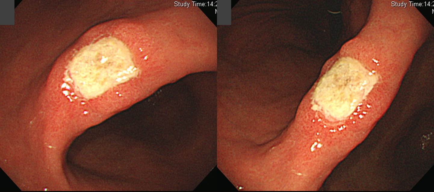
Findings: At the center of the angle, there was a 2 cm ulcer the sharp edge and edematous margin. The ulcer base was covered with thick white-yellow exudate. There was no fold change.
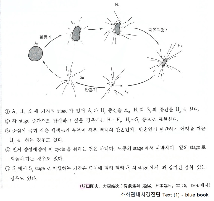
Impression: Benign gastric ulcer, active 2 stage
[Comment]
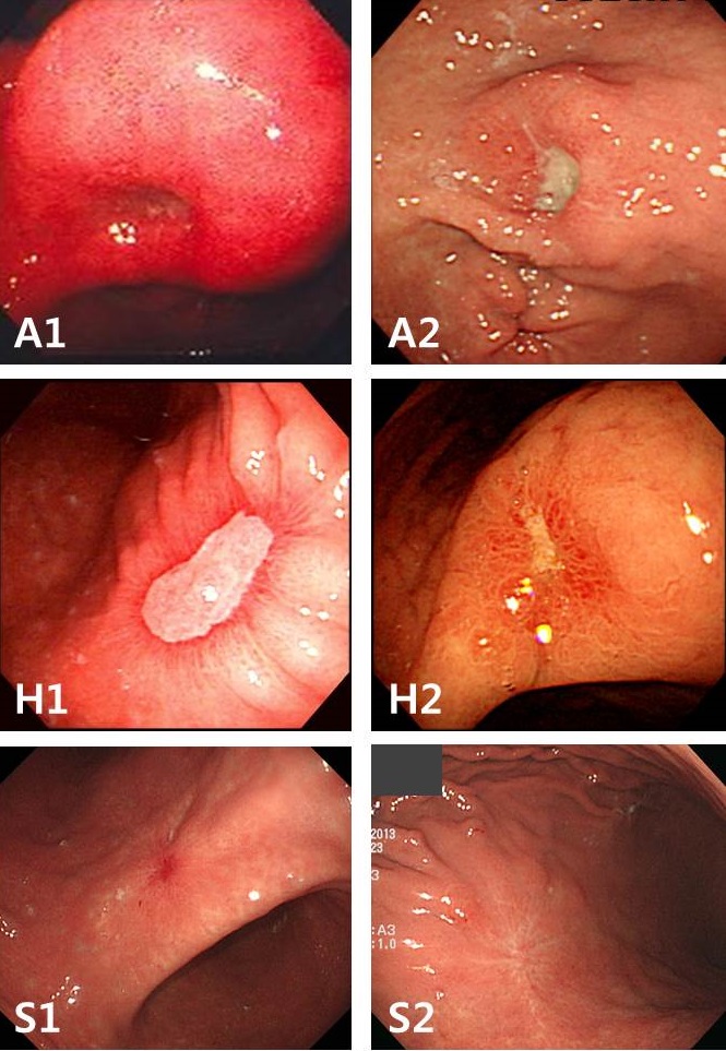
- Active stage 1 (A1): Active and blurred edge
- Active stage 2 (A2): Active and sharp edge
- Healing stage 1 (H1): Healing with regenerating epithelium
- Healing stage 2 (H2): Almost healed by regeneration
- Scar stage 1 (S1): Red scar
- Scar stage 2 (S2): White scar
![]() Case 5
Case 5
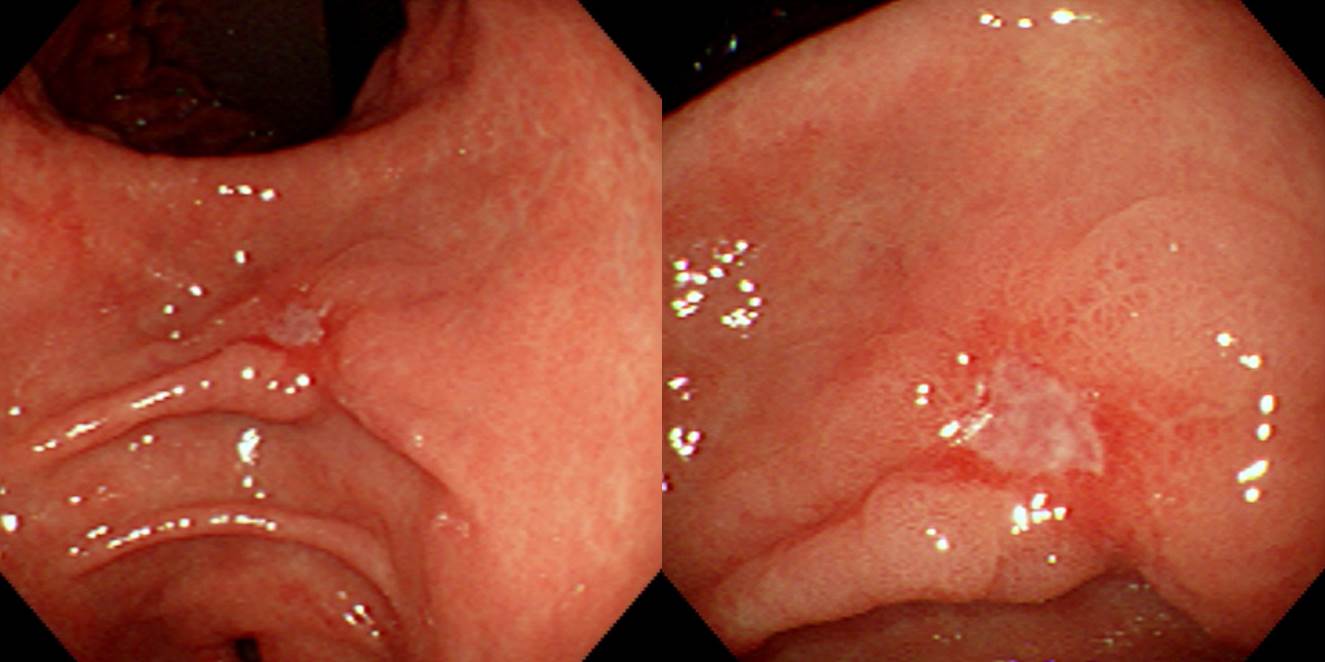
Findings: At the lesser curvature of the gastric antrum, a 1 cm sized depressed lesion was seen. The edge is relatively sharp, but there were some areas of spiculation. Multiple abnormal converging folds are seen (cutting and clubbing).
Impression: Early gastric cancer, type IIc
[Comment]
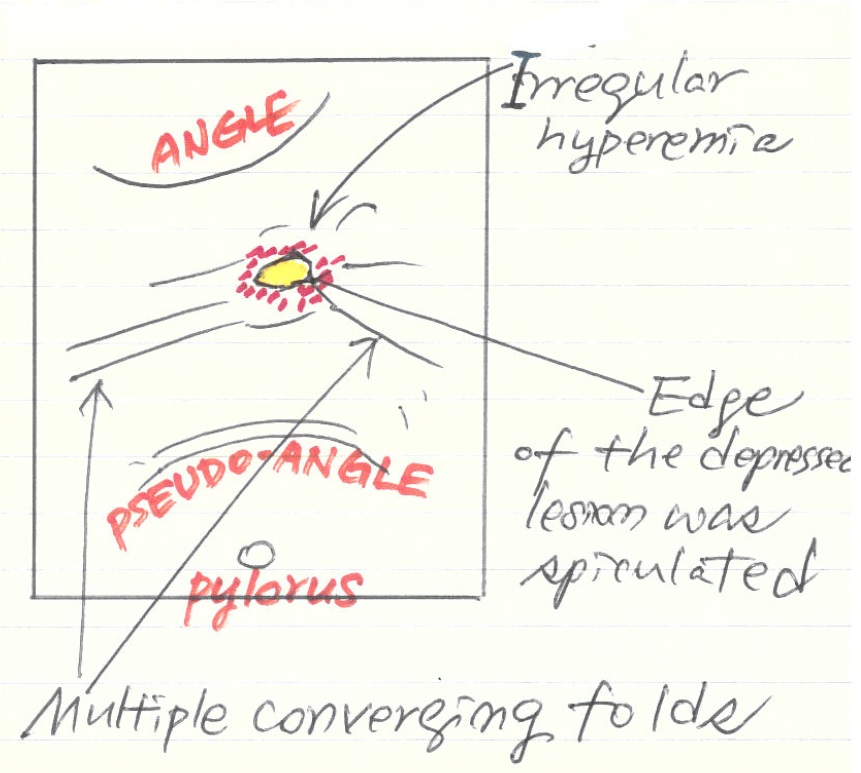
This is tricky. Because the center is depressed and margin is slightly elevated. We call it depressed lesion because marginal elevation is considered as secondary change. Actually, folds are formed only in the depressed lesions. So, the endoscopic diagnosis is EGC IIc (not IIa).
This is an old case, so surgery was done. In the current clinical practice, endoscopic submucosal dissection can be considered.
Stomach, subtotal gastrectomy:
Early gastric carcinoma
1. Location: middle third, center at body and posterior wall
2. Gross type: EGC type IIc+III
3. Histologic type: tubular adenocarcinoma, well differentiated
4. Histologic type of Lauren: intestinal
5. Size: 1.6x1.1 cm
6. Depth of invasion: extension to mucosa (muscularis mucosa) (pT1a)
7. Resection margin: free from carcinoma (safety margin: distal 6 cm, proximal 3.7 cm)
8. Lymph node metastasis: no metastasis in 31 regional lymph nodes (pN0)
9. Lymphatic invasion: not identified
10.Venous invasion: not identified
11.Perineural invasion: not identifiedRegarding the terminologies, the edge and the margin is confusing. Look at the following picture to see how we use the terms at SMC.

![]() Case 6
Case 6
Findings: There is a 5 cm sized mass at the lesser curvature side of the proximal antrum, just below the angle. The center was deeply ulcerated. The uneven ulcer base shows erythema, friability, and dirty exudate.
Impression: Advacned gastric cancer, Borrmann type II
[Comment]
We describe this kind of lesion using tricky expressions like 'mass with deep central ulceration (with narrow bank) or 'ulcerative mass'.
Stomach, subtotal gastrectomy:
Advanced gastric carcinoma
1. Location : lower third, Center at antrum and posterior wall
2. Gross type : Borrmann type 3
3. Histologic type : tubular adenocarcinoma, poorly differentiated
4. Histologic type by Lauren : intestinal
5. Size : 5.5x4.5x0.8 cm
6. Depth of invasion : penetrates serosa (pT3)
7. Resection margin: free from carcinoma, safety margin: distal 2.3 cm, proximal 4 cm
8. Lymph node metastasis : no metastasis in 37 regional lymph nodes (pN0)
9. Lymphatic invasion : present
10. Venous invasion : not identified
11. Perineural invasion : present
12. Stage by AJCC : II (T3, N0, MX)
![]() Case 7. Duodenal bulb.
Case 7. Duodenal bulb.
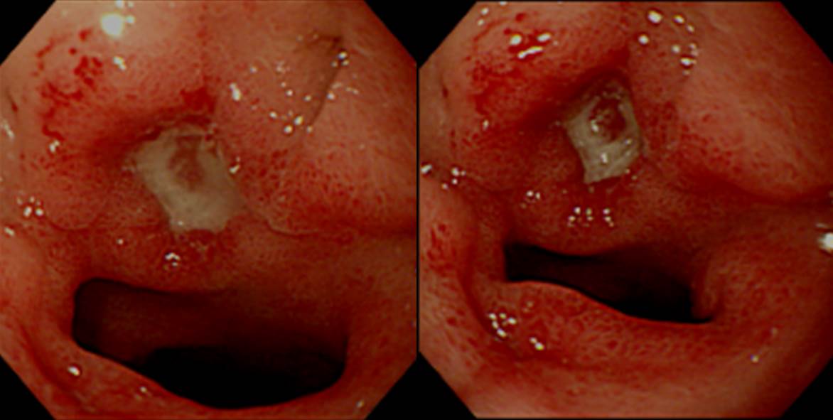
Findings: An active ulcer was seen. In the ulcer base, a red area was noticed. No exposed vessel was seen.
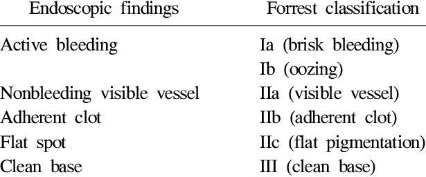
Impression: Duodenal ulcer scar, A1, Forrest classification IIc
[Comment]
In a patient with bleeding, Forrest classification is useful for (1) the evaluation of rebleeding rate, and (2) decision making about endoscopic hemostasis.

© EndoTODAY Endoscopy Learning Center. Lee Jun Haeng.