![]() [Description exercise 3]
[Description exercise 3]
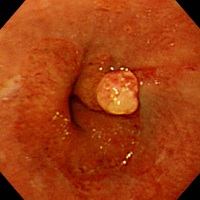
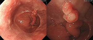
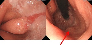
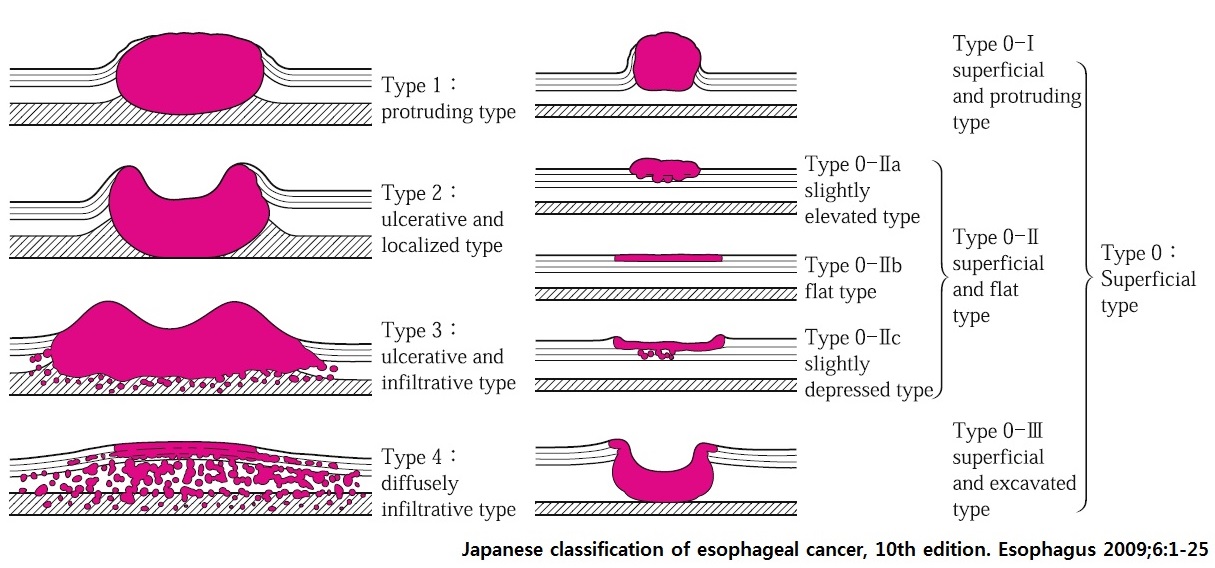
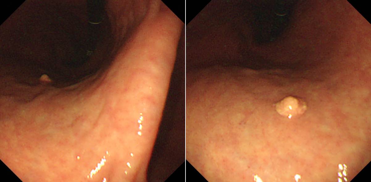
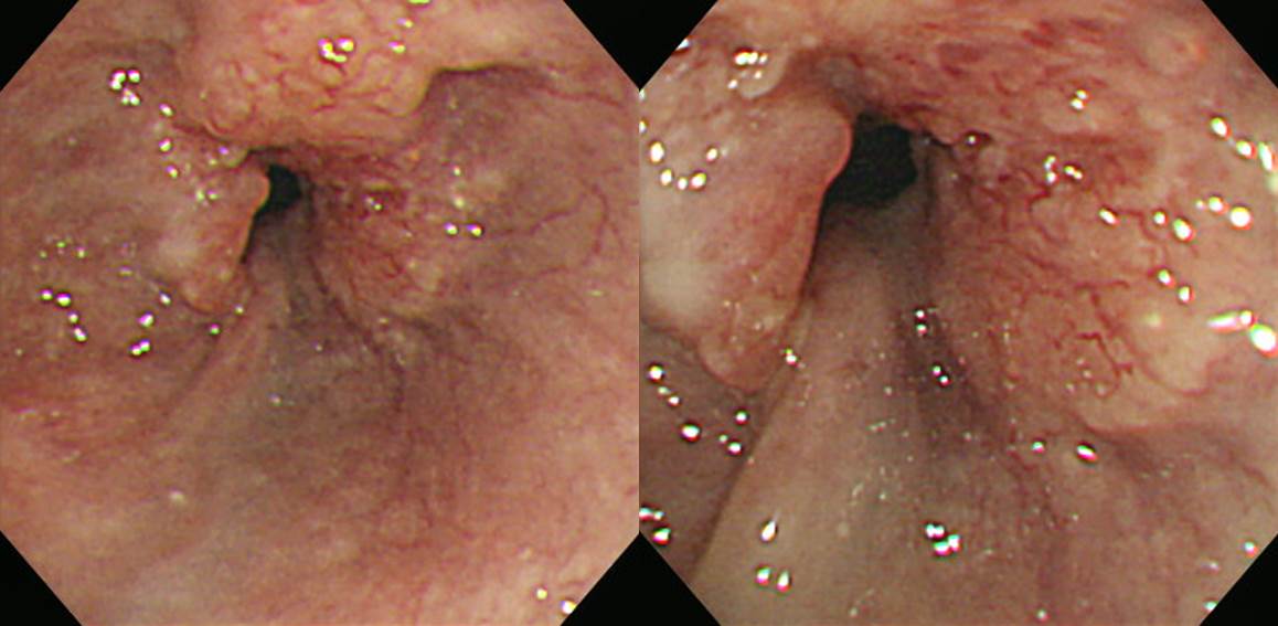
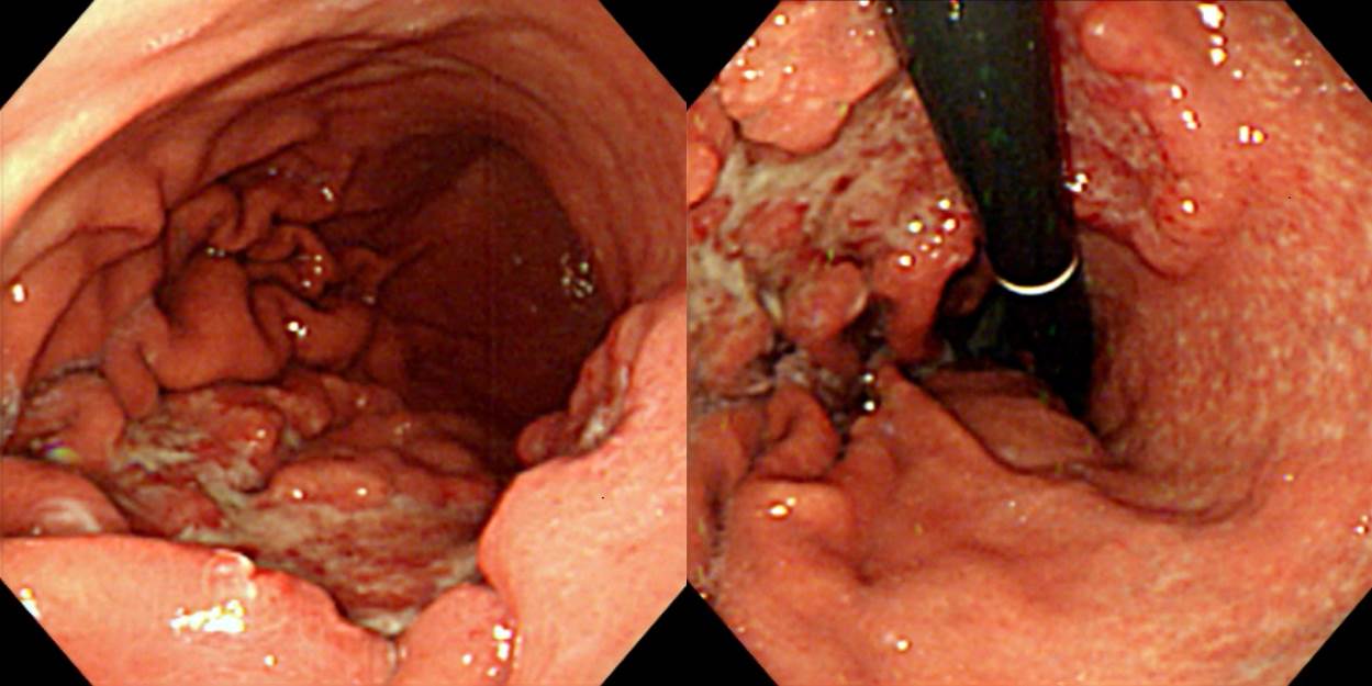
![]() Case 15. Near GE junction
Case 15. Near GE junction
Findings: Squamocolumnar junction is upward elevated from the hiatal opening. There is a 6-7mm sized sessile hyperemic polyp with short star-shaped shallow ulcer and mucosal breaks.
Impressions: (1) Reflux esophagitis, LA-A, (2) Sentinel polyp, (3) Sliding hiatal hernia
[Comments]
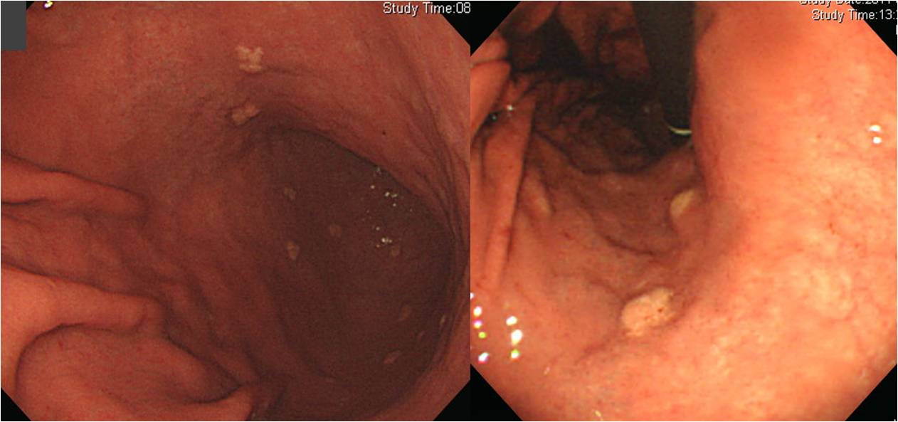
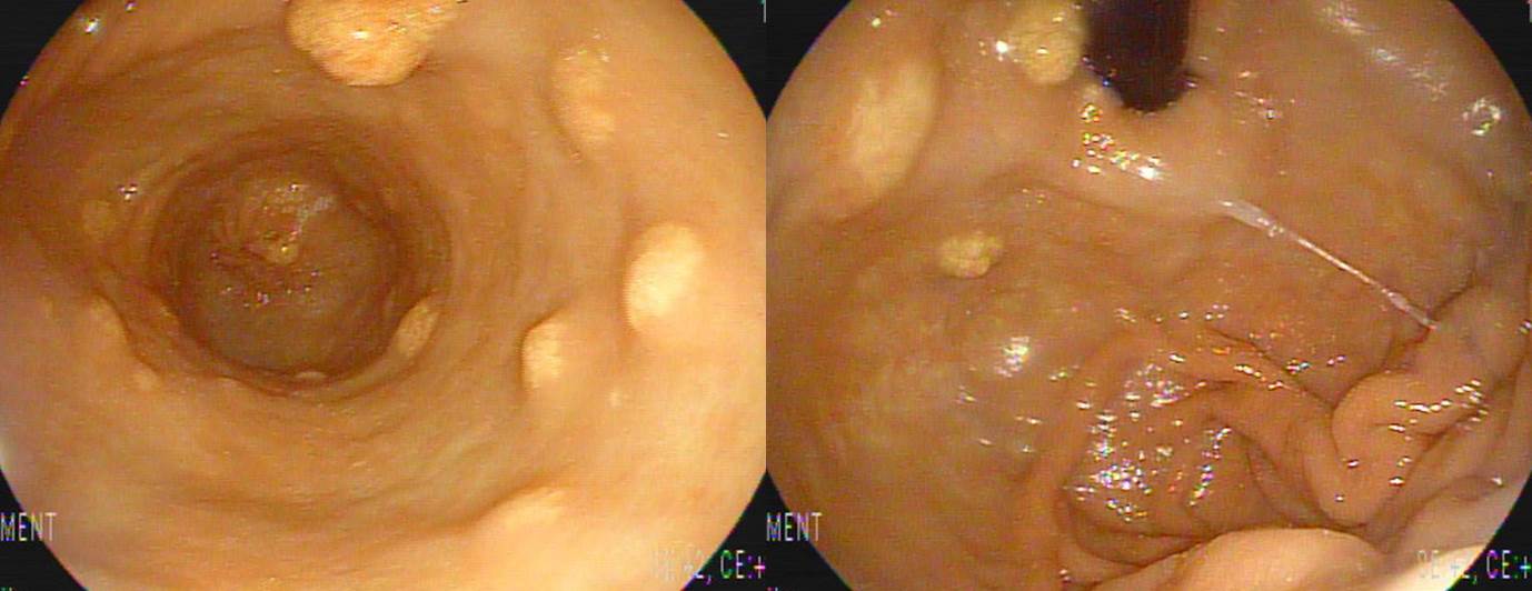
Followings are some more examples of sentinel polyp.
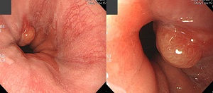
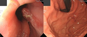
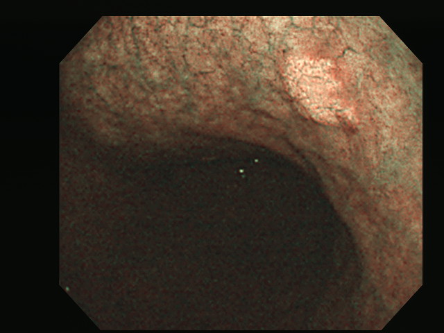
![]() Case 16. Mid to lower esophagus
Case 16. Mid to lower esophagus
Findings: At mid to lower esophagus, about 4-5cm sized mass with obstruction was seen. The surface of the mass lesion is uneven, focal hyperemic, and whitish discolorated.
Impression: Advanced esophageal cancer, type I
[Comments]
Classification of advanced esophageal cancer is tricky, but usually we use the same approach as advanced gastric cancer.
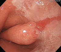
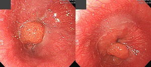
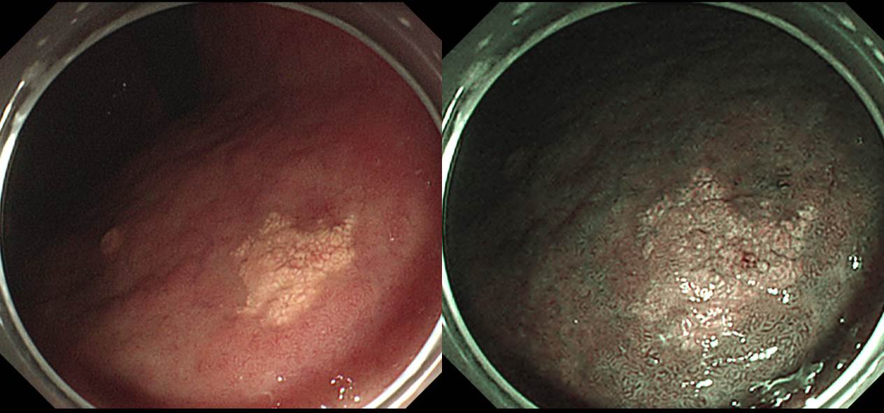
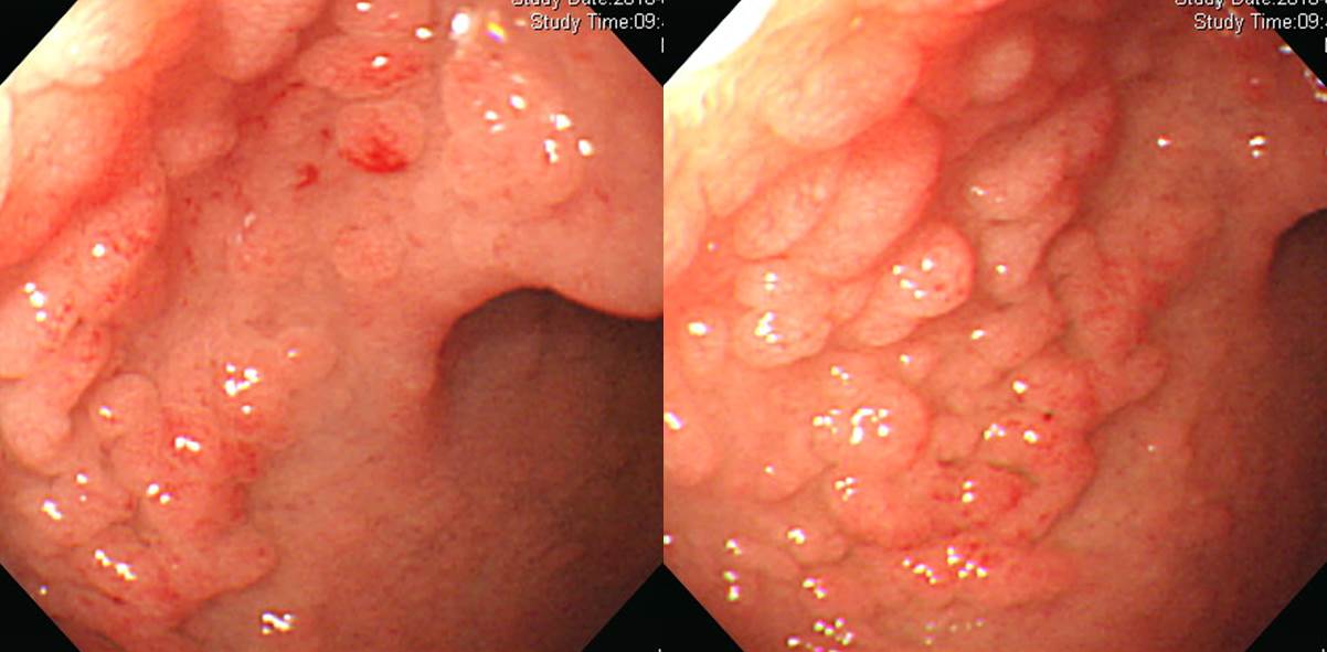
![]() Case 17
Case 17
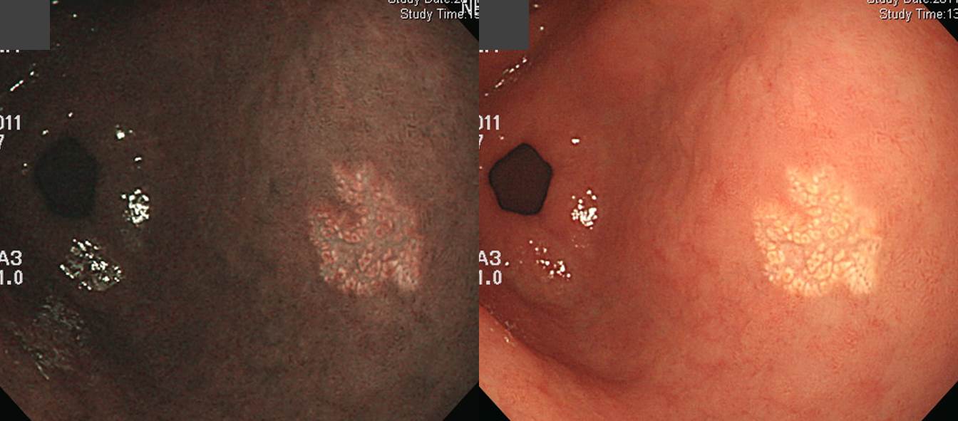
Findings: On the posterior wall aspect of the gastric cancer, a 0.5cm sized yellow flat lesion was found. The surface shows granullar pattern.
Impressions: Gastric xanthoma
[Comments]
Followings are some more examples of gastric xanthoma.
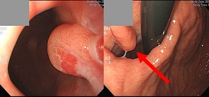
![]() Case 18 (melena)
Case 18 (melena)
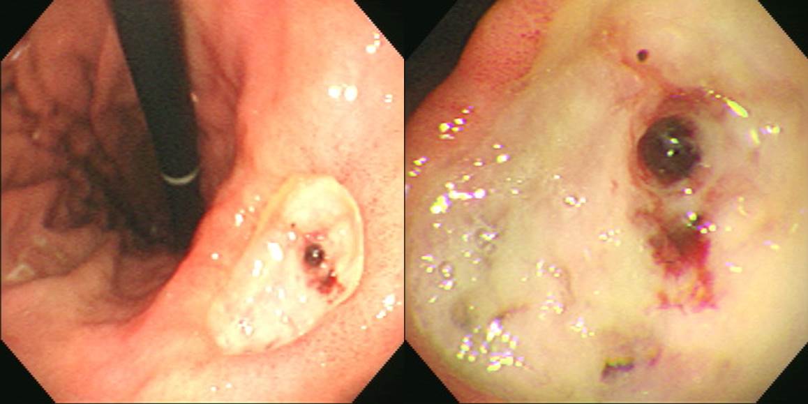
Findings: On the center of the angle, a 3x2cm sized ulcer with shart edege and edematous margin was found. There was no fold changes. The ulcer base was covered with dirty white exudate and there is a single exposed vessel.
Impression: Benign gastric ulcer, active 1 stage, Forrest classification IIa
[Comments]
Treatment plans for patients with peptic ulcer bleeding are based on endoscopic findings (Forrest classification).
SMC protocol
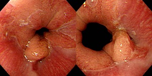
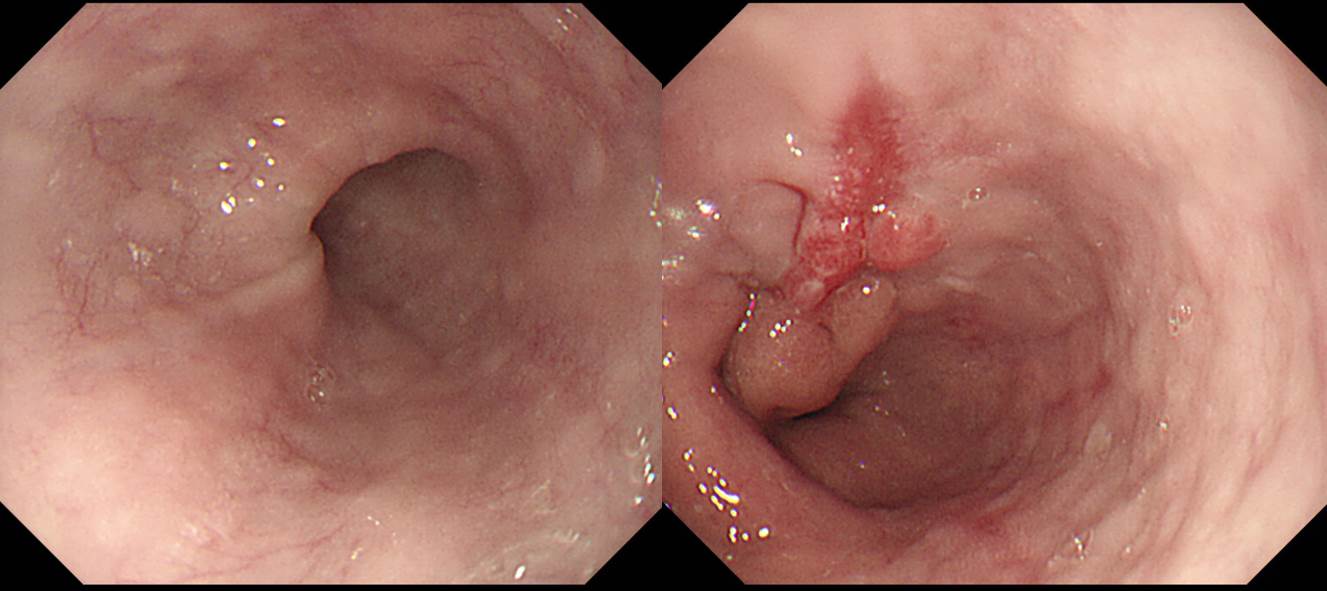
![]() Case 19
Case 19
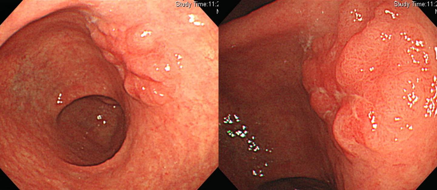
Findings: On the postero-LC side of the antrum, there is a 2cm sized nodular elevated lesion. The surface is slightly uneven, center of the lesion is slightly depressed, and the edge is clear. There is a focal hyperemia at 6 o'clock diresction.
Impression: EGC IIa+IIc
[Comments]
Endoscopic treatment was done.
Stomach, endoscopic submucosal dissection:
Early gastric carcinoma:
1. Location : antrum, posterior wall
2. Gross type : EGC type IIa+IIc
3. Histologic type : tubular adenocarcinoma, moderately differentiated
4. Histologic type by Lauren : intestinal
5. Size : 3.3x2.5 cm
6. Depth of invasion : invades mucosa (muscularis mucosa) (pT1a)
7. Resection margin: free from carcinoma, safety margin: distal 1 cm, proximal 1 cm, anterior 1 cm, posterior 2 cm
8. Lymphatic invasion : not identified
9. Venous invasion : not identified
10. Perineural invasion : not identified
11. Microscopic ulcer : absent
12. Histologic heterogeneity: absent
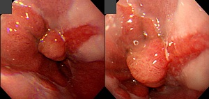
![]() Case 20
Case 20
Findings: There is a 7-8 cm sized ulceroinfiltrative lesion on the GC-PW of midbody. The ulcerative area is very uneven, and there are dirty exudates and hematins.
Impression: AGC, Borrmann type III
[Comments]
Surgery was done.
Stomach, total gastrectomy:
Advanced gastric carcinoma, Borrmann type III, mid body to high body of posterior wall and greater curvature,
Adenocarcinoma, poorly differentiated, diffuse type;
1) tumor size: 7x7 cm
2) extension to proper muscle layer
3) endolymphatic tumor emboli: present
4) peritumoral lymphoid follicles
5) negative resection margins (proximal: 5.5 cm; distal: 7.5 cm)
6) metastasis to 16 out of 48 regional lymph nodes (16/48: lesser curvature, 8/26; greater curvature, 3/10; "7", 5/6; "8", 0/6)
AJCC Stage IV (T2a, N3, MX)
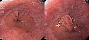
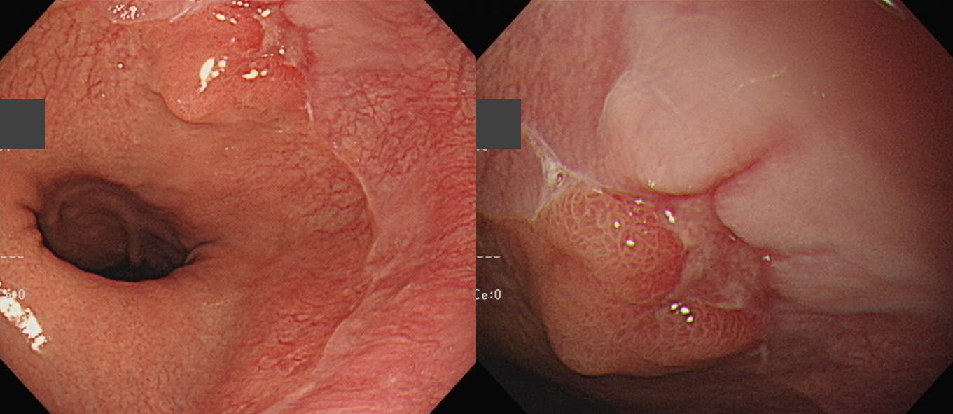
![]() Case 21. Duodenal bulb
Case 21. Duodenal bulb
Findings: In the duodenal bulb, multiple pale granular lesions were scattered.
Impression: Gastric heterotopia (= heterotopic gastric mucosa in the duodenum)
© EndoTODAY Endoscopy Learning Center. Lee Jun Haeng.