Previous | Home | EndoTODAY | List | Next
![]() [Case: PEG in a patient with esophageal stent]
[Case: PEG in a patient with esophageal stent]
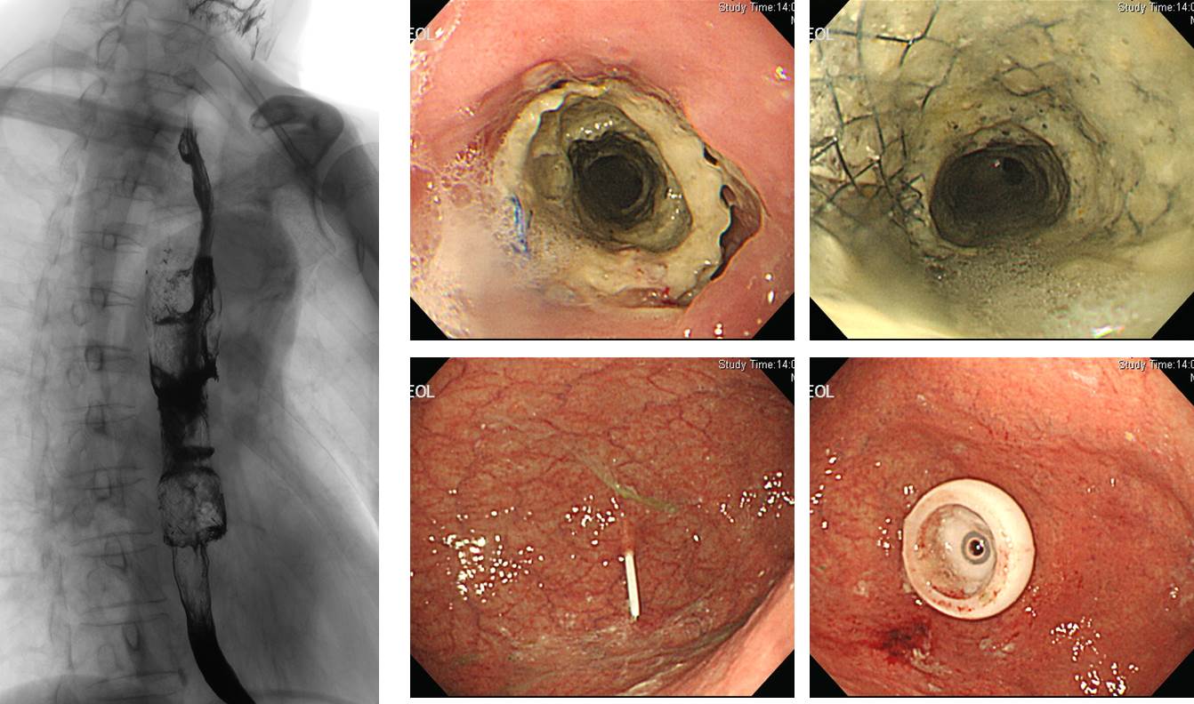
진행성 식도암으로 esophageal obstruction과 tracheo-esophageal fistula가 있어서 covered stent를 넣은 환자입니다. Stent 시술 후에도 지속적인 aspiration이 있어서 경구 식이섭취가 어렵다고 판단되어 PEG 삽입이 의뢰되었습니다. Stent로 인하여 식도가 충분히 넓어지지 않았다면 PEG tube가 식도를 통과하지 못할 가능성이 있는 상황이었습니다. 그러나 다행스럽게도 9.2 mm 외경의 내시경이 저항없이 식도를 통과할 수 있어서 통상적인 방법으로 PEG 시술이 가능하였습니다.
식도 stent에 dirty white한 exudate가 있었고 이 부위를 tube가 통과되었기 때문에 PEG tube의 contamination 정도는 통상보다 높을 수 밖에 없다고 판단되어 최대한 소독을 하였고 1-2일 정도의 IV 항생제 사용을 권하였습니다.
![]() [amebic colitis]
[amebic colitis]

Internet에서 가져온 자료로 기억되는데 현재 reference를 정확히 알 수 없습니다 (저작권자에게 양해를 구합니다). 진단은 amebic colitis입니다. Inflammatory bowel disease의 감별진단에 꼭 들어가는 기생충 질환입니다. 아직까지 제가 직접 진단내린 예는 없지만 중요한 병 중의 하나로 생각되므로 소개합니다.
이런 경우는 Ginsberg 내시경책의 일부를 옮기는 것이 가장 타당할 것 같습니다 (page 322). 내시경적으로는 flask-shaped ulcer를 만드는 것으로 알려져 있지만 아래 설명에 있듯이 사실 flask shaped ulcer를 보이는 경우는 없는 것 같습니다. Exudate가 많은 multiple dirty ulcers로 관찰되는 경우가 대부분이라고 생각합니다.
"Amebic colitis is a protozoan infection that primarily affects the large bowel. It is most often seen in patients who recently immigrated from developing countries and who recently traveled to developing countries. Symptoms can vary from none to explosive diarrhea, tenesmus, fever and abdominal cramps.
Colonoscopic appearance during the acute phase resembles ulcerative colitis, but in the chronic phase it appears more like Crohn’s disease. The most common segments involved are the cecum and right colon, with the rectum and sigmoid less often involved. Toxic megacolon may develop in severe cases of amebiasis.
Colonoscopy reveals granular, friable, and erythematous mucosa with discrete large ulcers covered by yellowish, mucopurulent exudates. Biopsies of the margins of the ulcers provide a 60% to 90% yield of trophozoites to make the diagnosis."
![]()