 [Lymphoepithelioma-like carcinoma (LELC). 림프양상피종양 암종 = Gastric carcinoma with lymphoid stroma] - 終
Previous | Home | EndoTODAY | List | Next
[Lymphoepithelioma-like carcinoma (LELC). 림프양상피종양 암종 = Gastric carcinoma with lymphoid stroma] - 終
2025-7-12. 잘 낫지 않는 궤양. MALT 림프종 의증으로 의뢰된 진행성 위암.
* LELC 증례 모음 위질환내시경아틀라스 8호 기고 (PDF)
3. EBV positive LELC of the stomach
4. EBV negative LELC of the stomach
5. 임상적 특징
6. Colon LELC
7. FAQs
8. References
 1. Introduction to lymphoepithelioma-like carcinoma (LELC)
정리: 임상강사 백남영
Lymphoepithelioma-like carcinoma(LELC)는 비인두암과 조직학적으로 유사하고, 림프구성 간질을 가진 미분화된 위종양입니다. Epstein-Barr 바이러스와의 연관성도 언급되고 있습니다. LELC은 식도, 위, 담관, 타액 선, 폐, 자궁, 피부 등에서 발생합니다.
위 LELC는 전체 위암의 l-4%를 차지하는데 조기위암의 경우 IIa + IIc형이 많으며 진행성 위암의 경우 Borrmann type IV 형이 많다고 합니다. 병리학자들은 medullary carcinoma로 부르기도 합니다. 조기위암의 형태로 나타날 경우는 일반적인 조기위암과 예후가 비슷하나 진행성 위암의 형태를 취할 경우 일반적인 진행성 위암에 비하여 예후가 훨씬 좋은 것으로 알려져 있습니다. 점막하 종양의 형태로 나타나는 경우도 있어 주의가 필요합니다. 85%는 EBV 양성인데, EBV 음성인 경우는 상대적으로 예후가 나쁩니다.
병리 소견 (https://www.pathologyoutlines.com/)
오래된 저널 하나 소개합니다.
Lymphoepithelioma-like carcinoma of the stomach: a subset of gastric carcinoma with distinct clinicopathological features and high prevalence of Epstein-Barr virus infection (Hepatogastroenterology 1999)
METHODOLOGY: Of 379 patients with gastric adenocarcinoma, from 1993 to 1996, 6 of them with lymphoepithelioma-like carcinoma of stomach were retrospectively studied.
RESULTS: Five patients were females and one patient was male. Their age ranged from 51-75 years with a mean age of 61.5 years. Endoscopically, 2 patients were initially diagnosed as early gastric cancer and the other 4 were diagnosed as advanced gastric cancer. Three patients had tumors located in the lower third of the stomach, while the other three tumors were located in the middle and upper third. Two tumors invaded into the serosal layer and the other four lesions were confined at submucosal and muscular layers. Using the in situ hybridization method, all 6 patients (100%) had positive nuclear Epstein-Barr virus-encoded small RNA signals in the tumor cells but not in the surrounding lymphoid stroma and non-neoplastic gastric mucosa. Helicobacter pylori was found in 4 (66.7%) of the cases. The mean follow-up period of the 6 patients was 27 months. Five patients were free of the disease. Lymph node involvement and mesenteric implantation was noted in one patient in which cancer recurred 1 year after gastrectomy.
CONCLUSIONS: Lymphoepithelioma-like carcinoma of stomach in this study revealed a female predominance, preferential localization in the proximal part of the stomach, better prognosis, and a high association with Epstein-Barr virus infection.
2. Esophageal LELC
Korean J Gastrointest Endosc 2010 - 삼성서울병원에서 보고하였던 증례
2019년 WHO 분류에 기술된 병리 소견: lymphoepithelioma-like carcinoma is considered to be a distinct subtype of undifferentiated carcinoma. It is characterized by a sheet-like arrangement of large epitheloid cells with prominent nuclei and indistinct borders. The tumor is surrounded by a characteristic inflammatory infiltrate that is rich in lymphocyte and plasma cells.
3. EBV positive lymphoepithelioma-like carcinoma. EBV 양성 림프양상피종양 암종

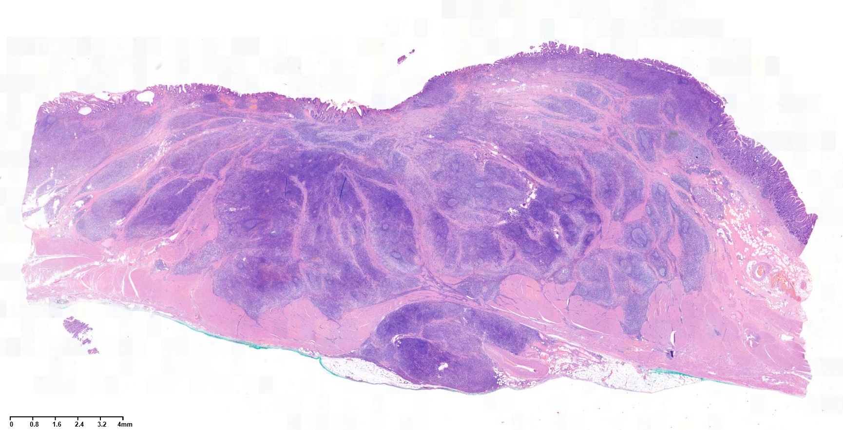

EBV in situ hybridization
Depth of invasion은 SM3였습니다.
EBV (+)

1. Location : upper third, center at cardia (Siewert III)
2. Gross type : Borrmann type 2
3. Histologic type : Medullary carcinoma (lymphoepithelioma-like carcinoma)
4. Histologic type by Lauren : indeterminate
5. Size : 2.2x2.2 cm
6. Depth of invasion : invades muscularis propria
7. Resection margin: free from carcinoma, safety margin: proximal 1.4 cm, distal 2 cm
8. Lymph node metastasis : no metastasis in 52 regional lymph nodes
9. Lymphatic invasion : not identified
10. Venous invasion : not identified
11. Perineural invasion : not identified
12. Result of in-situ hybridization: Epstein-Barr virus: Positive
조직검사에서는 poorly differentiated adenocarcinoma였고 EUS에서 SM invasion이 뚜렷했습니다.
Stomach, subtotal gastrectomy:
. Early gastric carcinoma
1. Location : lower third, Center at proximal antrum and anterior wall
2. Gross type : EGC type IIc
3. Histologic type : lymphoepithelioma-like carcinoma
4. Histologic type by Lauren : mixed
5. Size : 1.8x1.4 cm
6. Depth of invasion : invades submucosa (sm3) (pT1b)
7. Resection margin: free from carcinoma, safety margin: proximal 1 cm, distal 8.7 cm
8. Lymph node metastasis : no metastasis in 34 regional lymph nodes (pN0)
9. Lymphatic invasion : not identified
10. Venous invasion : not identified
11. Perineural invasion : not identified
12. AJCC stage by 7th edition: pT1b N0
13. Epstein-Barr virus : positive
Stomach, subtotal gastrectomy:
. Early gastric carcinoma
1. Location : middle third, Center at body and posterior wall
2. Gross type : EGC type IIb
3. Histologic type : carcinoma with lymphoid stroma (medullary carcinoma)
4. Histologic type by Lauren : indeterminate
5. Size : 1.8x1 cm
6. Depth of invasion : invades submucosa (sm3) (pT1b)
7. Resection margin: free from carcinoma, safety margin: proximal 1.2 cm, distal 13 cm
8. Lymph node metastasis : no metastasis in 33 regional lymph nodes (pN0)
9. Lymphatic invasion : not identified
10. Venous invasion : not identified
11. Perineural invasion : not identified
12. AJCC stage by 7th edition: pT1b N0
2달 간격으로 3번 내시경 검사를 받으셨고, 마지막 조직검사에서 signet ring cell carcinoma가 나왔습니다. 최종 병리는 carcinoma with lymphoid stroma로 나왔습니다. Lymphoepithelioma-like carcinoma라고 부르는 타입입니다. Lymphoid stroma 사이에 미분화조직형 위암이 박혀있는 형태이므로, 육안적으로는 SMT 비슷하게 보이고, 조직검사의 false negative가 많은 것 같습니다.
Early gastric carcinoma
1. Location : lower third, Center at antrum and greater curvature
2. Gross type : EGC type IIc
3. Histologic type : carcinoma with lymphoid stroma
4. Histologic type by Lauren : mixed
5. Size : 3.8x1.9 cm
6. Depth of invasion : invades mucosa (muscularis mucosae) (pT1a)
7. Resection margin: free from carcinoma
8. Lymph node metastasis : no metastasis in 17 regional lymph nodes (pN0)
9. Lymphatic invasion : not identified
10. Venous invasion : not identified
11. Perineural invasion : not identified
12. AJCC stage by 8th edition: pT1a N0
Nasopharyngeal cancer로 치료받던 중 PET를 시행하였는데 우연히 위암이 발견되어 수술을 받았음.
Stomach, subtotal gastrectomy: Early gastric carcinoma
1. Location : middle third, center at body and greater curvature
2. Gross type : EGC type I
3. Histologic type : lymphoepithelioma-like carcinoma
4. Histologic type by Lauren : intestinal
5. Size : 3.2x2.3x0.9 cm
6. Depth of invasion : extension to submucosa (sm2) (pT1b)
7. Resection margin: free from carcinoma, safety margin: proximal, 3.2 cm; distal, 8.8 cm
8. Lymph node metastasis : no metastasis in 36 regional lymph nodes (pN0) (0/36: lesser curvature, 0/10; greater curvature, 0/9; "1", 0/2; "3", 0/1; "4sb", 0/0; "5", 0/2; "6", 0/0; "7", 0/1; "8a", 0/2; "9", 0/6; "11p", 0/2; "12a", 0/1)
9. Lymphatic invasion : not identified
10.Venous invasion : not identified
11.Perineural invasion : not identified
4. EBV negative lymphoepithelioma-like carcinoma. EBV 음성 림프양상피종양 암종
임상상 및 내시경 소견
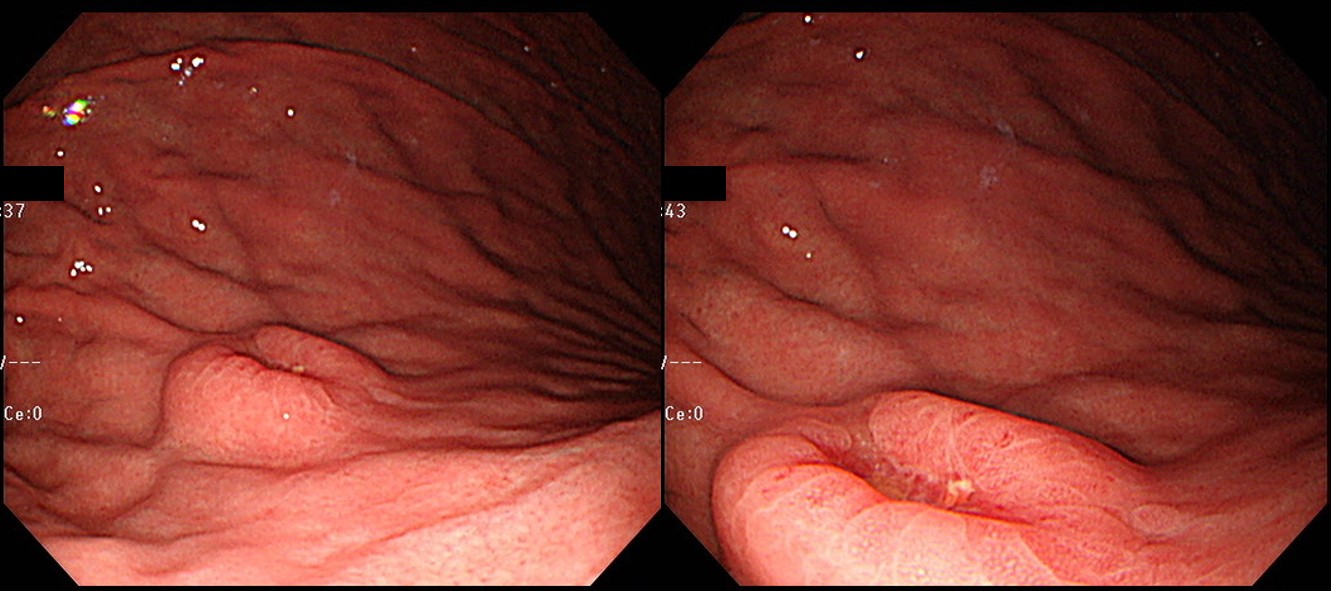
65세 남성이 건진내시경에서 발견된 위암 의심 소견으로 의뢰되었다. 조직검사 결과는 Atypical cells, suspicious for poorly differentiated adenocarcinoma였다. 내시경 재검(오른쪽 사진)에서 위체하부 소만에 4cm 크기의 융기된 병소가 있었고 중심부가 함몰되어 있었다. 이 병소의 근위부인 위체중부에 몇 개의 마치 무덤과 비슷한 1-3cm 크기의 몇 개의 융기부가 있었다. 위체하부 함몰을 동반한 융기부 조직검사는 tubular adenocarcinoma, moderately differentiated (cytokeratin AE1/AE3 positive) 였으며 이보다 근위부 점막하종양 유사한 융기부들의 조직검사는 gastritis 뿐이었다. 수술을 시행하였다.
진단 및 경과
EBV negative, lymphoepithelioma-like carcinoma. 수술장에서 subtotal gastrectomy 계획으로 위를 절제하였으나 검체 확인 시 proximal margin이 1cm 정도였으며 식도위접합부와 가까워 total gastrectomy로 전환하였다. 병리 결과 advanced gastric cancer, lymphoepithelioma-like carcinoma, Lauren indeterminate, 5x8x4.8cm, penetrated subserosal connective tissue, resection margin negative, no lymph node metastasis out of 40이었다 (AJCC 8th pT3NO). 궤양부 이외는 정상점막 아래로 넓게 침윤을 보였다. Epstein-Barr virus 음성이었다.
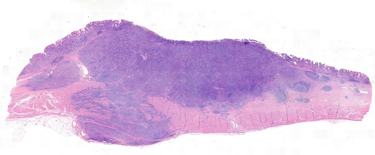
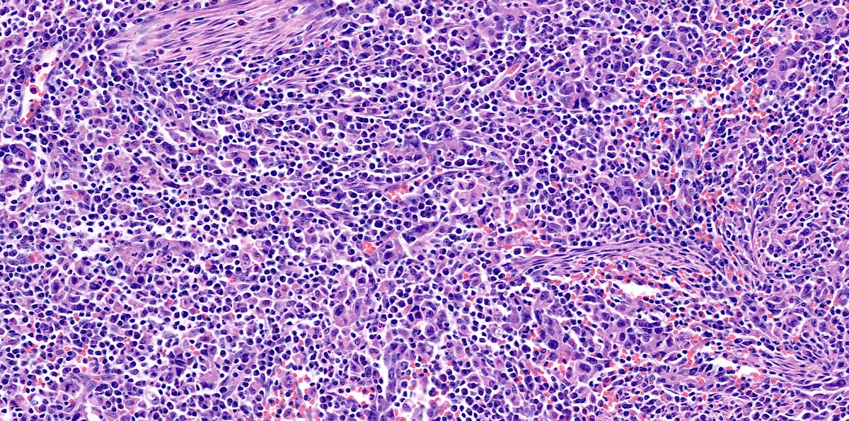
해설
Lymphoepithelioma-like carcinoma는 조직학적으로 비인두암과 유사하고 림프구성 간질을 가진 미분화된 위종양으로 SMT-like cancer 중 하나이다. 예후는 일반적인 위암보다 좋다. 본 증례처럼 EBV 음성인 예는 상대적으로 예후가 나쁘다 (Min. Gastric Cancer 2016).
5. 임상적 특징 - (1) refractory ulcer, (2) lymphoma와 구분이 어렵다
제가 느끼고 있는 몇 가지 특징을 소개합니다.
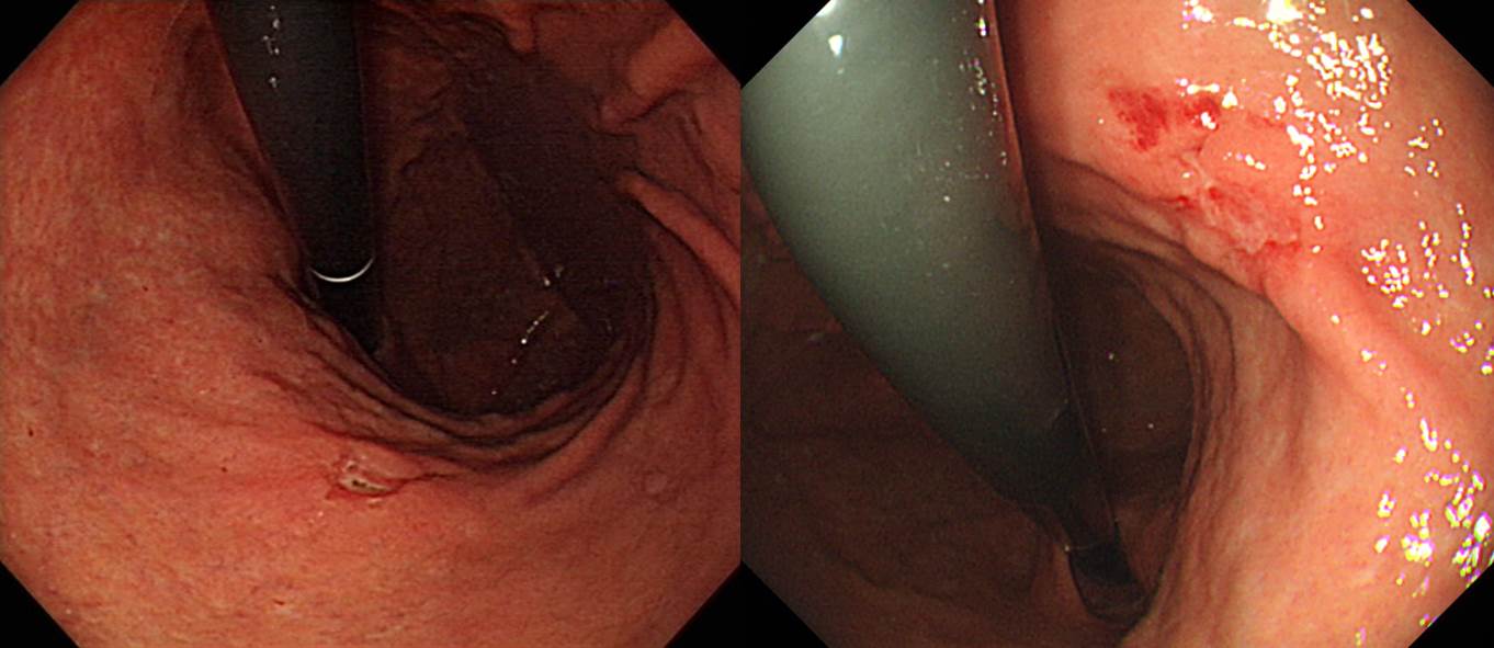
(1) Refractory ulcer의 임상상을 보일 수 있습니다. 조직검사의 false negative가 많다는 것과도 관련된 특성입니다. 아래 증례를 보십시오
Refractory ulcer로 의뢰된 분입니다. 의뢰 후 내시경 조직검사에서 signet ring cell carcinoma가 나왔습니다. 수술 후 최종 병리는 carcinoma with lymphoid stroma로 나왔습니다. Lymphoepithelioma-like carcinoma라고 부르는 타입입니다. Lymphoid stroma 사이에 미분화조직형 위암이 박혀있는 형태이므로, 육안적으로는 SMT 비슷하게 보이고, 조직검사의 false negative가 많은 것 같습니다.
Early gastric carcinoma
1. Location : lower third, Center at antrum and greater curvature
2. Gross type : EGC type IIc
3. Histologic type : carcinoma with lymphoid stroma
4. Histologic type by Lauren : mixed
5. Size : 3.8x1.9 cm
6. Depth of invasion : invades mucosa (muscularis mucosae) (pT1a)
7. Resection margin: free from carcinoma
8. Lymph node metastasis : no metastasis in 17 regional lymph nodes (pN0)
9. Lymphatic invasion : not identified
10. Venous invasion : not identified
11. Perineural invasion : not identified
12. AJCC stage by 8th edition: pT1a N0
(2) MALT 림프종 의증으로 의뢰되는 경우가 있습니다. 조직학적 특징이 미분화조직형인 암과 섞인 dense lymphoid infiltration이므로 작은 내시경 겸자 조직에서는 MALT 림프종과 혼동될 수 있는 것 같습니다. 드물게 겸자 조직에서 moderately differentiated인데 최종 병리결과 LELC로 나온 경우도 있습니다. 여하튼 겸자 조직에서 진단이 어려운 조직형입니다. 아래 동영상을 보시기 바랍니다.
6. Colon LELC
2022년 3월 KASID 교육자료. 설명 중 합포체는 syncytium을 말함 (세포 융합으로 나타나는 다핵성 세포)
2022년 3월 KASID 교육자료
[FAQs]
[2024-7-17. 애독자 질문]
Carcinoma with lymphoid stroma 라고 했을 때, lymphoid stroma가 얼마나 많으면 이 진단이 붙는 것인지 궁금합니다. 병리 의사의 주관적인 판단인 것인지 아니면 명확한 기준이 있는지요?
[2024-7-17. 한 병리 선생님의 답변]
Lymphoid stroma의 percentage, 양에 대한 정의는 없습니다. 병리의사의 주관적 판단이기는 하나, lymphoid stroma뿐 아니라 poorly formed glands 등 LELC의 병리 소견이 unique하기 때문에 병리의사간 진단 일치도는 높을것 같습니다. 단, LELC의 특징적 조직 소견은 submucosa이하에서 잘 보이기때문에 생검에서의 진단은 어렵습니다.
[References]
1. EndoTODAY EBV-associated gastric cancer
2. LELC 증례 모음 위질환내시경아틀라스 8호 기고 (PDF)
3. Epstein-Barr virus 관련 위암 이봉은. Korean J Helicobacter Up Gastrointest Res 2021 (PDF)
© 일원내시경교실 바른내시경연구소 이준행. EndoTODAY Endoscopy Learning Center. Lee Jun Haeng.
