Previous | Home | EndoTODAY | List | Next
![]() [EGC on top of SMT]
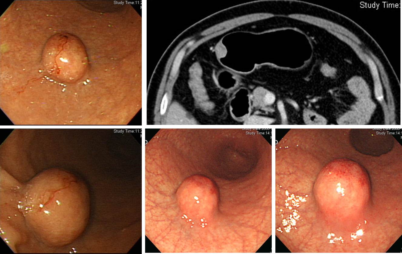
[EGC on top of SMT]
2 cm 크기의 위 SMT가 발견되어 시술 전 조직검사에서 암으로 나오지 않은 상태로 의뢰된 환자입니다. Wedge resection을 시행하였고 놀랍게도 아래와 같은 결과가 나왔습니다. 조직검사 위치를 바꾸어 보았다면 수술 전 위암을 진단할 수 있지 않았을까 생각해 보았습니다. 여하튼 herniated gastric mucosa로 인하여 SMT처럼 융기된 부위의 점막 top에서 발생한 위암이라고 결론지었습니다. 혹시 위암이 먼저이고 이로 인하여 gastric mucosa의 herniation이 발생한 것은 아닐까도 생각해 보았습니다. 마치 gastritis cystica profunda와 비슷하게 말입니다. 정답이 없는 질문을 혼자 해 보면서 웃었습니다.
- Histologic type : tubular adenocarcinoma, well differentiated
- Size : 1.5x1.5x1.0 cm
- Depth of invasion : extension to mucosa (muscularis mucosa) (pT1a)
- Resection margin: free from carcinoma
- Associated findings : herniated gastric mucosa in the submucosa
![]() 2013년 8월 9일 (금)부터 10일 (일)까지 강릉에서 대한상부위장관헬리코박터학회 Summer Workshop이 진행되었습니다.
2013년 8월 9일 (금)부터 10일 (일)까지 강릉에서 대한상부위장관헬리코박터학회 Summer Workshop이 진행되었습니다.
![]() 1) 일요일 특강연자이신 연세대학교 의과대학 의생명과학부 남기택 교수님께서 SPEM(Spasmoltic Polypeptide Expressing Metaplasia)을 소개해 주셨습니다. 오래전부터 SPEM이 chief cell에서 origin하지 않을까 추측하여 왔는데 lineage tracing(상피세포의 stem cell marker를 증명하는 방법임. Stem cell에 beta-Gal을 tagging하여 다른 세포로 분화해 가는 것을 증명하는 방법)을 통하여 증명할 수 있었다고 합니다.
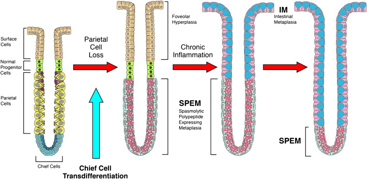
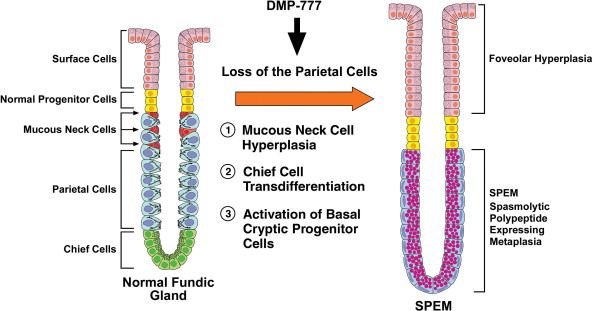
1) 일요일 특강연자이신 연세대학교 의과대학 의생명과학부 남기택 교수님께서 SPEM(Spasmoltic Polypeptide Expressing Metaplasia)을 소개해 주셨습니다. 오래전부터 SPEM이 chief cell에서 origin하지 않을까 추측하여 왔는데 lineage tracing(상피세포의 stem cell marker를 증명하는 방법임. Stem cell에 beta-Gal을 tagging하여 다른 세포로 분화해 가는 것을 증명하는 방법)을 통하여 증명할 수 있었다고 합니다.
Spasmoltic Polypeptide Expressing Metaplasia (SPEM)
- Marker: TFF2 (trefoil factor2), MUC 6 (참고로 intestinal metaplasia의 marker는 TFF3, MUC2)
- Present in fundic/oxyntic mucosa
- Associated with oxyntic atrophy (parietal cell loss)
- Arises from the base of the glands (antralization / pseudopyloric metaplasia)
- > 90% of cancer resection specimens contain SPEM in adjacent mucosa
- 52% of early gastric cancers stain for TFF2
참고문헌 1: Goldenring & Nam. Prog Mol Biol Transl Sci (2010) Review; Oxyntic atrophy, metaplasia, and gastric cancer - Gastric carcinogenesis involves the loss of parietal cells (oxyntic atrophy) and subsequent replacement of the normal gastric lineages with metaplastic cells. In humans, two metaplastic lineages develop as sequelae of chronic Helicobacter pylori infection: intestinal metaplasia and spasmolytic polypeptide-expressing metaplasia (SPEM). Mouse models of both chronic Helicobacter infection and acute pharmacological oxyntic atrophy have led to the discovery that SPEM arises from transdifferentiation of mature chief cells. The presence of inflammation promotes the expansion of SPEM in mice. Furthermore, studies in Mongolian gerbils as well as increasing evidence from human studies indicate that SPEM likely represents a precursor for the development of intestinal metaplasia. These findings suggest that loss of parietal cells, augmented by chronic inflammation, leads to a cascade of metaplastic events. Identification of specific biomarkers for SPEM and intestinal metaplasia hold promise for providing both early detection of preneoplasia and information on prognostic outcome following curative resection.
참고문헌 2: Nam. Gut (2012) Spasmolytic polypeptide-expressing metaplasia (SPEM) in the gastric oxyntic mucosa does not arise from Lgr5-expressing cells
참고문헌 3: Nam. Gastroenterology (2010) Mature chief cells are cryptic progenitors for metaplasia in the stomach (PDF)
참고문헌 4: Weis & Goldenring. Gastric Cancer (2009) Current understanding of SPEM and its standing in the preneoplastic process
![]() 2) 건국대학교 이선영 교수님께서 "일본 헬리코박터 치료의 최근 경향과 한일 가이드라인 point-to-point 비교"에 대하여 강의해 주셨습니다.
2) 건국대학교 이선영 교수님께서 "일본 헬리코박터 치료의 최근 경향과 한일 가이드라인 point-to-point 비교"에 대하여 강의해 주셨습니다.
PDF file, 0.8M
일본에서 최근 제안된 위염 내시경 진단에 대한 scoring 방법이 소개되었는데 흥미로웠습니다. Scoring을 통하여 위암 발생 위험을 guess하기 위한 노력이라고 생각됩니다. 우리나라에 적용하기는 어려운데요... 향후 어떻게 전개될지 관심을 가지고 지켜볼 필요는 있다고 생각됩니다.
쿄토 내시경적 위염 분류 (2013년 5월 일본 내시경학회 발표안)
| A (0,1, 2, 3점) | M (0, 2점) | H (0, 1점) | N (0, 2점) | AHHN ( 점) |
| 위축성위염* | 화생성위염 | 비대성위염 | 결절성위염 | 총점 |
0점: CAG가 없거나 closed type 1 1점: closed type 2, 3 2점: open type 1, 2 3점: open type 3 | 장상피화생 변화가 보이면 2점 Imaging enhanced endoscopy 사용시 (IM)으로 표시 | 두꺼워진 위 주름이 보이면 1점 | 결절성 변화가 보이면 2점 | 제균치료 성공 후 비감염 상태라면 뒤에 E-1 표시 후 1점을 차감 |
![]() 3) 한양대학교 김정목 교수님께서 "변화하는 환경에서 연구비 획득에 대한 조언"이라는 제목의 유익한 강의를 해 주셨습니다. 비단 연구계획서 작성뿐만 아니라 모든 글쓰기에 참고할만한 좋은 내용이었습니다. 맺음글을 옮깁니다.
3) 한양대학교 김정목 교수님께서 "변화하는 환경에서 연구비 획득에 대한 조언"이라는 제목의 유익한 강의를 해 주셨습니다. 비단 연구계획서 작성뿐만 아니라 모든 글쓰기에 참고할만한 좋은 내용이었습니다. 맺음글을 옮깁니다.
PDF file, 0.5M
'연구비 선순환(virtuous cycle)'이라는 과정은 지속적인 연구를 위해 반드시 필요하다. 이를 위한 전통적인 방법은 연구계획서 작성, 연구비 획득, 그리고 연구 수행과 논문발표라는 단계를 되풀이하는 것이다. 그렇지만 먼저 연구를 수행하고, 이 결과를 바탕으로 연구계획서를 작성하는 것이 바람직하다. 이런 과정을 통해 "이 연구분야는 내가 최고"라고 자부할만한 핵심 연구분야를 만들어야 한다. 그리고 새로운 연구영역을 위한 끊임없는 변신이 필요하다. 무엇보다 연구 계획서를 신청했음에도 불구하고 선정되지 못했다고 좌절해서는 안 된다. 도전하지 않으면 실패가 없지만, 실패가 없으면 발전도 없다는 말을 명심하고 본 강의가 여러분의 연구비 획득에 도움이 되길 바란다.
[Home]