Previous | Home | EndoTODAY | List | Next
![]() [Appendiceal mucocele. 충수 점액낭]
[Appendiceal mucocele. 충수 점액낭]
충수의 점액낭(mucocle)은 국소적으로 또는 광범위하게 확장된 충수의 내강에 다량의 점액이 채워진 경우이다. 샘종, 샘암종, 자궁내막증, 신경내분비종양 등 다양한 원인에 의해 내강이 폐쇄된 경우에 점액이 축적되어 발생하는 것으로 생각한다. 하지만 '점액낭 mucocele'이라는 용어는 그 원인에 대해 어떠한 정보도 주지 못하므로 병리진단명으로 사용하기에는 부적절하다. (병리학 제8판 619쪽)
[2013-11. 애독자 질문]
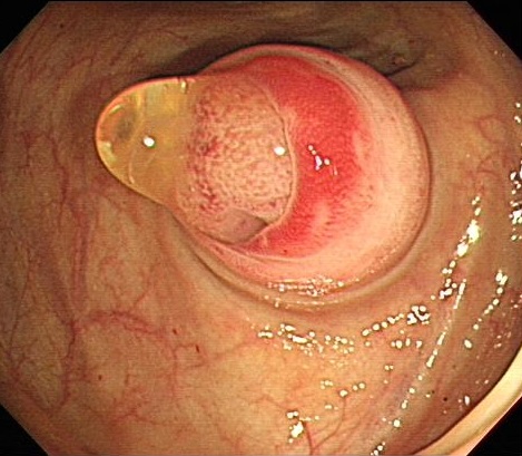
선생님 제가 대장내시경을 시행한 환자 중 appendix orfice에 점액 같은 것이 있었습니다. 1 cm 크기의 SMT로 보였습니다. Appendiceal mucocele에서 점액이 나오는 것은 처음 본 것 같습니다. Air inflation 시키니 orfice에 있는 SMT는 orfice에서 이동하는 것 처럼 보였습니다.

[2013-11. 홍성노 교수님 답변]
말씀하신 소견은 모두 가능합니다. 흔히 보는 것은 appendiceal orfice 혹은 주위 cecal base에 점막하종양 형태로 관찰되는 것이 많지만
1. Gelatin같은 점액 분비가 가능하고 (병리적으로 단순한 mucosa hyperplasia도 있지만, cystadenoma, retention cyst 가 많은 부분을 차지하며, cystadenoma of the appendix가 터져서 복강내 intraperitoneal mucinous spread 및 mucinous implants가 되는 경우가 pseudomyxoma peritonei인 것은 잘 아실 것입니다.)
2. 장관 외 병변이기 때문에 호흡, 자세변화 또는 공기 주입에 따라 점막하 종양의 융기 형태의 변화가 가능합니다. Uptodate의 일부를 옮깁니다.
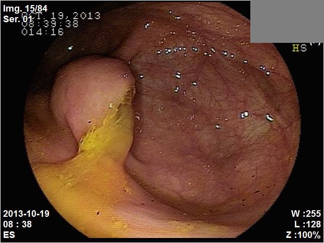
At endoscopy, an extrinsic or submucosal lesion may be suspected based upon smooth indentation into the cecal lumen, which may be the only sign of a mucocele. When poked with the biopsy forceps, the lesion may be firm in consistency or soft with a cushion sign. The appearance of the appendiceal orifice in the center of the mound has been labeled as the "volcano sign". An occasional and highly suggestive endoscopic finding is that of a glossy, rounded, balloon-shaped mass protruding from the appendiceal orifice and moving in and out of the latter with respiration. An outpouring of inflammatory exudate from the appendiceal orifice may be seen. As a general rule, the overlying mucosa is normal and mucosal biopsies are not usually done. If available, an endosonographic probe can disclose the cystic nature of the mucocele at the time of colonoscopy. Solid lesions such as carcinoids, lipomas, and lymphangiomas can thus be ruled out. Moreover, stromal invasion may perhaps be detected, which would predict the malignant behavior of the mucocele.
[2013-6-26. 애독자 질문]
엔도투데이 독자입니다. 상의드리고 싶은 케이스가 있습니다. 건강검진으로 대장내시경하는 분인데, cecal base 에 polypoid lesion 보이고, 점액질 같은것이 묻어있고, 똑똑 떨어지는 듯 보였습니다. Forcep으로 조직 약간 채취해서 나온 biopsy 에서는 hyperplastic polyp 으로만 나왔습니다. 이런 소견 무엇인지요?
[2013-6-26. 홍성노 교수님 답변]
Mucocele로 생각됩니다. CT를 찍고 수술적 절제가 필요할 것으로 생각됩니다. Hyperplastic polyp은 조직학적으로 정상 조직과 병리적으로 잘 구분됩니다만, 본 사진에서는 congestion이 있지만, pit pattern등은 정상으로 보여져 조직검체가 적고 폴립이라 병리 슬립에 기술하셔서 병리의사가 그렇게 판독하셨을 가능성도 있을 것으로 생각됩니다
[Cases]
2022-5. 삼성서울병원 구내식당 게시판
대장내시경에서 appendiceal opening은 다소 애매하였으나 CT에서 appendiceal mucocele 소견이 현저하여 수술을 하였습니다.
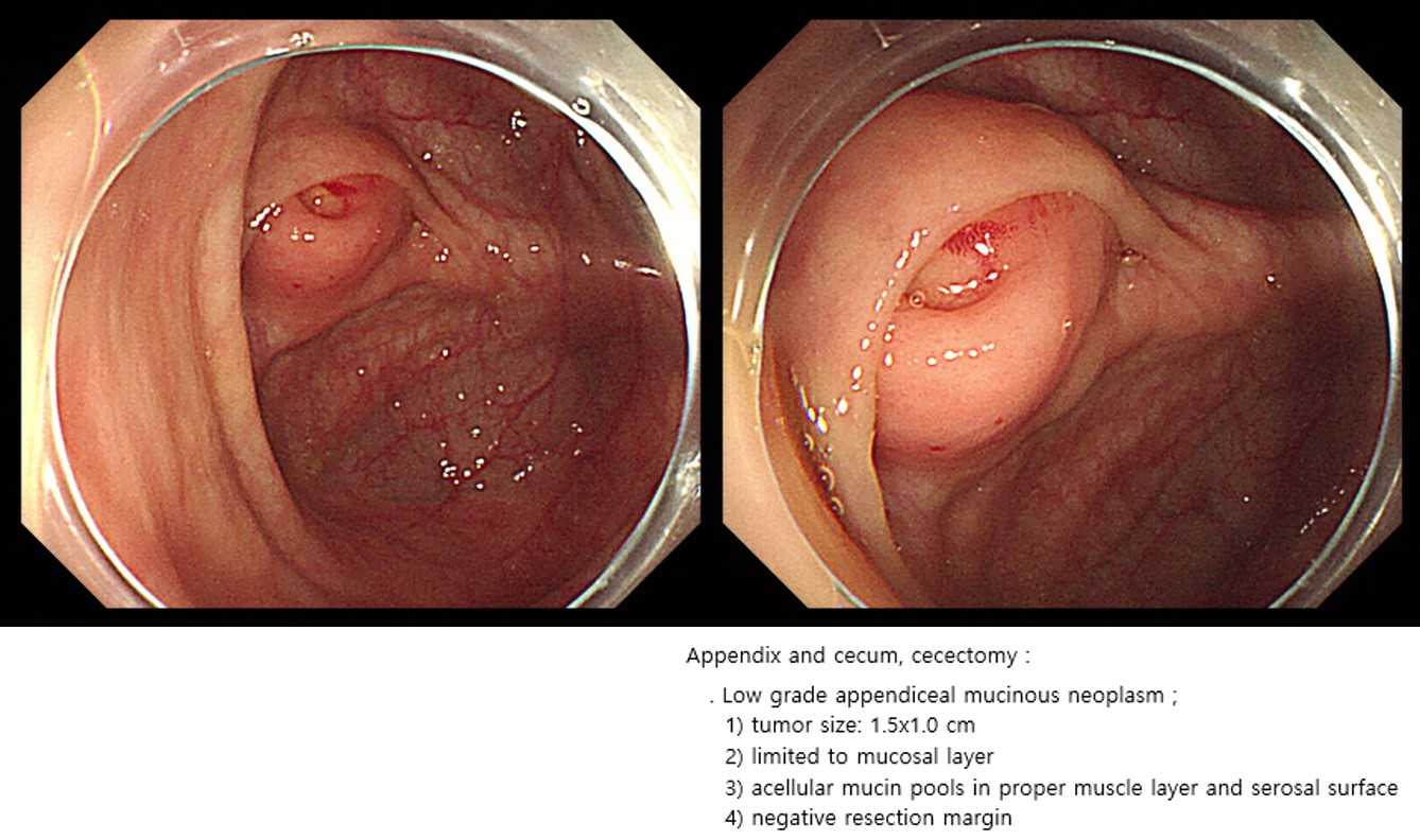
Cecum and appendix, cecectomy:
Low grade appendiceal mucinous neoplasm ;
1) size: 3.7x2 cm
2) confined to mucosa
3) no mucin spillage
4) negative resection margins (proximal, 4.5 cm; distal, 3.2 cm)
@ 일원내시경교실 바른내시경연구소 이준행. EndoTODAY Endoscopy Learning Center. Lee Jun Haeng