EndoTODAY 내시경 교실
Beginner | ESA | Schedule | OPD
Seminars | Atlas | Recent | Links
 [Peripheral T cell lymphoma of the GI tract] - 終
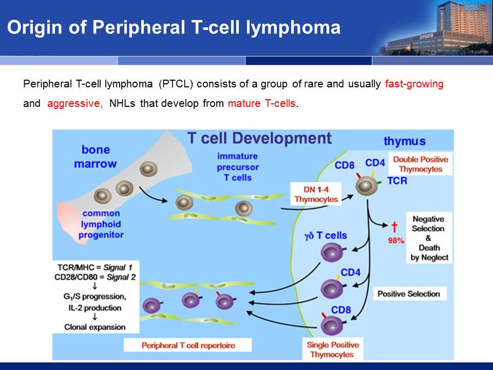
 1. Origin of T cell and PTCL
- The T-cells are born from stem cells, similar to that of B-cells, but mature in the thymus.
- They help the immune system work in a coordinated fashion.
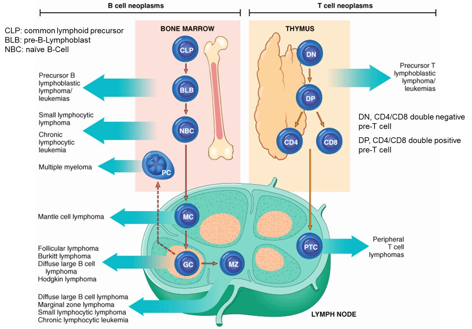
2016 WHO 분류
2. Gastrointestinal T cell lymphoma
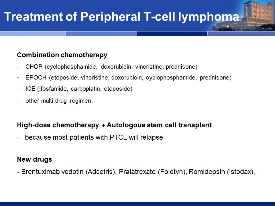
- Owing to the rarity the ideal treatment remains controversial.
- Primary surgery followed by CCT is best.
- A higher frequency of intestinal perforation and bleeding noted by some if Sx is omitted.
- In the largest series by Daum et al (n = 35, J Clin Oncol 21:2740-2746), 2yr OS was 28% only
- In another large series by Gale et al (n = 31, J Clin Oncol 2000;18:795), 5-year OS was 19.7%,and 5-year failure-free survival rate was only 3.2%.
PTCL: chronic diarrhea, loss of weight (15 kg), and night sweat

PERIPHERAL T-CELL LYMPHOMA, NOT OTHERWISE SPECIFIED, with TFH phenotype
CD 4, (Leu-3a) : Positive in tumor cells ^
CD 8, (Leu-2a) : Negative in tumor cells ^
CD 30 : Positive in tumor cells (2+, 100%) ^
β- F 1 : Positive in tumor cells ^
TCR-Delta : Negative in tumor cells ^
PD-1 : Positive in tumor cells ^
CD 21, B-CELL : Negative in tumor cells ^
CXCL 13 : Positive in some tumor cells ^
ALK : Negative in tumor cells ^
T cell lymphoma of the terminal ileum
Colon T cell lymphoma
Colon T cell lymphoma
만성복통. 수개월 검사 후 jejunal obstruction으로 수술하여 진단한 PTCL
Gastric PTCL의 예후가 나쁘다는 것은 잘 알려져 있습니다. 그러나 완치되는 환자가 없는 것은 아닙니다. 아래 증례를 보십시오. 힘냅시다.
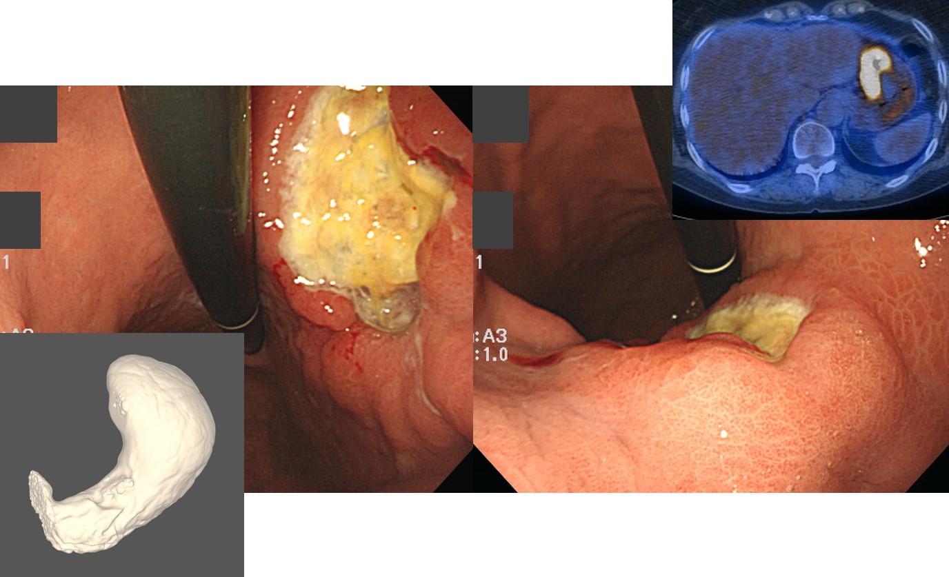
위 환자의 13년 후 추적내시경입니다.
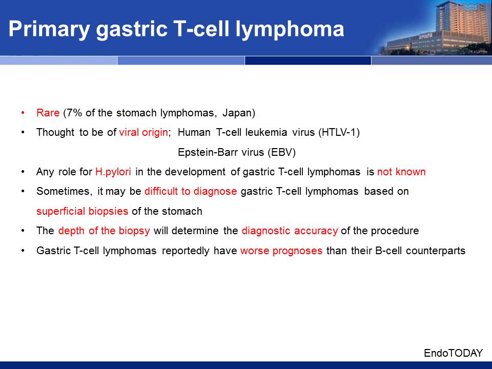
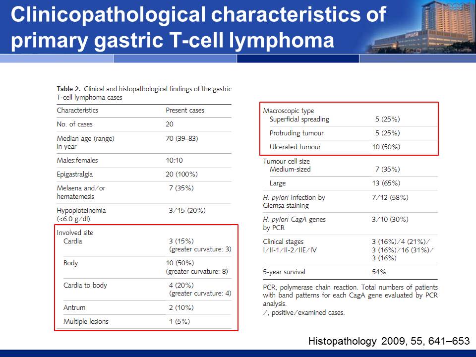
3. PTCL of the stomach
- Rare (7% of the stomach lymphomas, from Japan)
- Thought to be of viral origin
- Human T-cell leukemia virus (HTLV-1) and Epstein-Barr virus (EBV)
- Any role for HP in the development of gastric T-cell lymphomas is not known
- Sometimes, it may be difficult to diagnose gastric T-cell lymphomas based on superficial biopsies of the stomach
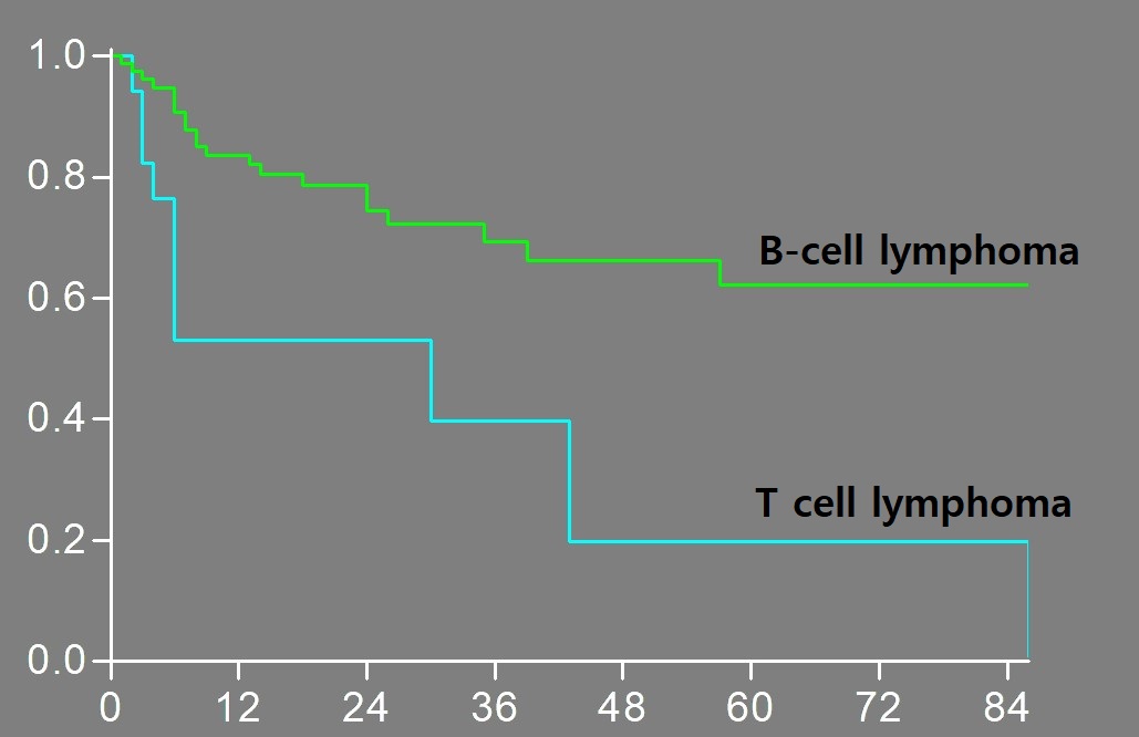
- The depth of the biopsy will determine the diagnostic accuracy of the procedure Gastric T-cell lymphomas reportedly have worse prognoses than their B-cell counterparts
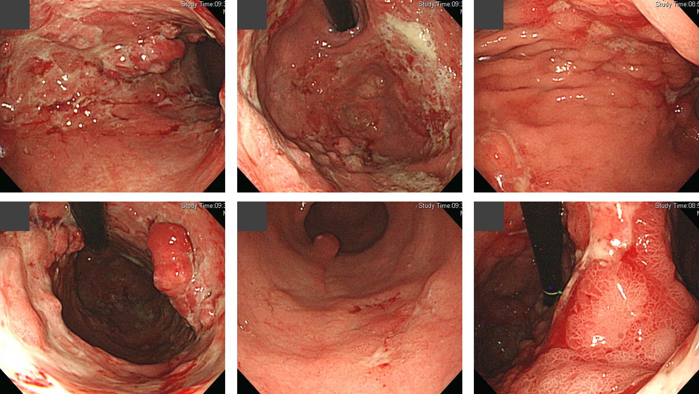
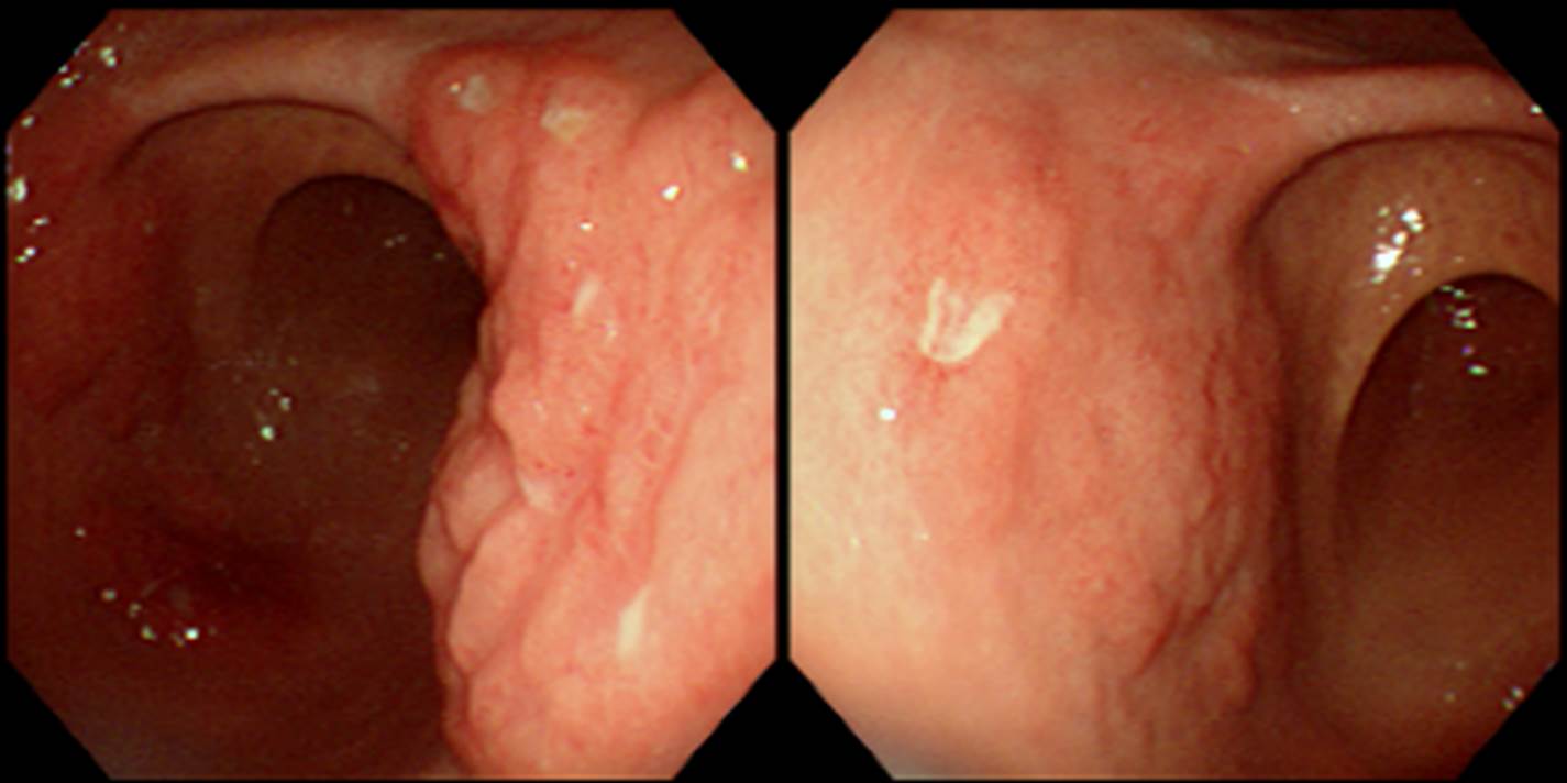
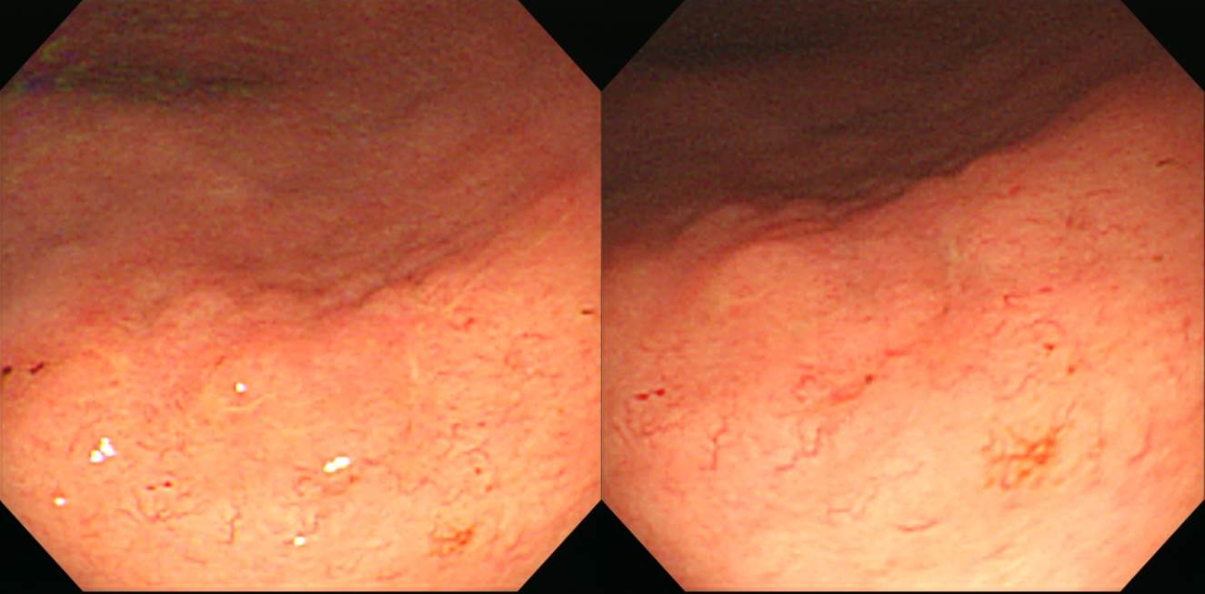
4. Endoscopic findings of PTCL of the stomach
5. Screening endoscopy에서 발견된 gastric PTCL
6. Rapidly growing stomach T cell lymphoma
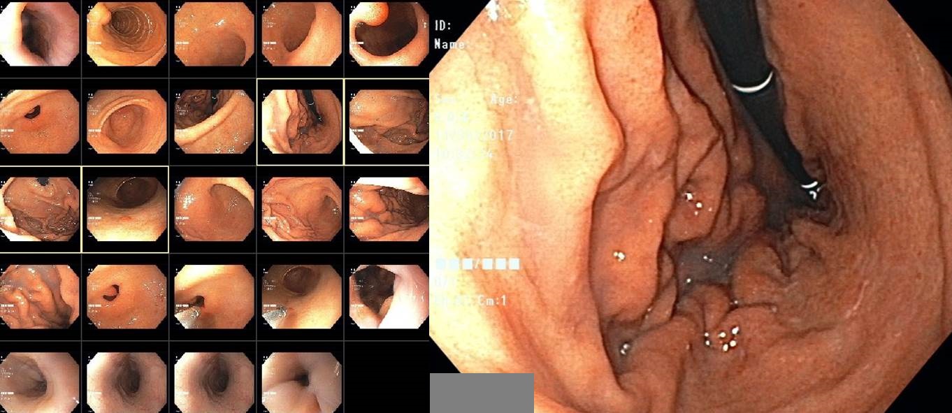
위내시경과 대장내시경을 동시에 받은 분입니다. 대장용종절제술을 하였고 deep SM invasion을 보여 수술적 치료를 위하여 의뢰되었습니다.
대장암에 대한 workup으로 시행한 CT에서 위벽 비후와 림프절 전이가 발견되어 첫 내시경 4주 후 다시 위내시경을 시행하였고 위주름 비후와 깊은 궤양이 발견되었습니다.
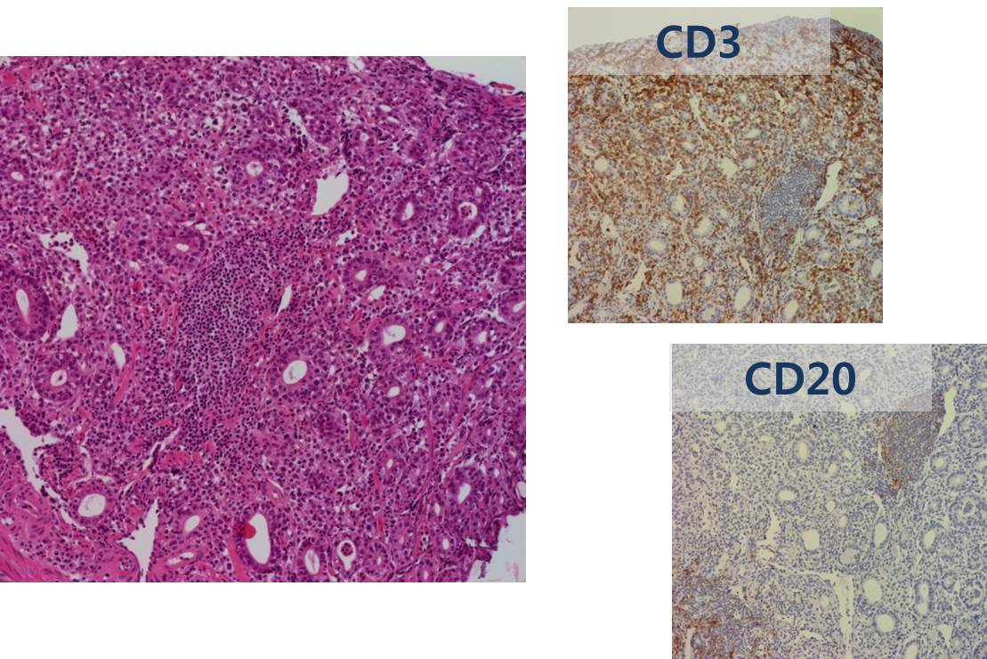
위내시경 조직검사에서 T cell lymphoma로 나왔습니다. 4주 간격의 내시경에서 큰 모양의 변화를 보였는데, 이는 매우 빨리 자랄 수 있다는 lymphoma의 특징 중 하나입니다. 사실 첫 내시경에서 약간의 위주름 비후가 있었던 것 같기도 합니다.
조직 진단: Peripheral T-cell lymphoma, not otherwise specified, CD 30-positive
. CD 30 : Positive in tumor cells (>80%)
. CD 4, (Leu-3a) : Positive in tumor cells
. β- F 1 : Positive in tumor cells
. TIA-1, T cell : Positive in tumor cells
. CD 8, (Leu-2a) : Negative in tumor cells
. CD 20, L26 : Negative in tumor cells
. Ki-67 : Positive in about 50% of tumor cells
. CD 3, T-CELL : Positive in tumor cells
. Epstein-Barr virus : Negative
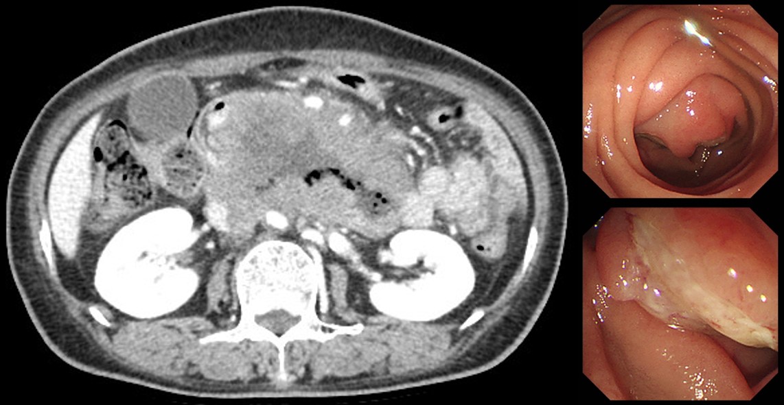
7. Duodenal PTCL
8. 소장 T 세포 림프종. Small intestinal T cell lymphoma
monomorphic epitheliotropic intestinal T-cell lymphoma. 장연구학회 집담회
monomorphic epitheliotropic intestinal T-cell lymphoma. 장연구학회 집담회
9. 대장 T 세포 림프종. Colon T cell lymphoma
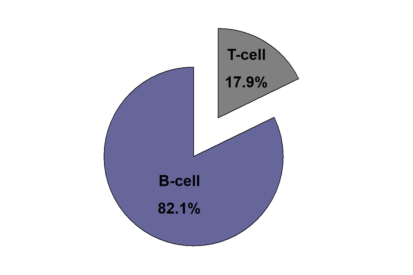
우리나라 대장 림프종의 약 15%는 T 세포 기원입니다. 서구에서는 매우 드문 질환이지만 우리나라에서는 가끔 볼 수 있습니다. Mass를 형성하지 않고 염증처럼 diffuse한 모양을 가지고 있어서 진단이 늦어지는 예가 많습니다.
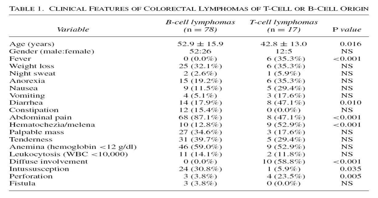
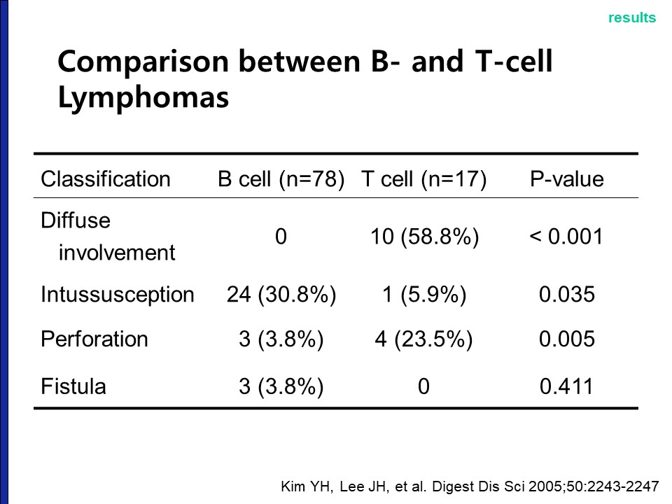
T cell lymphoma는 focal mass lesion보다는 diffuse한 점막 병변처럼 보이는 경우가 많습니다.
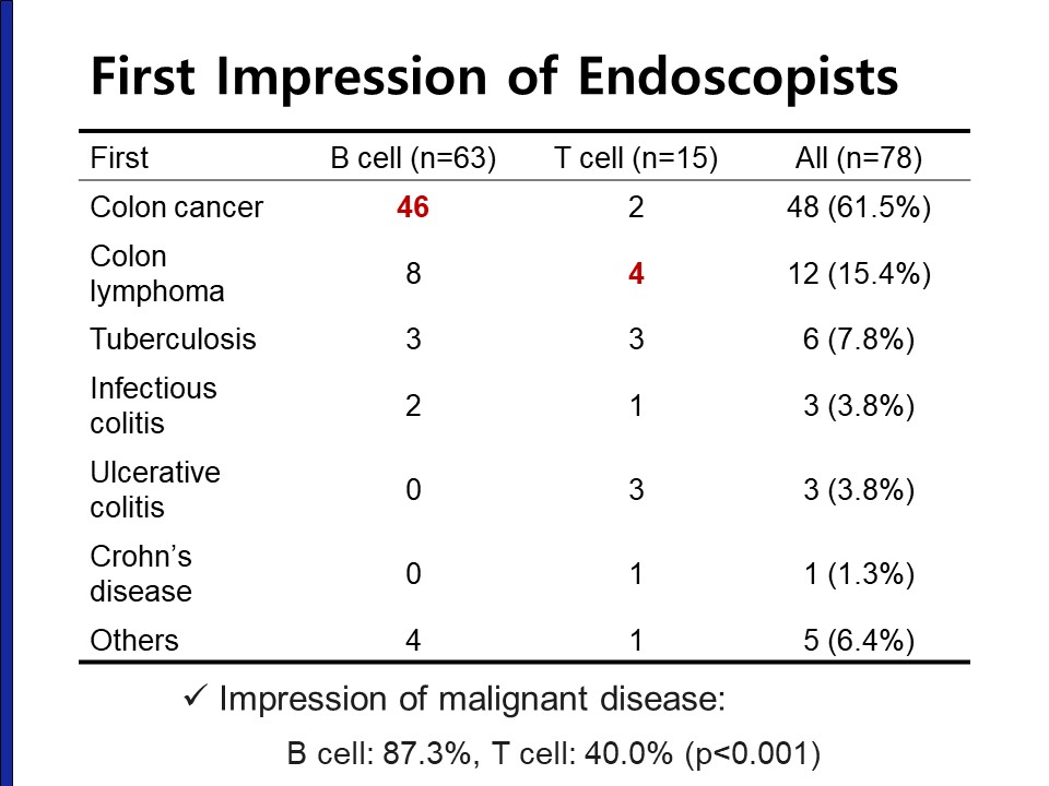
T cell lymphoma는 내시경 및 조직검사 후에도 림프종으로 진단되지 못하고 UC나 TB나 Crohn으로 치료받다가 뒤늦게 진단되는 경우가 있습니다. 진단 후에는 예후가 매우 나쁩니다.
10. Indolent type GI T cell lymphoproliferative disease
© 일원내시경교실 바른내시경연구소 이준행. EndoTODAY Endoscopy Learning Center. Lee Jun Haeng.