EndoTODAY 내시경 교실
Beginner | ESA | Schedule | OPD
Seminars | Atlas | Recent | Links
![]() [Delayed perforation] - 終
[Delayed perforation] - 終
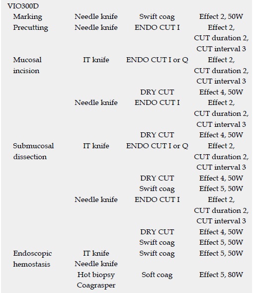
![]() 1. 2015년 동경암센터에서 ESD 후 지연 천공에 대한 자료를 발표하였습니다(Suzuki H. WJG 2015).
1. 2015년 동경암센터에서 ESD 후 지연 천공에 대한 자료를 발표하였습니다(Suzuki H. WJG 2015).
고주파 발생장치 setting은 일반적이었습니다.
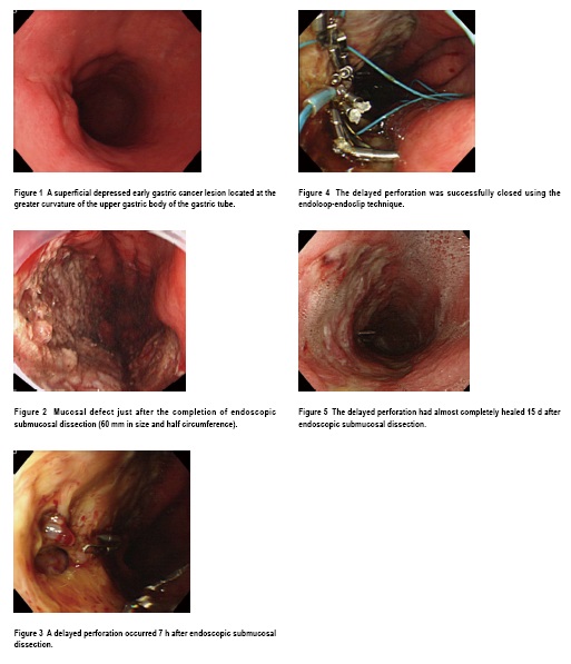
4,943명 중 7명에서 지연 천공이 발생하였는데 그 임상상은 아래와 같습니다. Gastric tube cancer가 둘 있었으며, 7명 중 3명은 응급수술을 했습니다.
Gastric tube ESD 후 지연 천공이 발생하여 endoloop-endoclip technique으로 치료한 예가 소개되었습니다.
![]() [Seminars/symposiums]
[Seminars/symposiums]
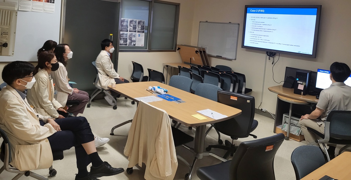
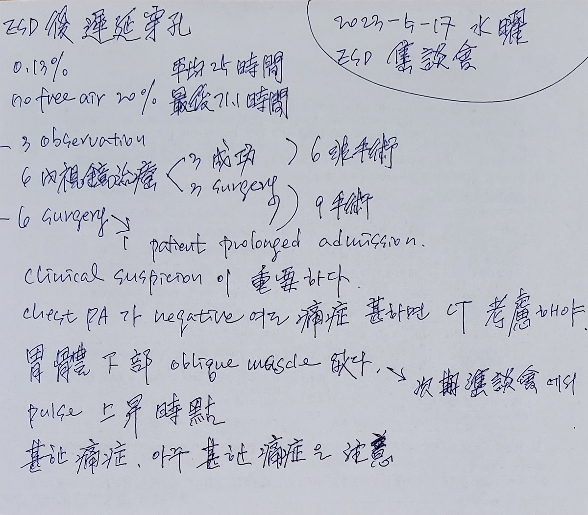
[2023-5-17] 수요 아침 ESD conference. Delayed perforation after ESD
![]() [Cases]
[Cases]
60대 여성입니다. ESD indication을 초과하는 환자인데 환자의 강력한 요구로 ESD가 시행되었음. 시술 직후 가슴 X-ray에서는 천공이 보이지 않았으나 증상이 지속되어 시행한 CT에서는 뚜렷한 천공이 있었음. 병리결과는 수술이 필요한 상황이었음.
Early gastric carcinoma
1. Location : high body, anterior wall
2. Gross type : EGC type IIa
3. Histologic type : tubular adenocarcinoma, moderately differentiated
4. Histologic type by Lauren : intestinal
5. Size of carcinoma : (1) longest diameter, 32 mm (2) vertical diameter, 25 mm
6. Depth of invasion : invades submucosa, (depth of sm invasion : 3000 ㎛) (pT1b)
7. Resection margin : involved deep margin by carcinoma with cauterized artifacts, safety margin : distal 6 mm, proximal 10 mm, anterior 6 mm, posterior 6 mm, deep 0 mm (sm only)
8. Lymphatic invasion : present (+++)
9. Venous invasion : present
10. Perineural invasion : present
11. Microscopic ulcer : absent
12. Histologic heterogeneity: absent
시술 직후 chest PA에는 문제가 없었는데 2일 후 복통이 있어 시행한 추적검사에서 free air가 관찰되었습니다. 다행스럽게 endoscopic clipping으로 호전되었습니다.
Early gastric carcinoma
1. Location : antrum, greater curvature
2. Gross type : EGC type IIc
3. Histologic type : tubular adenocarcinoma, well differentiated
4. Histologic type by Lauren : intestinal
5. Size of carcinoma : (1) longest diameter, 8 mm (2) vertical diameter, 4 mm
6. Depth of invasion : invades mucosa (lamina propria) (pT1a)
7. Resection margin : free from carcinoma(N), safety margin : distal 9 mm, proximal 12 mm, anterior 12 mm, posterior 12 mm, deep 200 ㎛
8. Lymphatic invasion : not identified(N)
9. Venous invasion : not identified(N)
10. Perineural invasion : not identified(N)
11. Microscopic ulcer : present
12. Histologic heterogeneity: absent
2016년 여자 70세. 위암 3개에 대한 ESD 후 다음 날 복통이 있어 천공 발견
![]() [References]
[References]
1) Delayed Perforation Occurring after Gastric Endoscopic Submucosal Dissection 김태세 Gut Liver 2024
© 일원내시경교실 바른내시경연구소 이준행. EndoTODAY Endoscopy Learning Center. Lee Jun Haeng