EndoTODAY ГЛНУАц БГНЧ
Beginner | ESA | Schedule | OPD
Seminars | Atlas | Recent | Links
![]() [Dignostic group classification before and after resection. Pathological discrepancy. Pathological upgrading] - №ћ
[Dignostic group classification before and after resection. Pathological discrepancy. Pathological upgrading] - №ћ
![]() 1. Diagnostic group classification (ЛяМКМПяКДПј 2016)
1. Diagnostic group classification (ЛяМКМПяКДПј 2016)
2012Гт ФЁЗс АсАњИІ КаМЎЧЯПЉ 'Diagnostic group classifications of gastric neoplasms by endoscopic resection criteria before and after treatment: real world experience'ЖѓДТ СІИёРИЗЮ КаМЎЧЯПДНРДЯДй (Lee JH. Surg Endosc 2016 / PDF). Absolute indication EGC, Expanded indication EGC, Beyond expanded indication EGC ЕюРЛ 'СјДмИэ'РЬЖѓАэ КйРЯ Мі ОјОюМ 'diagnostic group classification'РЬЖѓДТ ИЛРЛ ИИЕщОю Нс КИОвНРДЯДй.
Background and study aims: There are often discrepancies between the pretreatment evaluation of gastric neoplasms by endoscopy with biopsy and the final diagnosis of resected specimen in terms of pathology and depth of invasion. We evaluated the spectrum of discrepancies between pretreatment and posttreatment diagnosis which may deliver significant differences on clinical practice.
Patients and Methods: A total of 2,041 patients with gastric dysplasia or cancer who underwent curative endoscopic resections or surgeries in 2012 were enrolled. Patients were classified into five different diagnostic groups; low-grade dysplasia (LGD), high-grade dysplasia (HGD), absolute indication early gastric cancer (AI-EGC), beyond absolute indication early gastric cancer (BAI-EGC), and advanced gastric cancer (AGC). The choice of initial treatment and final pathologic diagnosis was analyzed.
Results: The study patients belonged to the following pretreatment diagnostic groups; LGDs in 162, HGDs in 164, AI-EGCs in 396, BAI-EGCs in 824, and AGCs in 495 cases. Posttreatment diagnostic groups were LGDs in 140, HGDs in 121, AI-EGCs in 322, BAI-EGCs in 947, AGCs in 505, and no residual tumor in 6 cases. In general, 6.9% (141/2,041) of cases were down-graded, and 15.9% (324/2,041) were up-graded. Thirty-four percent of pretreatment HGDs (56/164) were changed to cancers after endoscopic resection. Thirty-three percent of pretreatment AI-EGCs (131/396) were re-grouped as posttreatment BAI-EGCs. The additional surgery rate in each pretreatment group was 0.6% in LGD, 4.3% in HGD, 15.7% in AI-EGC, 23.6% in BAI-EGC among the patients with initial endoscopic resection (p < 0.01).
Conclusions: Twenty-three percent of gastric neoplasms changed in their final diagnostic group after endoscopic resection or surgery. This discrepancy should be considered when the initial treatment strategy is being selected.
ПьИЎГЊЖѓПЁМ ESD ПЕПЊРК ЙЋУД ШЅЖѕНКЗДНРДЯДй. НУМњ РќШФ КДИЎ АсАњАЁ ЙйВюДТ ПЙАЁ ГЪЙЋ ИЙБт ЖЇЙЎРдДЯДй. РЯКЛРК СЖБнИИ РЬЛѓЧЯИщ Дй ОЯРИЗЮ СјДмРЛ КйПЉЙіИЎЙЧЗЮ ESD НУЧр ШЏРкРЧ ДыКЮКаРЬ УГРНКЮХЭ РЇОЯРдДЯДй. БзЗБЕЅ ПьИЎГЊЖѓПЁМДТ ЛѓДчМіАЁ НУМњ Рќ adenoma, НУМњ ШФ adenocarcinomaРдДЯДй. РЬ КЮКаРЛ frank ЧЯАд КИАэЧб ГэЙЎРЬ ОјОюМ ИЖРН ИдАэ ЧбЙј СЄИЎЧб АЭРдДЯДй.
РќУМРћРИЗЮ 6.9% (141/2,041)АЁ down-grade ЕЧАэ 15.9% (324/2,041)АЁ up-grade ЕЧОњНРДЯДй. Diagnostic group classificationРЬ БзЗИАд ЙйВюОњДйДТ РЧЙЬРдДЯДй.
Absolute indicationРИЗЮ ЦЧДмЕШ ШЏРкРЧ 89.6%АЁ УЙ ФЁЗсЗЮ ESDАЁ МБХУЕЧАэ РжНРДЯДй.
РЬ АсАњИІ ЙйХСРИЗЮ ESD ШФ МіМњРЬ ЧЪПфЧв ШЎЗќРЬ 15%ЖѓАэ МГИэЧЯАэ РжНРДЯДй.
ФЁЗс Рќ КаЗљПЁ ЕћЖѓ АсАњИІ КИПЉСжДТ АЭАњ ФЁЗс ШФ КаЗљПЁ ЕћЖѓ АсАњИІ КИПЉСжДТ АЭРК ИХПь ХЋ ТїРЬАЁ РжНРДЯДй. Expanded indictionРЧ АцПь Бз ТїРЬАЁ АЁРх ЧіРњЧеДЯДй.
АЂ diagnostic groupПЁ ДыЧЯПЉ РЬПЭ КёНСЧб diagramРЛ ИИЕщОюКИИщ РчЙЬРжРЛ АЭ ААНРДЯДй. Real worldПЁМДТ РЬЗИАд КЙРтЧб РЯРЬ ЙњОюСіАэ РжДТ АЭРдДЯДй.
РЇ ЕЕЧЅПЁМ ESD юё absolute indication EGCЗЮ ЦЧДмЕЧОњРИГЊ УжСОРћРИЗЮ AGCЗЮ ГЊПТ ШЏРкАЁ 1Иэ РжОњНРДЯДй. ИХПь ЕхЙЎ АцПьПДБтПЁ МвАГЧеДЯДй. ESDАЁ НУЕЕЕЧОњДТЕЅ submucosal adhesionРИЗЮ ESDИІ ИЖФЅ Мі ОјОњАэ МіМњРЛ НУЧрЧЯПЉ AGCЗЮ ШЎРЮЕШ ШЏРкПДНРДЯДй. ESDЗЮ РЮЧб РЮАјБЫОчЖЇЙЎПЁ КИИИ 3ЧќРИЗЮ КаЗљЕЧОњСіИИ ГЛНУАцРћРИЗЮДТ EGC-like AGC Ся Borrmann type unclassifiedАЁ АЁРх РћЧеЧб КаЗљЖѓАэ Л§АЂЧеДЯДй.
Stomach, total gastrectomy:
Status post endoscopic submucosal dissection (incomplete)
Advanced gastric carcinoma
1. Location : upper third, Center at body and lesser curvature
2. Gross type : Borrmann type 3
3. Histologic type : tubular adenocarcinoma, moderately differentiated
4. Histologic type by Lauren : intestinal
5. Size : 3x1.5 cm
6. Depth of invasion : invades muscularis propria (pT2)
7. Resection margin: free from carcinoma, safety margin: proximal 2 cm, distal 11.4 cm
8. Lymph node metastasis : no metastasis in 38 regional lymph nodes (pN0)
9. Lymphatic invasion : present
10. Venous invasion : not identified
11. Perineural invasion : present
12. Peritoneal cytology : negative
[ТќАэ РкЗс]
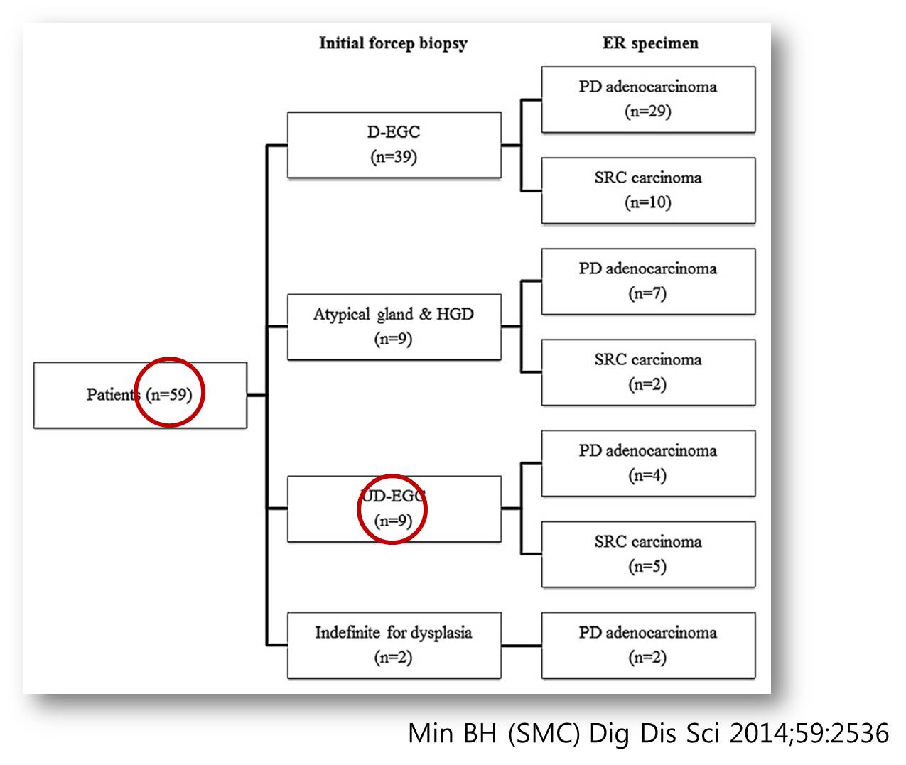
1) 2014Гт ЛяМКМПяКДПј ГэЙЎРдДЯДй (Dig Dis Sci 2014). Ор 9ГтАЃ 2,194ИэРЧ ESDИІ ФЁЗсЧпДТЕЅ Бз Сп ФЁЗс ШФ undifferentiated-typeРЬ 59ПЙ(2.7%)ПДНРДЯДй. Undifferentiated-type 59ПЙ Сп 50ПЙ(84.7%)АЁ ФЁЗс РќПЁДТ differentiated-type, atypical gland, indefinite for dysplasiaПДНРДЯДй. ФЁЗс Рќ СЖСїАЫЛчПЁМЕЕ undifferentiated-typeРИЗЮ ГЊПТ АцПьДТ 9ПЙ (15.2%)ПЁ КвАњЧЯПДНРДЯДй. Undifferentited-typeРЛ ESDЧЯДТ АцПьАЁ АХРЧ ОјДТ КДПјПЁМ КИРЬДТ АцЧтРЛ ДыЧЅЧбДйАэ Чв Мі РжНРДЯДй (ТќАэ: 2014Гт РЬШФКЮХЭДТ РлРК undifferentiated typeПЁ ЧбЧЯПЉ СОСО ESDИІ ЧЯАэ РжНРДЯДй). Posttreatment КаМЎПЁМДТ undifferentiated-typeРЬЖѓЕЕ pretreatment КаМЎРЛ ЧиКИИщ differentiated-typeРЬ РћСі ОЪДйДТ СЁРЛ АСЖЧЯПДНРДЯДй.
2) 2014Гт ПЌММДыЧаБГ АГВММКъЖѕНККДПј ГэЙЎРдДЯДй (Pathol Res Pract 2014). Ор 9Гт АЃ СЖБтРЇОЯ 289ПЙИІ ФЁЗсЧпДТЕЅ Бз Сп ФЁЗс ШФ undifferentiated-typeРЬ 38ПЙ(13.1%)ПДНРДЯДй. ФЁЗс ШФ undifferentiated-type 38ПЙ Сп 7ПЙ(18.4%)АЁ ФЁЗс РќПЁДТ differentiated-typeРЬОњНРДЯДй. Undifferentited-typeПЁ ДыЧб ESDПЁ ДыЧЯПЉ КёБГРћ РћБиРћРЮ mindИІ АЁСј КДПјРЧ АцЧтРЛ ДыЧЅЧЯДйАэ Чв Мі РжНРДЯДй.
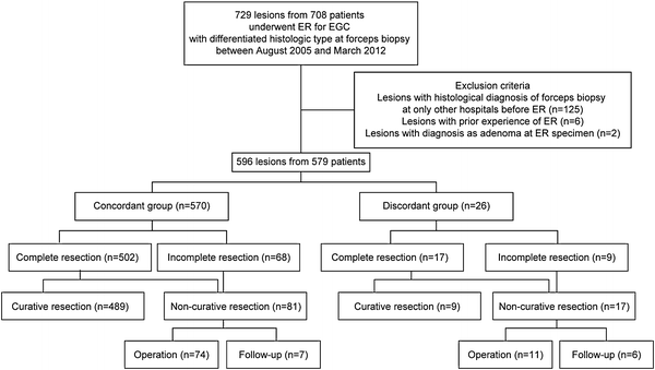
3) 2014Гт ПЌММДыЧаБГ НХУЬММКъЖѕНККДПј ГэЙЎРдДЯДй (Surg Endosc). Ор 7Гт АЃ ФЁЗс Рќ СЖСїАЫЛч differentiated-type 596КДМвИІ ФЁЗсЧпДТЕЅ, Бз Сп ФЁЗс ШФ undifferentiated-typeРИЗЮ ЙйВя АцПьАЁ 26ПЙ (4.3%)ПДНРДЯДй. ААРК БтАЃ ФЁЗс Рќ СЖСїАЫЛч undifferentiated-typeРК Ию ПЙАЁ РжОњРКСіДТ ЙрШїСі ОЪОвНРДЯДй.
4) 2016Гт МПяДыЧаБГКДПј ГэЙЎРдДЯДй (Choi JM. Surg Endosc 2016). Biopsy-proven differentiated type EGC 1,641 КДМвИІ ESD ЧЯПДРЛ ЖЇ 5.2% (85 КДМв)АЁ undifferentiatedЗЮ ЙйВюОњНРДЯДй. АќЗУЕШ РЮРкДТ female sex, age < 65 years, large endoscopic size, depressed morphology, surface nodularity, whitish discoloration ПДНРДЯДй. ААРК БтАЃ ФЁЗс Рќ СЖСїАЫЛч undifferentiated-typeРК Ию ПЙАЁ РжОњРКСіДТ ЙрШїСі ОЪОвНРДЯДй.
Undifferentiated type histologyЗЮ ЙйВя 85ПЙРЧ curative resection rateДТ 36.5%(31/85)ПДНРДЯДй. КёЗЯ undifferentiate type histologyЖѓАэ ЧЯДѕЖѓЕЕ УГРНПЁ differentiate typeРЬОњАэ ESD РћРРСѕПЁ МгЧпДйИщ curative resectionРЬ ГЊПдРЛ ЖЇПЁДТ ПЙШФАЁ ССДйДТ АЭРЛ ОЫ Мі РжНРДЯДй.
5) 2015Гт 11Пљ Gastric CancerСіПЁ NECA ПЌБИРЧ КДИЎ part АсАњАЁ E-pubРИЗЮ ЙпЧЅЕЧОњНРДЯДй (Kim JM. GC 2015 - Epub). ЙЎЕц ПО РЯРЬ Л§АЂГЕНРДЯДй. NECA ПЌБИРЧ БтШЙ ДмАшПЁ ТќПЉЧпДйАЁ СпАЃПЁ КќСЎМ ПЉЗЏ МБЛ§ДдВВ НЩЧЯАд ВйСпРЛ ЕщОњДј ОЦЧТ БтОяРЬ РжНРДЯДй.
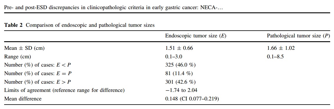
РЬЙј ПЌБИПЁМ beyond expanded indication СѕЗЪАЁ 20.1%ПДНРДЯДй. ДйМв ГєРК ЦэРЬ ОЦДв Мі ОјНРДЯДй. ДйБтАќ ПЌБИПДДј АЭАњ АќЗУЕШ АЭРИЗЮ УпСЄЕЫДЯДй.
ПЉЗЏ КЏМіАЁ КаМЎЕЧОњДТЕЅ СІ АќНЩРЛ Ві АЭРК ХЉБт ТїРЬПДНРДЯДй. Л§АЂКИДй ЦэТїАЁ РлОвНРДЯДй. ГЛНУАцРИЗЮ УјСЄЧб ХЉБтПЭ КДИЎ ХЉБтРЧ ЦђБе ТїРЬАЁ 1.5 mm ЙлПЁ ЕЧСі ОЪОвРИДЯБюПф. 2 cmЗЮ Л§АЂЧЯАэ НУМњЧпДТЕЅ 8.5cmАЁ ГЊПТ АЭЕЕ РжСіИИ...
![]() 2. Diagnostic group classification at National Cancer Center (БЙИГОЯМОХЭ 2016)
2. Diagnostic group classification at National Cancer Center (БЙИГОЯМОХЭ 2016)
Diagnostic group classificationРЬЖѓДТ ПыОюИІ ЛчПыЧЯСіДТ ОЪОвРИГЊ БЙИГОЯМОХЭПЁМЕЕ ESD РќШФ КДИЎ СјДмРЧ ТїРЬПЁ ДыЧб ССРК РкЗсИІ ЙпЧЅЧЯПДНРДЯДй.
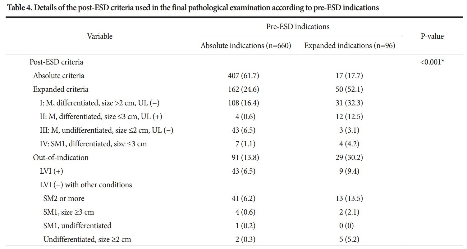
ESD Рќ absolute indicationРИЗЮ ЦђАЁЧЯПДДј ШЏРкРЧ 13.8%АЁ out-of-indicationРИЗЮ, Ся МіМњРЬ ЧЪПфЧб АЭРИЗЮ ГЊПдАэ, ESD Рќ expanded indicationРИЗЮ ЦђАЁЧЯПДДј ШЏРкРЧ 30.2%АЁ out-of-indicationРИЗЮ ГЊПдДйДТ РЬОпБтРдДЯДй.
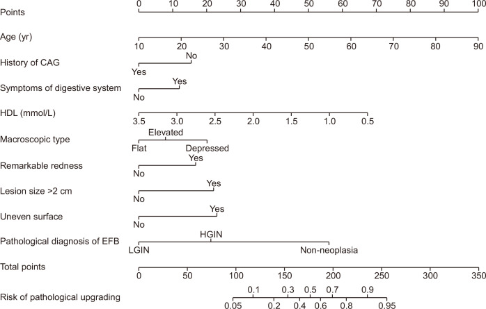
![]() 3. A prediction model for pathological upgrading (Nanjing Medical University 2023)
3. A prediction model for pathological upgrading (Nanjing Medical University 2023)
![]() [FAQ]
[FAQ]
[2017-12-21]
КДИЎАњПЭ ШИРЧИІ ЧпНРДЯДй. ESD ШФ Р§СІЧЅКЛПЁ ДыЧб КДИЎ МіАЁПЁ ДыЧЯПЉ СЄКЮ АэНУАЁ РжОњДйАэ ЧеДЯДй. ESD ЧѲ ЧЅКЛРЛ ОЧМК СњШЏАњ ОчМК СњШЏПЁ ЕћЖѓ ФкЕхИІ ДоИЎЧЯАкДйДТ ГЛПыРЬОњНРДЯДй. ESD ШФ ОЯРЮСі ОЦДбСіДТ АсАњАЁ ГЊПЭ КСОп ОЦДТ АЭРЮЕЅ ЖЧ ESD Рќ НУМњПЁ ЕћЖѓ АЁАнРЛ ДоИЎЧЯДТ РЬЛѓЧб СЄУЅРЬ ГЊПдНРДЯДй. О№СІБюСі РЬЗБ РЬЛѓЧб СЄУЅРЛ АшМг ИИЕщ АЭРЮСі ЧбНЩЧЯБт БзСі ОјНРДЯДй.

![]() [References]
[References]
1)
© РЯПјГЛНУАцБГНЧ ЙйИЅГЛНУАцПЌБИМв РЬСиЧр. EndoTODAY Endoscopy Learning Center. Lee Jun Haeng.