EndoTODAY 내시경 교실
Beginner | ESA | Schedule | OPD
Seminars | Atlas | Recent | Links
![]() [Glomus tumor. 사구종양] - 終
[Glomus tumor. 사구종양] - 終
![]() 1. Introduction to glomus tumor
1. Introduction to glomus tumor
Miettinen M. Am J Surg Oncol 2002를 요약합니다.
Mesenchymal tumor composed of modified smooth muscle cells representing a neoplastic counterpart of the perivascular glomus bodies
Most commonly occur in the peripheral soft tissues, especially in the distal parts of extremities
In adults of all ages
Commonly in the gastric antrum
Symptoms: GI bleeding, ulcer-like symptoms
A small possibility of malignant behavior cannot be ruled out. (tumor size > 5 cm, nuclear atypia)
DDx: GIST, carcinoid tumor, paraganglioma, hemangiopericytoma, lymphomaGlomus tumors usually occur in the peripheral soft tissues, but similar tumors have also been reported in the stomach and occasionally in the intestines. However, the relationship of these tumors to peripheral glomus tumors and gastrointestinal stromal tumors has not been fully clarified because previous series of gastrointestinal glomus tumors predate availability of immunohistochemistry. This clinicopathologic study examined 32 gastrointestinal glomus tumors. All but one of the tumors were located in the stomach and the remaining tumor was from the cecum. The tumors occurred with a strong female predominance (23 females and 9 males) and a median age of 55 years (range 19-90 years). The gastric tumors typically presented with gastrointestinal bleeding or ulcer-like symptoms, and 14 tumors had mucosal ulceration. Five tumors were incidental findings. The tumor sizes varied from 1.1 to 7 cm (median 2 cm), and most were located in the antrum. Histologically, the tumors typically had a solid pattern of sharply demarcated, round glomus cells with prominent, mildly dilated pericytoma-like vessels. Vascular invasion and focal atypia were relatively common (seen in 11 and 13 cases, respectively), and low mitotic activity (1-4 per 50 high power fields), was seen in 10 cases. Immunohistochemically, all tumors were positive for alpha-smooth muscle actin and calponin, and nearly all had a net-like pericellular laminin and collagen type IV positivity. All tumors were negative for desmin and S-100 protein. Three tumors had focal synaptophysin positivity, but none was positive for chromogranin. All tumors lacked KIT expression and the GIST-specific mutations in the c-kit gene. Follow-up revealed one patient death of metastatic disease to liver at 50 months; this tumor had 1 mitosis per 50 high power fields, but had spindle cell foci, mild atypia, and vascular invasion. Thirteen patients were well and alive after long-term follow-up. Gastrointestinal glomus tumors occur almost exclusively in the stomach, and they have a good overall prognosis, but a small, unpredictable potential for malignant behavior exists. These tumors are phenotypically similar to peripheral glomus tumors and differ from epithelioid GISTs.
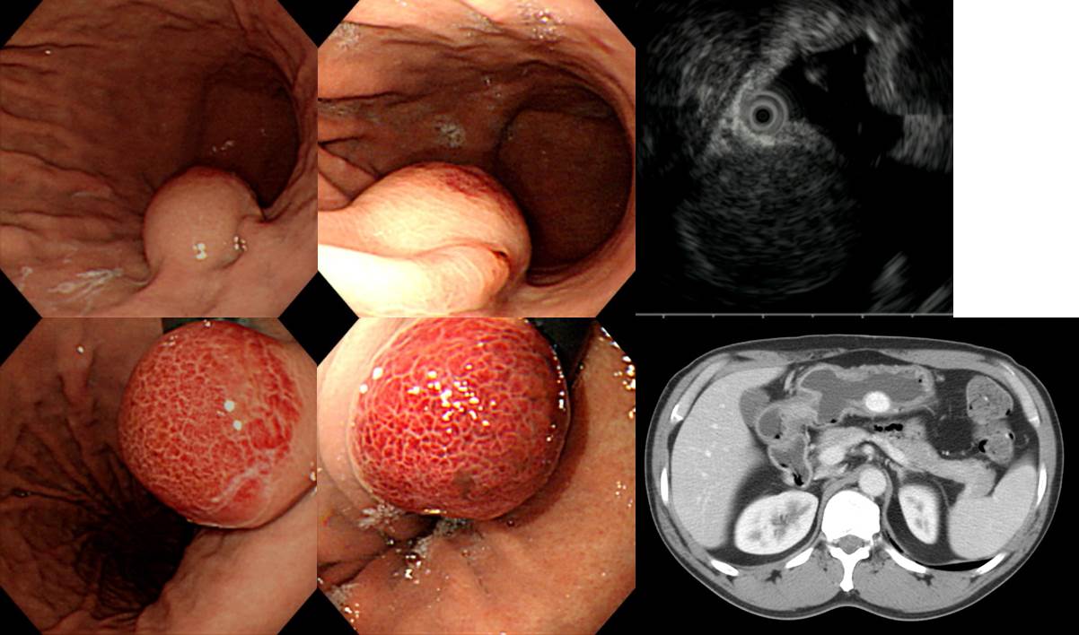
![]() 2. Gastric glomus tumor
2. Gastric glomus tumor
전형적인 gastric glomus tumor입니다. CT에서 enhancement가 아주 잘 됩니다. Wedge resection으로 치료하였고 조직과 면역 염색에서 VT(vimentin) 과 SMA(smooth muscle actin) 이 양성인 소견이고 감별진단에서 다른 혈관 기원 종양과의 감별은 CD31(-), Factor VIII(-) 로 확인했습니다.
. c-kit: Negative
. CD31: Negative
. Factor VIII: Negative
![]() 3. Gastric glomus tumor (mimicking gastric neuroendocrine carcinoma)
3. Gastric glomus tumor (mimicking gastric neuroendocrine carcinoma)
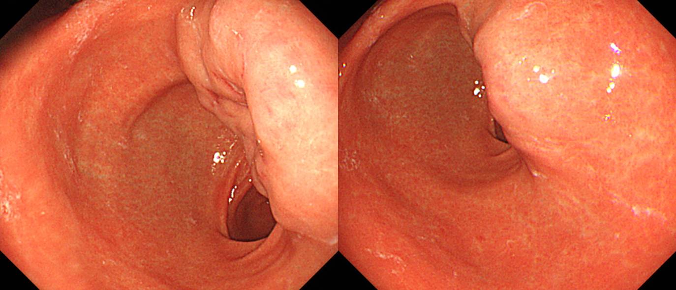
Stomach, subtotal gastrectomy:
Glomus tumor of unknown malignant potential
1) tumor site: antrum and posterior wall
2) tumor size: 3x2 cm
3) mitosis: 0/10 HPFs
4) necrosis: absent
5) cellularity: intermediate
6) cellular atypia: mild
7) invasion into mucosa and serosa
8) infiltrative growth
9) lymphovascular invasion: not identified
10) perineural invasion: present
11) resection margin: negative (safety margin: proximal, 4 cm; distal, 5 cm)
12) no metastasis in 19 regional lymph nodesChromogranin : Negative in tumor cells
Synaptophysin : Positive in tumor cells
Ki-67 : Positive in 5 % of tumor cells
SMA: Diffusely positive in tumor cells
CD 31 : No tumor emboli
D2-40 Podoplanin : No tumor emboli
c-erbB-2(HER2): Negative
Epstein-Barr virus : Negative
병리과에서 처음에는 neuroendocrine carcinoma를 고려하였으나 mitotic index가 너무 낮은 점이 이상하여 여러 추가검사를 한 후 최종적으로 glomus로 진단한 흥미로운 예입니다.
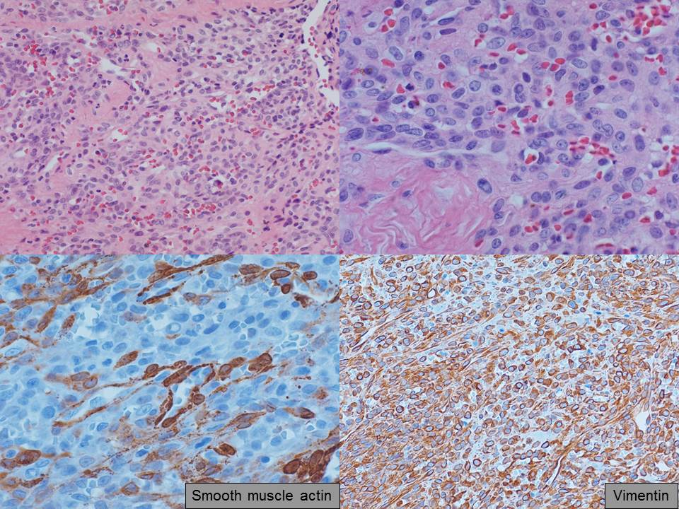
![]() 4. Pathology of glomus tumor
4. Pathology of glomus tumor
YouTube
© 일원내시경교실 바른내시경연구소 이준행. EndoTODAY Endoscopy Learning Center. Lee Jun Haeng.