 EndoTODAY 내시경 교실
Beginner | ESA | Schedule | OPD
Seminars | Atlas | Recent | Links
![]() [Pentax Imagina Virtual Atlas (PIVA) project] - End of document
[Pentax Imagina Virtual Atlas (PIVA) project] - End of document
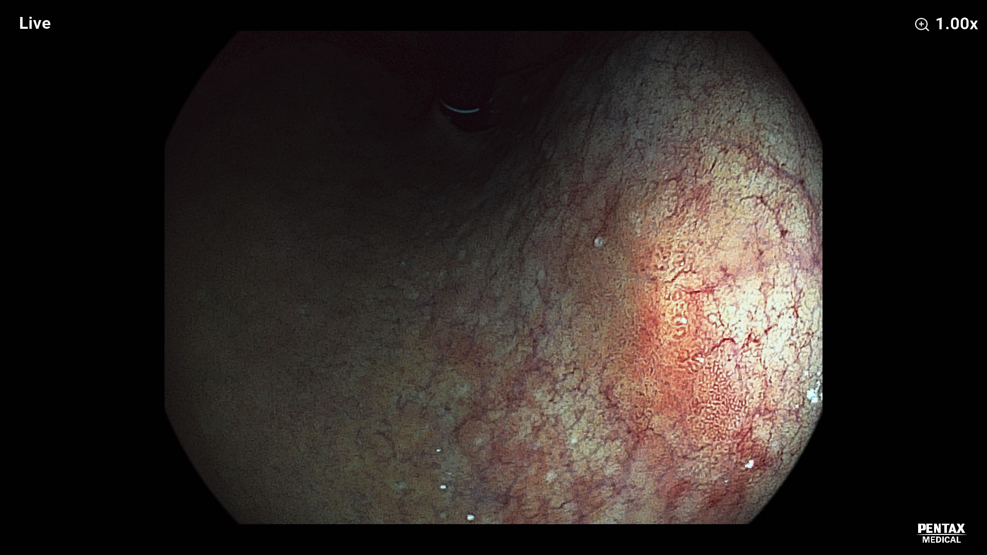
Case 1. Advanced gastric cancer, Borrmann type IV
Sex/age: F/49
Symptoms and signs: early fullness, weight loss
Endoscopic images:
Video clips:
Diagnosis and treatment: The endoscopic forceps biopsy result was poorly cohesive carcinoma. Abdominal CT images showed diffuse wall thickening mass of the stomach upper body and fundus with mild perigastric infiltation. The preoperative diagnosis was advanced gastric cancer, Borrmann type IV. Surgical consultation was done for exploratory laparotomy. However, there were multiple seeding nodules were found in the laparoscopy. Palliative chemotherapy was done.
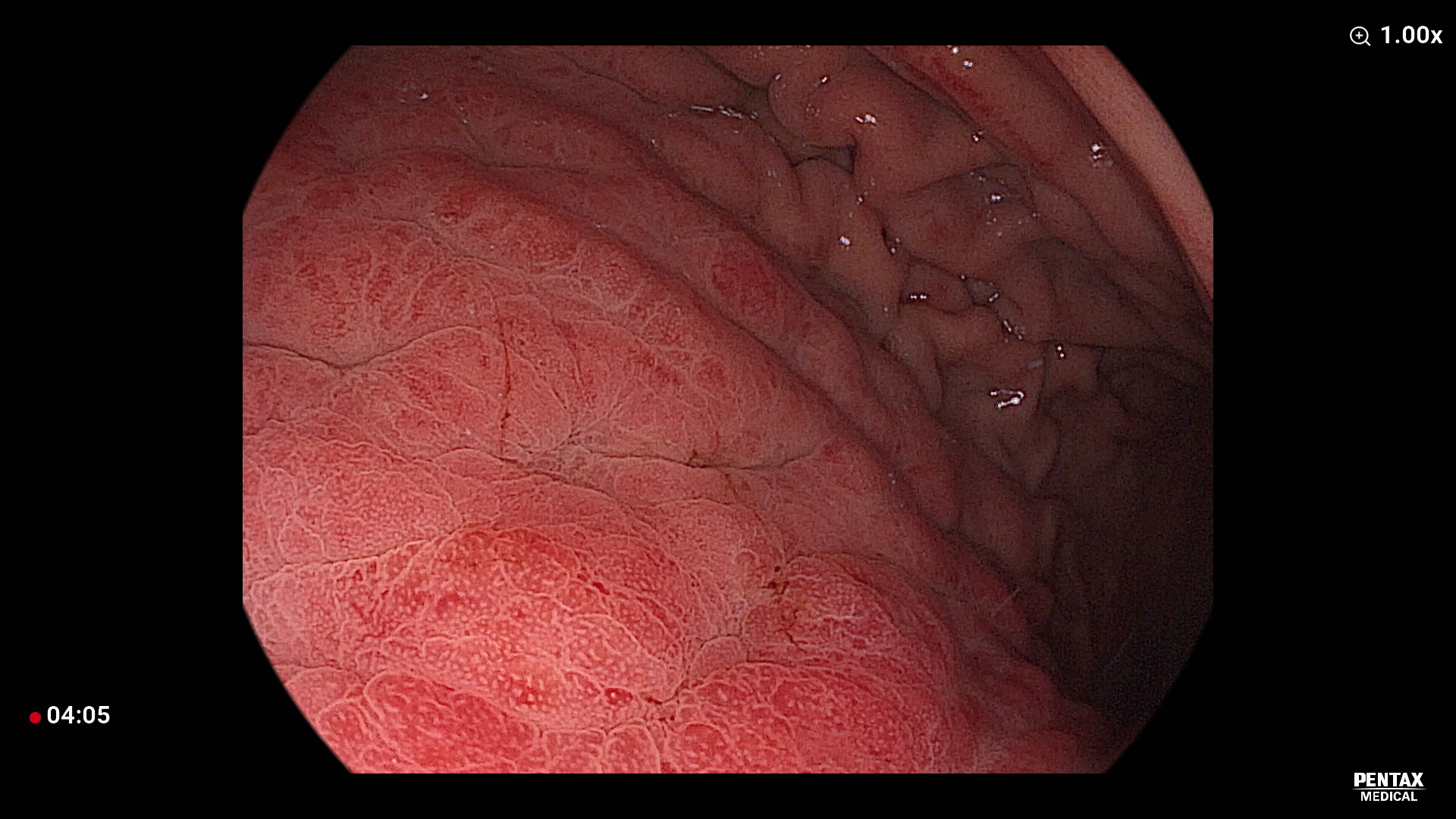
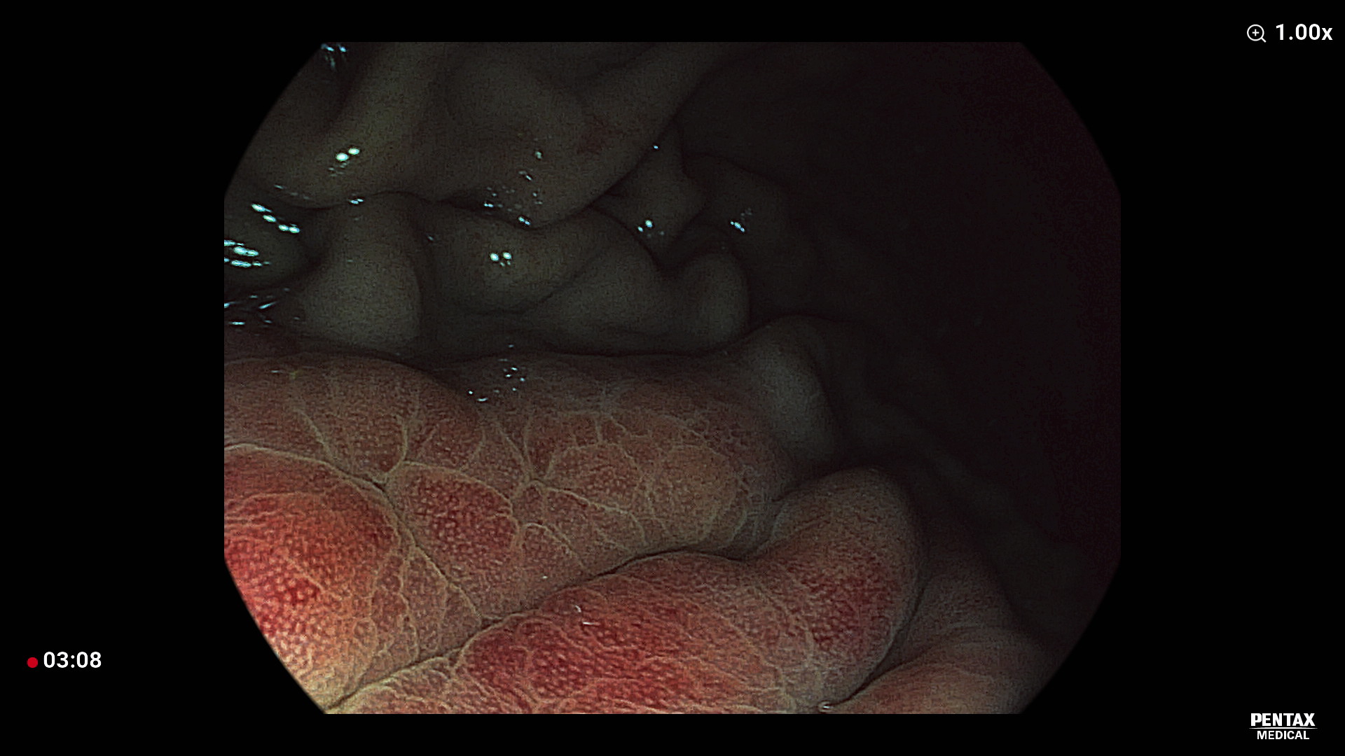
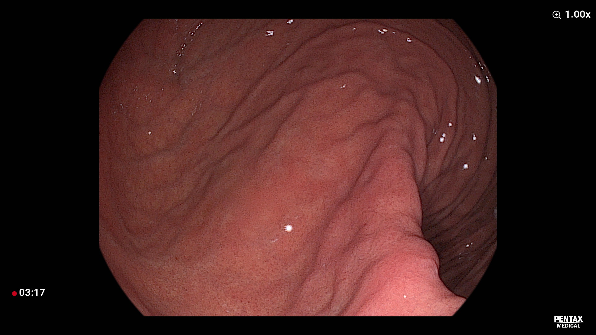
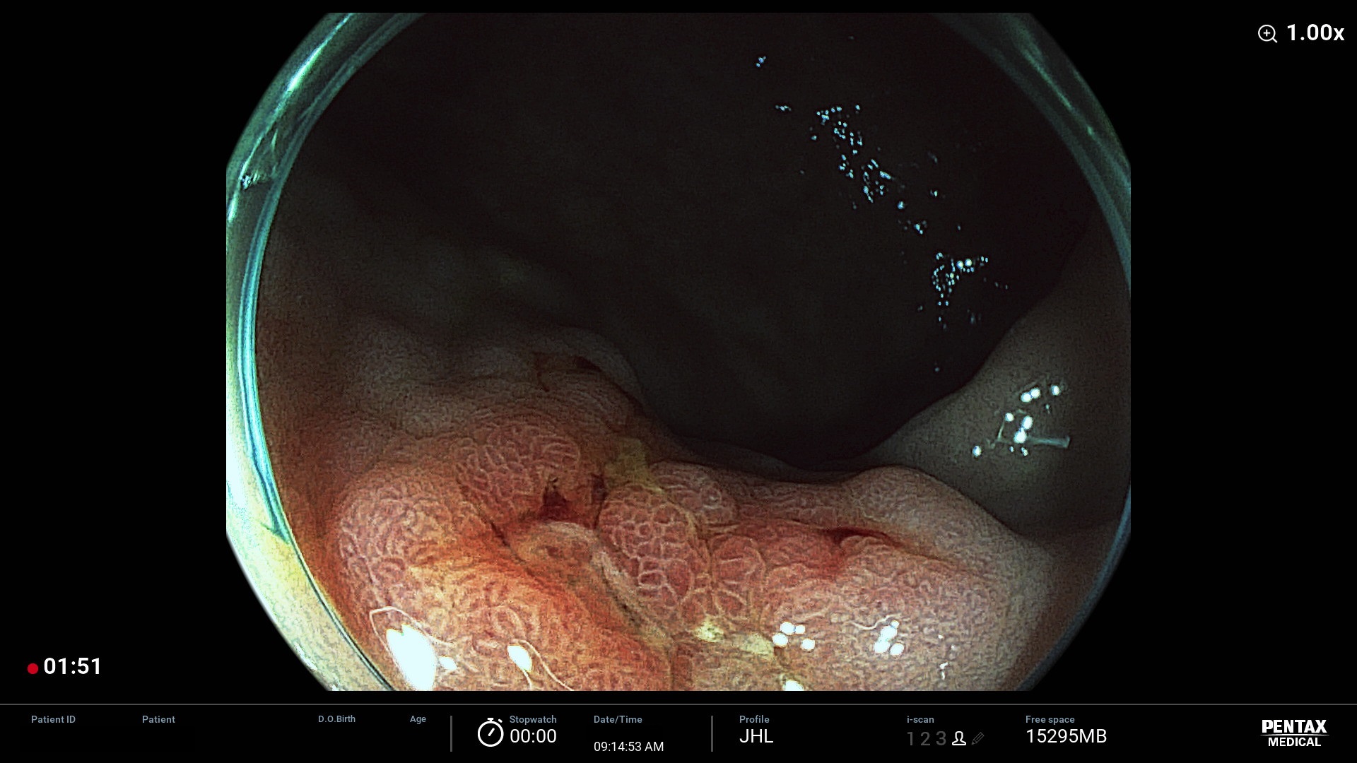
Case 2. Cascade stomach
Sex/age: F/48
Symptoms and signs: no (screening endoscopy)
Endoscopic images:
Video clips:
Diagnosis and treatment: When the endoscope was introduced into the stomach, the lumen of the gastric body was seen at the upper right corner. After infusion of some air into the stomach, a ridge between the gastric fundus and upper body can be seen. The ridge runs from the posterior wall of the cardia toward the anterior wall of the stomach, crossing the greater curvature. When the stomach has a prominent ridge between the fundus and high body, it is called as the cascade stomach (CS). The clinical significance of CS is still uncertain.
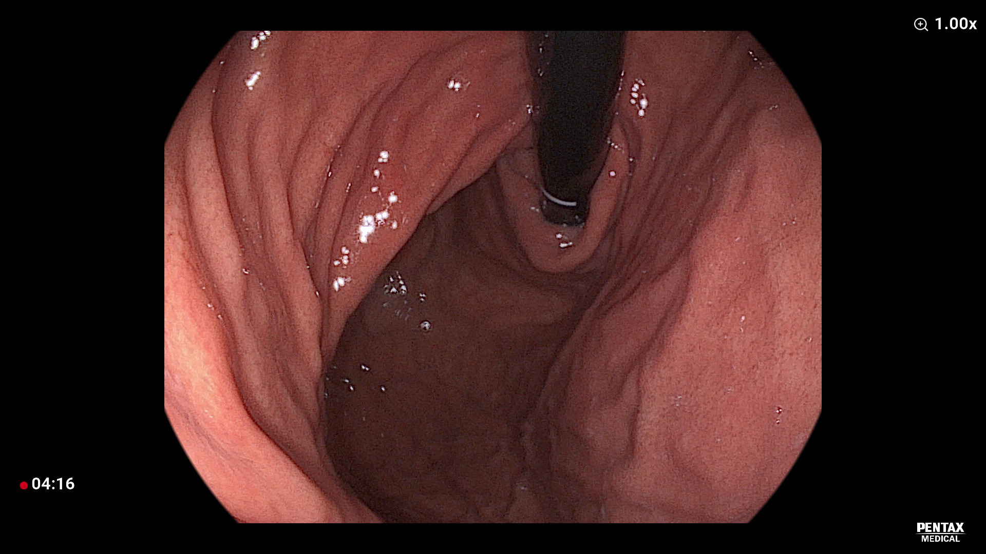
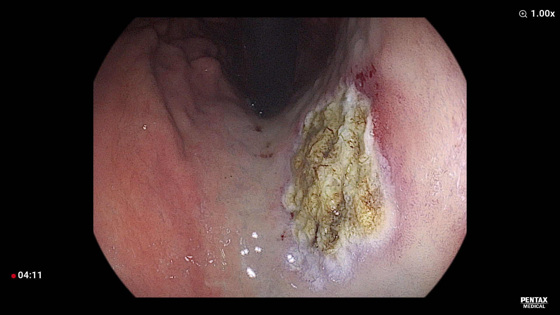
Case 3. APC ablation for adenoma
Sex/age: M/68
Symptoms and signs: no (referred for the treatment of incidental gastric adenoma found in the screening endoscopy)
Endoscopic images:
Video clips:
Diagnosis and treatment: In the screening endoscopy, about 1.5cm sized ill-defined mucosal irregularity was found in the lesser curvature of the midbody. Background gastric mucosal was atrophic. The biopsy showed adenoma with low grade dysplasia. Ablation treatment using argon plasma coagulation apparatus was done. After the treatment, PPI was given for two weeks.
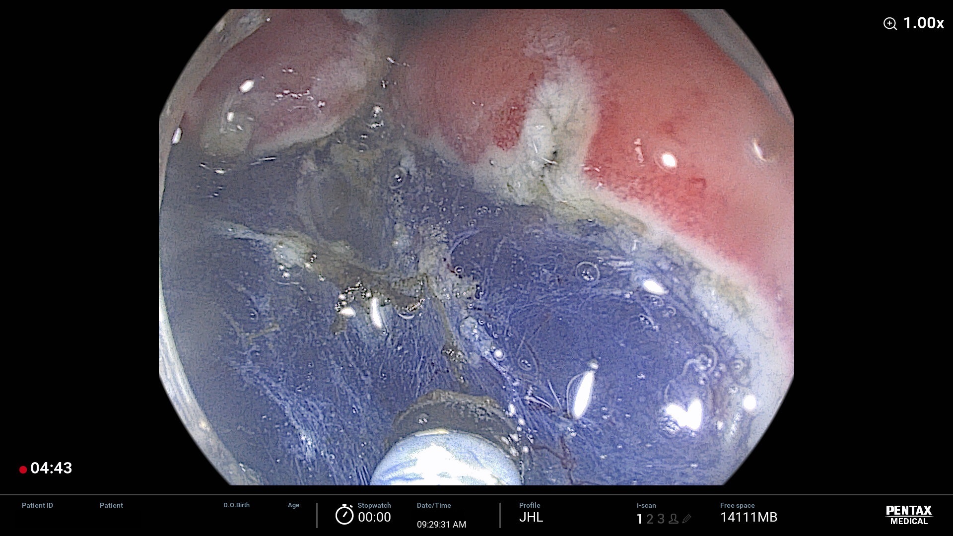
Case 10. ESD for EGC using Pentax M knife only
Sex/age: M/62
Symptoms and signs: A 62 years old male patient visited my clinic due to a small gastric lesion found in the screening endoscopy. Outside pathology report was 'tubular adenoma with high grade dysplasia, suspicious of adenocarcinoma'. Outside pathology slide was reviewed by an in-house pathologist of my hospital and the final pathology was 'adenocarcinoma, moderatly differentiated'. The CT scan was negative for metastasis. ESD was done with Pentax Splash M knife.
- System: Pentax EPK-i5500c Imagina
- Endoscope: Pentax EC29-i10c gastroscope
- Injector: Finemedix 23G-0.1T-1800mm
- ESD knife: Pentax Splash M knife
- ESU: ERBE VIO 300D (mainly EndoCut I 3-3-3)
- Bleeding control: Pentax Splash M knife (Hemostatic forceps was not used.)
- Sedation: Pethidine 25mg + Midazolam 5 mg IV
Endoscopic images:
Video clips:
Diagnosis and treatment: Final ESD pathology was as followings;
Stomach, GC of proximal antrum, ESD: Early gastric carcinoma
1. Location : antrum, greater curvature
2. Gross type : EGC type IIc+IIa
3. Histologic type : tubular adenocarcinoma, moderately differentiated
4. Histologic type by Lauren : intestinal
5. Size of carcinoma : (1) longest diameter, 11 mm (2) vertical diameter, 10 mm
6. Depth of invasion : invades mucosa (muscularis mucosa) (pT1a)
7. Resection margin : free from carcinoma(N); safety margin : distal 14 mm, proximal 10 mm, anterior 10 mm, posterior 10 mm, deep 200 ㎛
8. Lymphatic invasion : not identified(N)
9. Venous invasion : not identified(N)
10. Perineural invasion : not identified(N)
11. Microscopic ulcer : absent
12. Histologic heterogeneity: absent
![]() © 일원내시경교실 바른내시경연구소 이준행. EndoTODAY Endoscopy Learning Center. Lee Jun Haeng. (since 1999-8-23)
© 일원내시경교실 바른내시경연구소 이준행. EndoTODAY Endoscopy Learning Center. Lee Jun Haeng. (since 1999-8-23)