EndoTODAY 내시경 교실
Beginner | ESA | Schedule | OPD
Seminars | Atlas | Recent | Links
 [StomachTODAY 061. Lanthaum deposition]
만성 신부전 환자의 고인산혈증의 치료에 사용되는 lanthanum carbonate는 비교적 안전하다고 알려져 있지만 위장관 점막에 침착할 수 있습니다. 한 리뷰 (World J Gastroenterol. 2020;26:1439)에서 가지고 온 사진입니다.
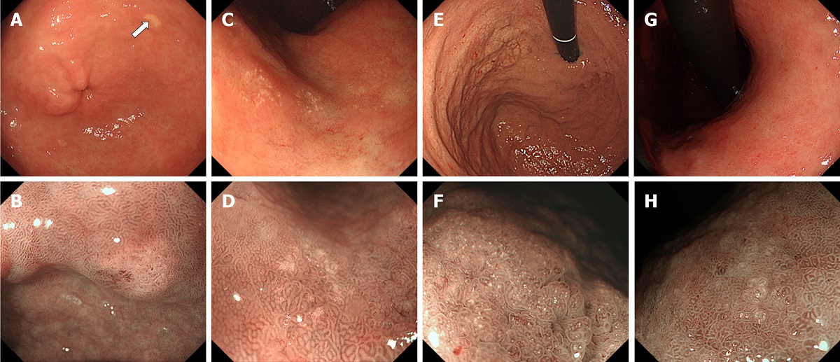
Typical endoscopic features of gastric lanthanum deposition during conventional white-light (upper row) and magnified observation with narrow-band imaging (lower row). A, B: “Whitish spot” is defined as a whitish lesion ≤ 20 mm in diameter with a uniform white color; C, D: “Annular whitish mucosa” are lesion (s) ≤ 20 mm in diameter with white color in the periphery; E, F: “Diffuse whitish mucosa” appears with a white area > 20 mm in diameter; G, H: “Fine granular whitish deposition” is a tiny or faint whitish lesion (s) ≤ 1 mm in diameter.
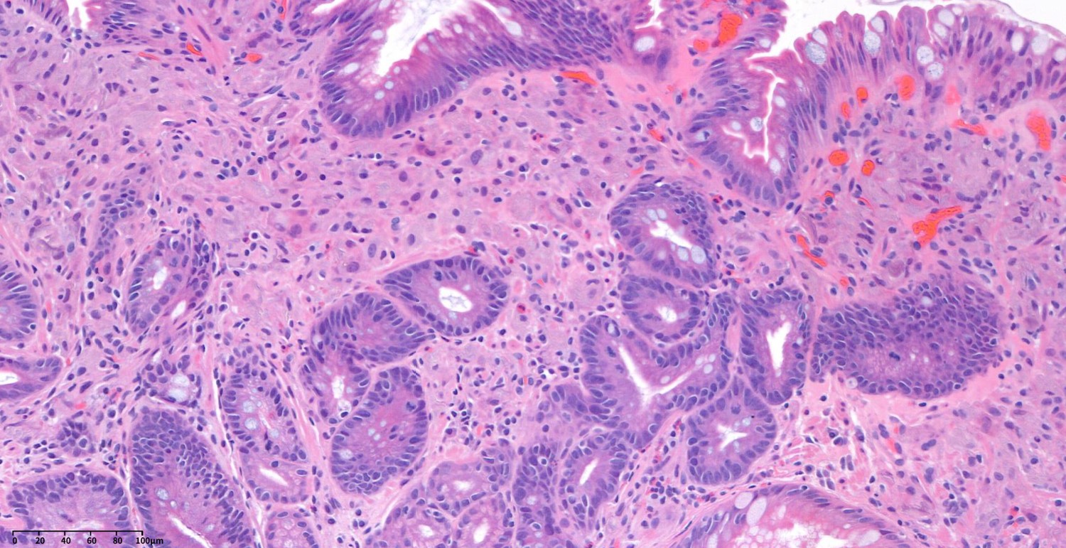
저희가 경험한 환자의 병리 사진입니다.
Google AI의 설명입니다. Lanthanum carbonate, a phosphate binder used in kidney disease, can cause gastritis and other gastrointestinal issues. These issues include nonspecific gastritis, erosions, ulcerations, and gastric polyps, which are visible during endoscopic examination. While lanthanum carbonate is known for its poor systemic absorption, it can still deposit in the gastric mucosa, leading to these changes.
© 일원내시경교실 바른내시경연구소 이준행. EndoTODAY Endoscopy Learning Center. Lee Jun Haeng (2025-6-19)