 EndoTODAY 내시경 교실
Beginner | ESA | Schedule | OPD
Seminars | Atlas | Recent | Links
 [EsoTODAY 065 - Squamous cell carcinoma over leiomyoma. 근종 위 식도암] - 終
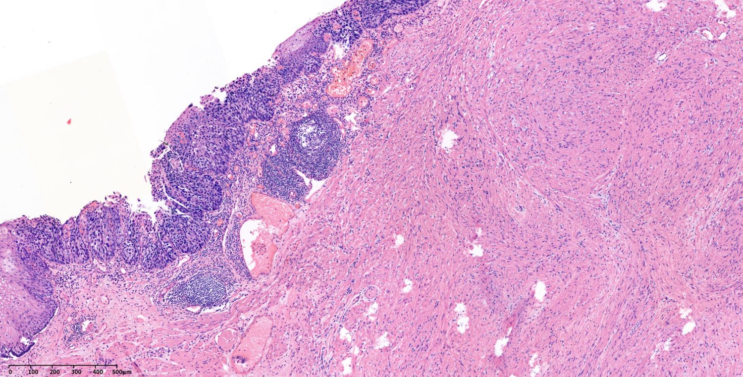
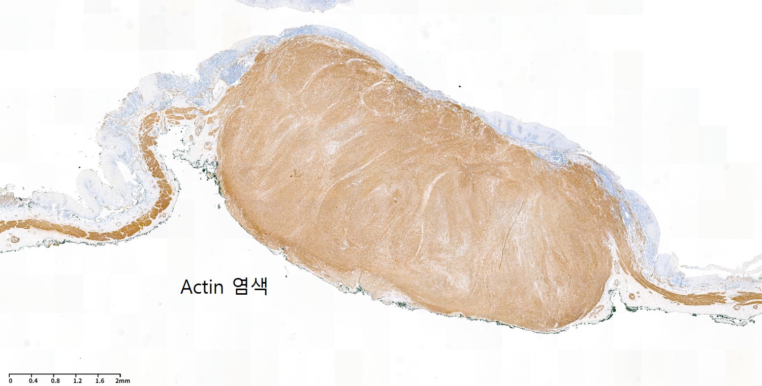
식도 근종 위의 표면이 이상하여 조직검사하여 식도암 의심으로 의뢰된 분입니다. ESD를 하였습니다.
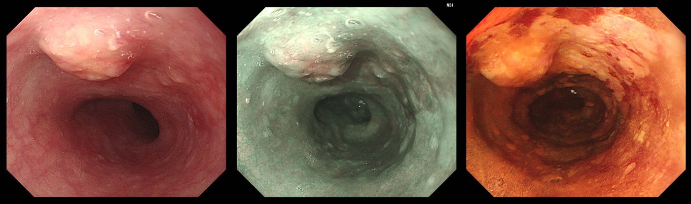
IT 27-29 cm, biopsy(ESD) : Squamous cell carcinoma, moderately differentiated
1. Location : 27-29 cm from incisor
2. Size of invasive carcinoma : (1) longest diameter, 3 mm (2) vertical diameter, 2 mm
3. Size of in situ lesion : (1) longest diameter, 12 mm (2) vertical diameter, 11 mm
4. Tumor multicentricity : single
5. Depth of invasion : invades mucosa (lamina propria) (pT1a)
6. Resection margin : free from carcinoma(N), safety margin : distal 3 mm, proximal 3 mm, anterior 2 mm, posterior 4 mm, deep 500 ㎛
7. Lymphatic invasion : not identified(N)
8. Venous invasion : not identified(N)
9. Perineural invasion : not identified(N)
10. Peritumoral lymphoid follicle: not identified(N)
[2023-4-2. Fellow 질문]
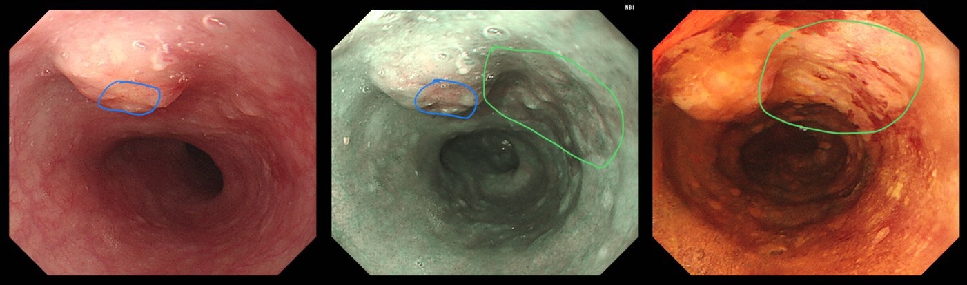
1. 파란색 부분이 근종 위의 암 부위인지요? 처음 보았을 때 SET 인것 같았으나 저 부위때문에 암인가...? 했었습니다
2. 초록색 부위는 병변이 아닌것인가요? 색소내시경을 보는걸 잘 모르겠습니다ㅠㅠ 기초가 너무 부족한 것 같으니 열심히 공부해야겠습니다 ㅎㅎ
[2023-4-2. 이준행 답변]
좋은 질문입니다. 아래 그림처럼 점막하종양 표면과 주변 점막이 모두 식도암이었습니다.
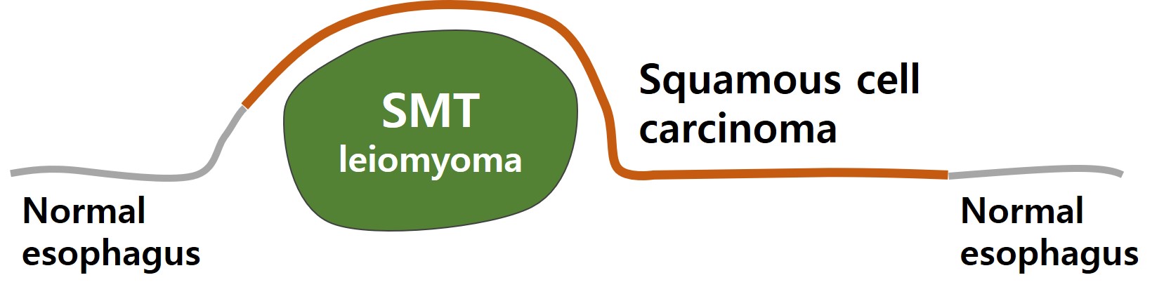
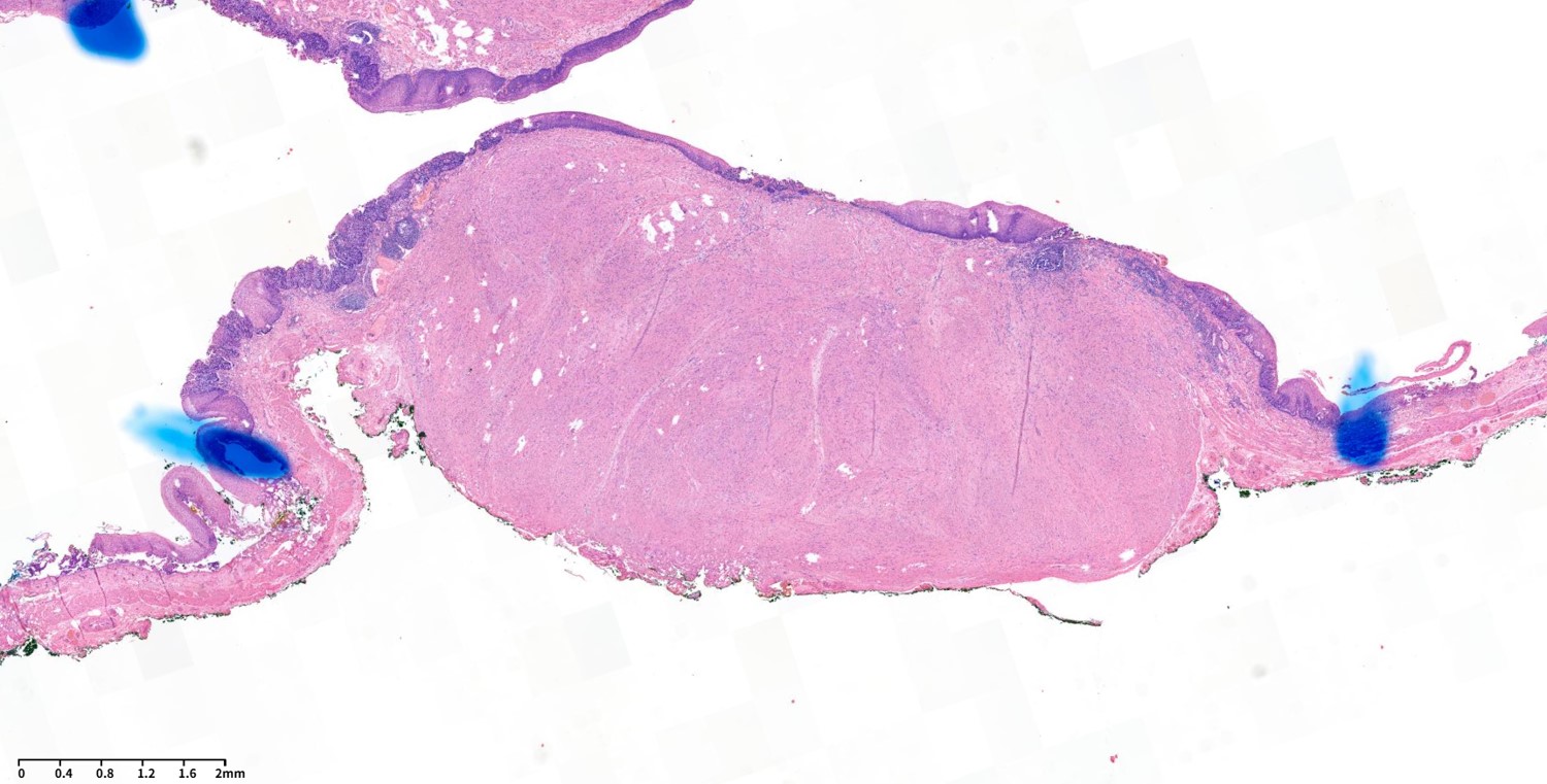
처음 진단하신 분이 병소의 범위를 이렇게 넓게 평가하셨는지는 불명확합니다. SMT가 보였고 처음 발견된 SMT는 조직검사를 한다는 원칙을 따른 것인지, SMT 표면이 정상이 아니라고 느껴 조직검사를 한 것인지 모르겠으나 조직검사 결과가 좋지 않아 의뢰된 경우이고 NBI와 Lugol을 이용하여 자세히 관찰한 바 병소의 범위를 정확히 알 수 있었던 증례입니다.
운이 좋은 환자였다고 생각합니다. 우리 병원 한 선생님께서 esophageal ESD로 치료하셨고 최근 추적검사에서 재발 소견이 없었습니다.
 © 일원내시경교실 바른내시경연구소 이준행. EndoTODAY Endoscopy Learning Center. Lee Jun Haeng. (2022-11-26)