 EndoTODAY 내시경 교실
Beginner | ESA | Schedule | OPD
Seminars | Atlas | Recent | Links
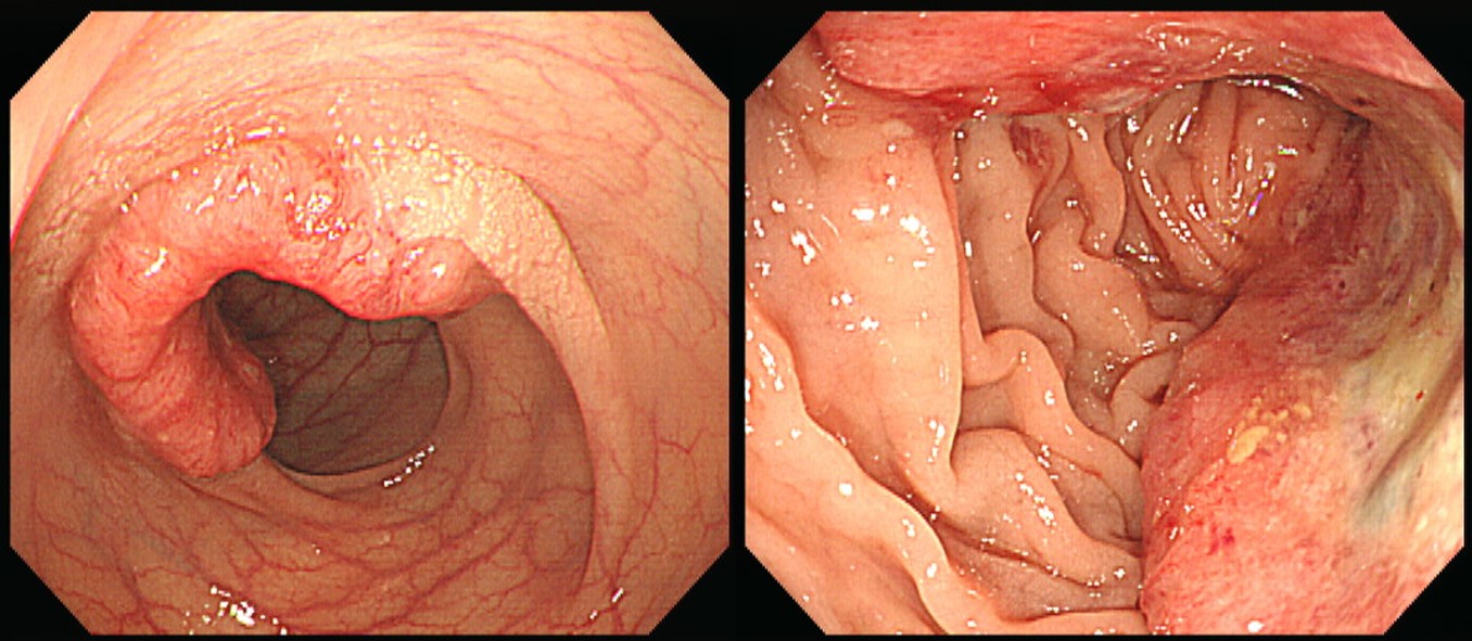
![]() [ColonTODAY 093 - White spot, chicken skin marking, chicken skin mucosa]
[ColonTODAY 093 - White spot, chicken skin marking, chicken skin mucosa]
[2018-8-3. 애독자 질문]
직장에 있는 sessile polyp 으로 bx 에서 High grade dysplasia 나와서, 대학병원에 의뢰할 예정입니다. High grade 또는 cancer 의심되는 병변일 때는, 폴립 주위에, 마치 xanthoma 처럼 노란 점들로 점막 색조 변화 있는 경우가 많았습니다.
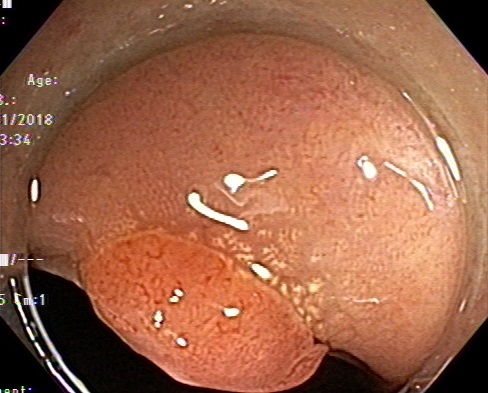
이렇게 노란 점들이 악성 암 또는 High grade 를 시사하는 소견인지요? 발생 이유가 무엇인지 궁금합니다.
[2018-8-3. 이준행 답변]
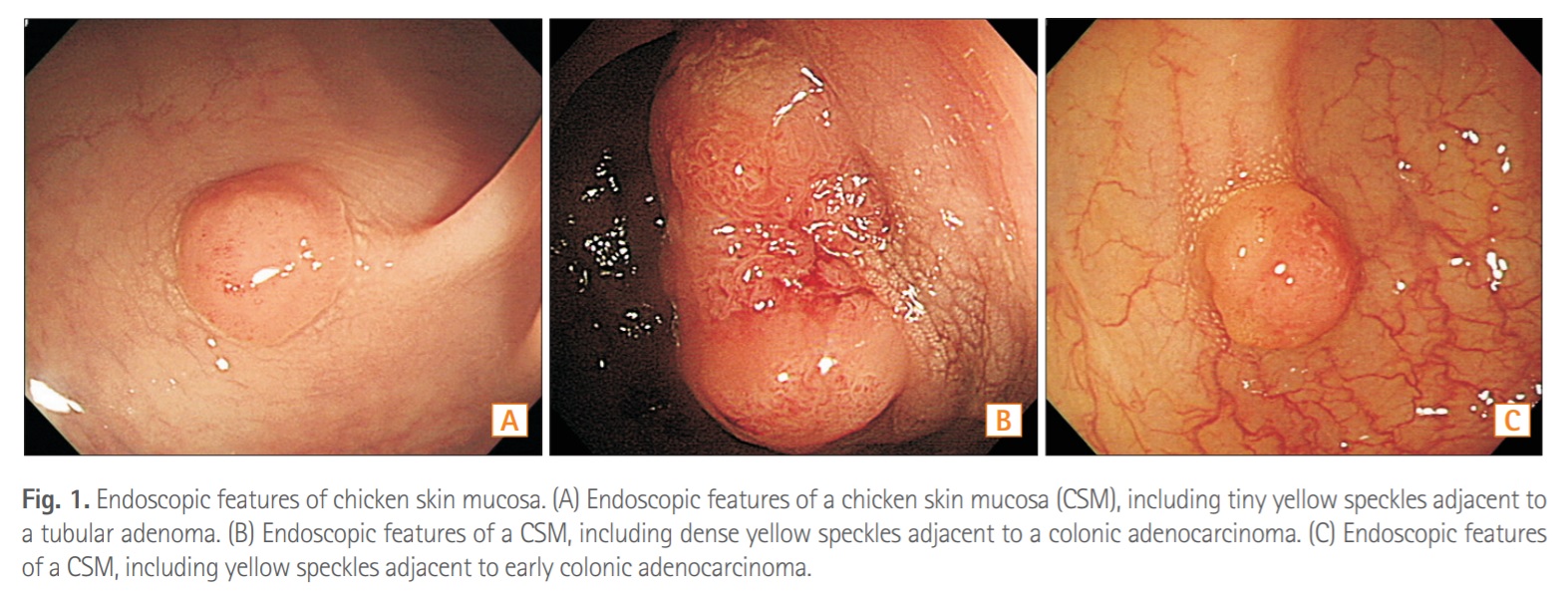
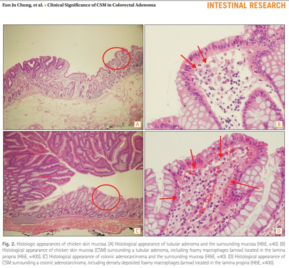
예, 맞습니다. White spot 혹은 chicken skin marking 혹은 chicken skin mucosa라고 하는 것입니다. 선생님 말씀대로 xanthoma와 비슷합니다. 조직학적으로는 lipid-laden macrophage이니까요.
1984년 한 논문의 초록은 아래와 같습니다.
White spots were observed on the mucosa immediately adjacent to polyps and carcinomas; the majority of the polyps proved to be carcinoma in situ or had invasive carcinoma. The white spots consisted of accumulations of foamy cells with features similar to muciphage.
발생 원인은 불명확합니다. Juvenile polyp 주변의 chicken skin marking에 대한 연구(Acta Gastroenterol Belg. 2011)에서 아래와 같이 결론을 맺고 있는데 저는 타당성이 있다고 보았습니다.
The polyp showed the most intense mucosal inflammatory reaction. CSM with the unique thickening of muscularis mucosae especially around larger polyps almost disappeared after polypectomy. So these results suggest that CSM is a benign compensatory reaction induced by the mechanical effect of the polyp.
2015년 대한장연구학회에서 발간하는 학술지(Intest Res)에 실린 아산병원 논문(Intest Res 2015)에서 이에 대하여 좀 더 자세히 설명하고 있으니 참고하시기 바랍니다. Log in 없이 보실 수 있는 open journal입니다 (PDF). 조직학적으로는 foamy macrophage라고 합니다.
저자들은 아래와 같이 결론을 맺고 있습니다.
In conclusion, the present retrospective analyses showed that CSM was associated with tumor multiplicity and advanced histology in distally located colon adenomas. Obesity, metabolic syndrome, and old age were not associated with CSM. It is uncertain whether CSM surrounding colorectal adenoma is a risk factor for colon carcinogenesis; however, CSM is a distinctive marker of advanced pathology of colorectal adenoma.
제 의견으로는 임상적 의의는 여전히 불분명하고 현 시점에서는 무시해도 좋은 소견 아닌가 싶습니다.
[Cases]
Sigmoid colon, anterior resection: Adenocarcinoma, moderately differentiated
1. Location: sigmoid colon
2. Gross type: fungating
3. Size: 2.7x2.5 cm
4. Depth of invasion: invades muscularis propria(pT2)
5. Resection margin: free from carcinoma, safety margin: proximal, 5 cm ; distal, 3 cm ; radial, > 10 mm
6. Regional lymph node metastasis :
- No metastasis in all 13 regional lymph nodes(pN0) (0/13: pericolic, 0/13)
- Number of Extramural Tumor Deposits: 0
7. Lymphatic invasion: present
8. Venous invasion: not identified
9. Perineural invasion: not identified
10. Tumor budding : negative
11. Micropapillary component: yes (10%)
12. Tumor border: pushing
13. Pathologic staging: pT2 N0
Colon, anterior resection : Adenocarcinoma, moderately differentiated
1. Location: sigmoid colon
2. Gross type: ulceroinfiltrative
3. Size: 4.8x3.7 cm
4. Depth of invasion: invades pericolic adipose tissue(pT3)
5. Resection margin: free from carcinoma, safety margin: proximal, 4 cm ; distal, 7 cm ; circumferential, > 10 mm
6. Regional lymph node metastasis : Metastasis to 2 out of 18 regional lymph nodes(pN1b) (2/18: pericolic, 2/18), Number of Extramural Tumor Deposits: 1
7. Lymphatic invasion: present
8. Venous invasion: not identified
9. Perineural invasion: not identified
10. Tumor budding : positive (5-9)
11. Micropapillary component: no
12. Tumor border: infiltrative
13. Pathologic staging: pT3 N1b
© 일원내시경교실 바른내시경연구소 이준행. EndoTODAY Endoscopy Learning Center. Lee Jun Haeng. (2018-8-6)