EndoTODAY 내시경 교실
Beginner | ESA | Schedule | OPD
Seminars | Atlas | Recent | Links
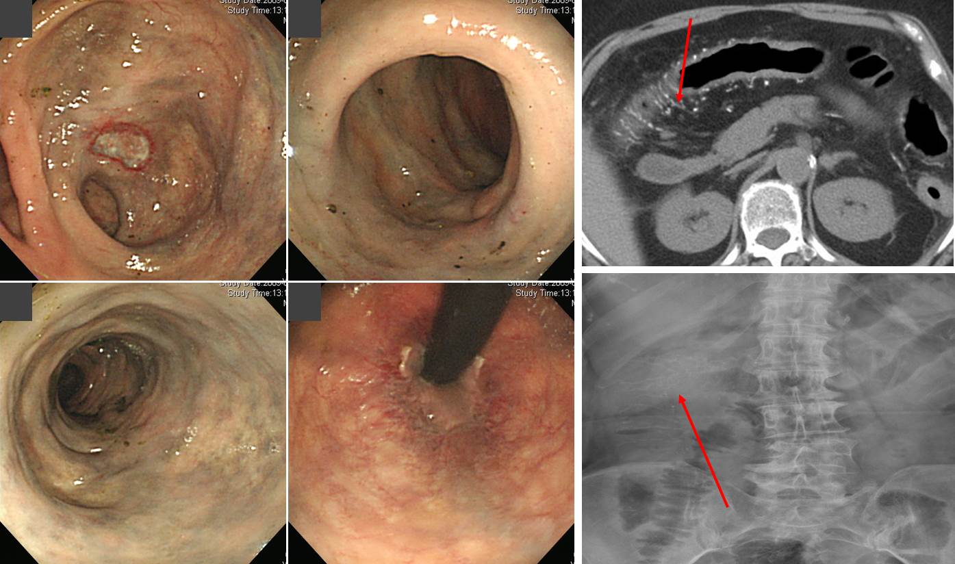
![]() [ColonTODAY 100 - Mesenteric phlebosclerosis]
[ColonTODAY 100 - Mesenteric phlebosclerosis]
만성복통으로 내원하신 70대 여자입니다. 내시경과 CT 후 mesenteric phlebosclerosis라는 chronic ischemic colitis를 일으키는 원인미상의 드문 질환으로 진단되었습니다. 내시경에서는 궤양과 함께 꺼무접접한 점막으로 관찰되었습니다 (Rectum쪽은 정상이었음). Imaging 에서는 right colon의 vessel에 linear calcification이 보였습니다.
Dis Colon Rectum 2003;46:209-20에 보고된 7 증례에 대한 기술을 요약하면 아래와 같습니다.
All seven patients had calcifications in the small mesenteric veins and their intramural branches. No evidence of vasculitis or portal hypertension was recognized. Clinical findings included abdominal pain and diarrhea of a gradual onset and chronic course. A positive fecal occult blood test and mild anemia were often found. The patients had linear calcifications and stenosis in the right colon, which were discovered by plain abdominal radiography and barium enema, respectively. Endoscopic findings included edematous, dark colored mucosa and ulcerations. Four patients underwent a subtotal colectomy because of persistent abdominal pain or ileus. The histopathologic findings were macroscopically characterized by a dark purple or dark brown colored colonic surface, the swelling and disappearance of plicae semilunares coli, and marked thickening of the colonic wall, while they were microscopically characterized by marked fibrous thickening of the venous walls with calcifications, marked submucosal fibrosis, deposition of collagen in the mucosa, and foamy macrophages within the vessel walls.
2019년 3월 소화기학회지에 같은 증례(정맥경화성 대장염)가 소개되었기에 반가운 마음으로 옮깁니다. 내시경 소견이 앞에 소개한 증례보다 조금 심하였습니다. Mucosal lesion이 제 증례보다 현저하였거든요... 그러나 마지막 사진은 위에 소개한 제 증례와 동일한 모습이었습니다. 검푸른 점막 말입니다.
PDF 1.1M
© 일원내시경교실 바른내시경연구소 이준행. EndoTODAY Endoscopy Learning Center. Lee Jun Haeng.