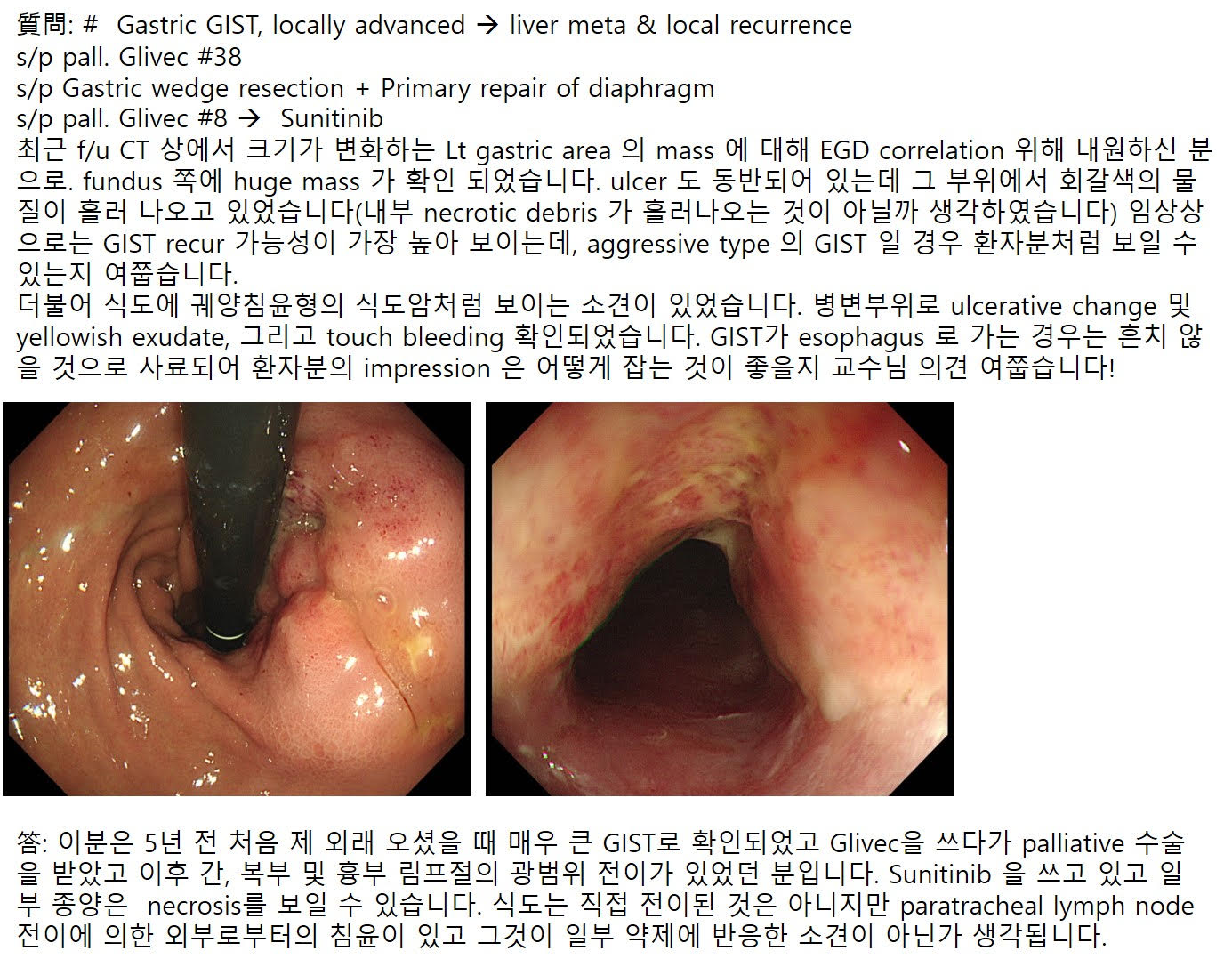
 EndoTODAY 내시경 교실
Beginner | ESA | Schedule | OPD
Seminars | Atlas | Recent | Links
![]() [GIST (gastrointestinal stromal tumor). 위장관 간질성 종양. 위장관 기질종양] - 終
[GIST (gastrointestinal stromal tumor). 위장관 간질성 종양. 위장관 기질종양] - 終
1. SET(SMT)와 GIST - 무엇이 같고 무엇이 다른가?
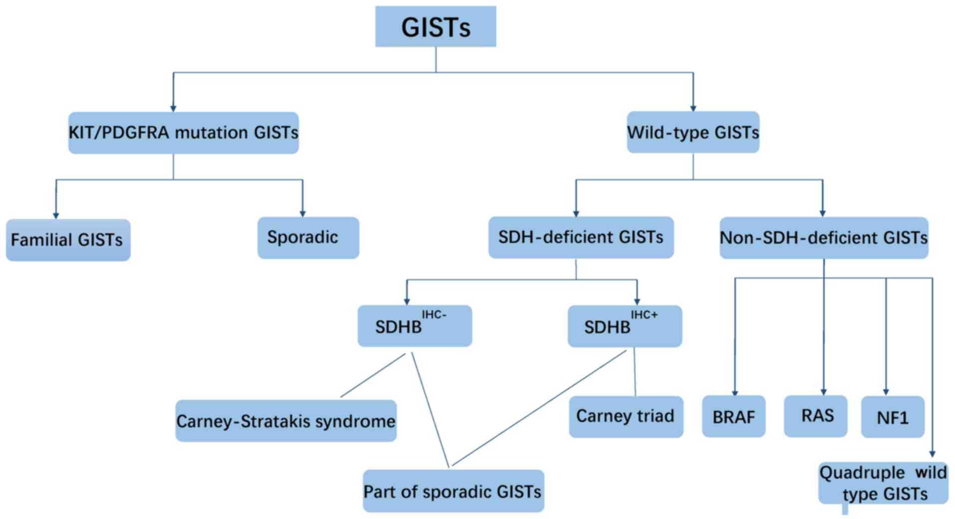
2. GIST 병리진단 - D842V variant, SDH-deficient GIST
4. GIST 치료
10. 수술을 권유받았으나 거부하고 지내다가 간전이 상태로 의뢰된 환자
11. More cases of gastric GIST
12. 소장 GIST 증례
13. 대장 GIST 증례
14. 다른 장기 GIST 증례
15. Code
16. FAQ
17. References
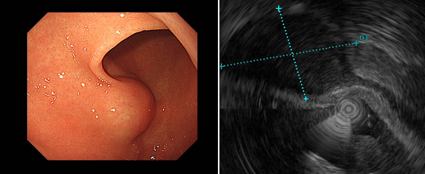
![]() 1. SMT와 GIST - 무엇이 같고 무엇이 다른가?
1. SMT와 GIST - 무엇이 같고 무엇이 다른가?
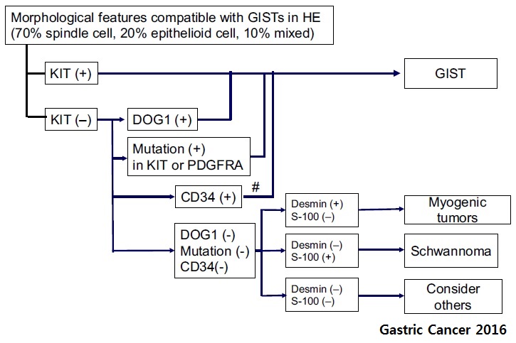
GIST는 하나의 진단명입니다. GIST도 heterogeneous하여 Kit (+) GIST, Kit (-) GIST가 섞여 있으며 다양한 유전적 아형분류가 가능합니다. 그래도 GIST는 GIST입니다. 하나의 조직학적 진단명입니다.
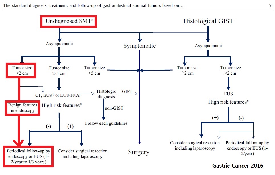
SMT는 아직 진단이 붙지 않은 어떠한 finding입니다. GIST일 수도 있지만 아닐 수도 있습니다. SMT는 조직학적 진단명이 아닙니다. SMT에 대한 진단을 찾아나가면 일부가 GIST이지만, 특정 시점에서 GIST인 SMT와 GIST가 아닌 SMT를 정확히 구분할 수 없습니다. 2016년 Gastric Cancer지 1월호의 그림은 이런 상황을 잘 보여줍니다. 즉 꼭지가 둘입니다.
여기서 첫 꼭지가 중요합니다. Undiagnosed SET(SMT)와 histological GIST라는 두 꼭지 중 적당한 곳에서 시작해야 합니다. 무증상 성인의 검진 내시경에서 만나는 대부분의 SMT는 undiagnosed SMT일 뿐, histological GIST는 아닙니다. 무증상 성인의 2 cm 미만 undiagnossed SMT에 대한 표준 접근법은 high risk features (ulceration, irregular border, increase in size)가 없는 한 CT, EUS, EUS-FNA를 하는 것이 아니고 내시경 추적관찰입니다. 1-2 cm 미만의 SMT에 대하여 모두 EUS를 권하는 현재의 관행은 꼭지를 틀리게 선택한 결과일 뿐입니다.
![]() 2. GIST 병리진단
2. GIST 병리진단
증례 1

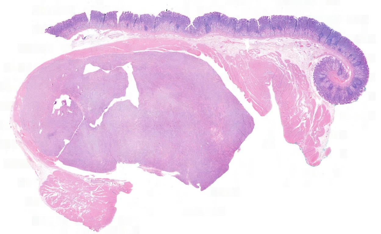

c-kit
증례 2

Gastrointestinal stromal tumor of high risk of malignant potential by proposed modification for adjuvant therapy (2008) (see note);
1) tumor size: 5.2x5.0 cm
2) mitosis: 6/50 HPF (high powered fields)
3) histological type: spindle
4) necrosis: absent
5) cellularity: intermediate
6) cellular atypia: mild
7) invasion into mucosa: absent
8) resection margin involvement: absent
9) No metastasis in two regional lymph nodes (0/2: "Lymph node", 0/2)
[Histological classification]
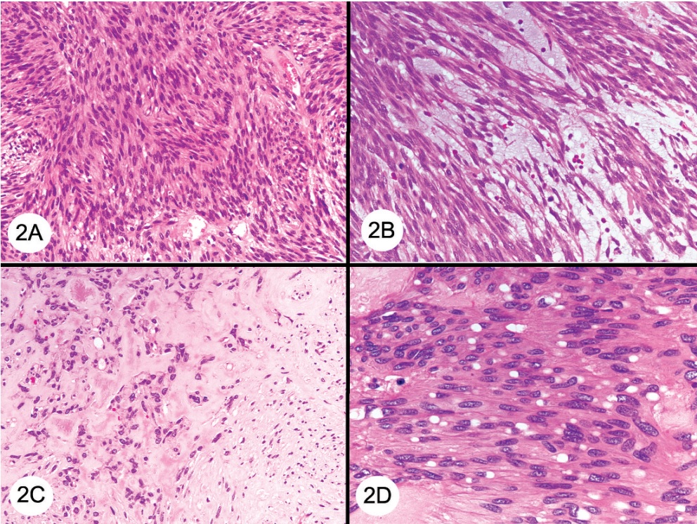
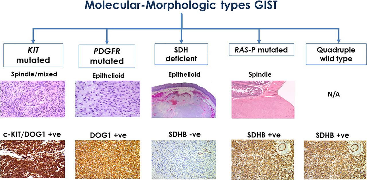
3 morphologic types: spindle (70%), epithelioid (20%), mixed (10%)
(1) Spindle: Bland spindle cells with faintly eosinophilic cytoplasm in a syncytial pattern; elongated nuclei with inconspicuous nucleoli; artifactual paranuclear vacuoles common in stomach GIST.
- Subtypes: sclerosing, palisaded, vacuolated, diffuse hypercellular, sarcomatoid features with significant nuclear atypia and mitotic activity
(2) Epithelioid: Round cells with clear to eosinophilic cytoplasm in sheets or nests; increased tendency for pleomorphism versus spindle type
- Subtypes: sclerosing, discohesive, hypercellular, sarcomatous with significant atypia and mitotic activity
Epitheliod GIST
위 환자의 병리. Epithelioid GIST with mitosis (circle)
(3) Mixed: Tumor is composed of cells with spindle and epithelioid morphology
(4) SDH deficient: epithelioid or mixed epithelioid / spindle cell morphology, multinodular pattern, minimal nuclear pleomorphism, occasional atypical mitoses
(5) Dedifferentiated: anaplastic appearance with an unusual phenotype (may lose expression of KIT or may aberrantly express other markers such as cytokeratin)

Histologic features and patterns of gastrointestinal stromal tumor (GIST). (Arch Pathol Lab Med 2011)
A, Spindle cell GIST composed of fascicles of uniform, bland cells with pale, eosinophilic cytoplasm.
B, Spindle cell GIST with myxoid change.
C, Another GIST with foci of osseous metaplasia.
D, Spindle cell GIST with prominent paranuclear vacuoles.
E, Spindle cell GIST with nuclear palisading that is reminiscent of Antoni A areas encountered in a schwannoma.
F, Spindle cell GIST arising in the small bowel, displaying numerous bundles of deeply eosinophilic ‘‘skeinoid fibers.’’
G, Epithelioid GIST arising in the stomach, composed of cells with abundant, eosinophilic cytoplasm and distinct cell borders.
H, Dedifferentiated GIST composed of atypical epithelioid and spindle cells
Risk assessment: not applied to SDH-deficient GIST, GISTs in patients with syndrome, following neoadjuvant therapy, metastatic setting
Mitosis should be determined in the most mitotically active area of the tumor, and should be reported per 5 mm2.
Molecular test in biopsy specimen: adequate.
[Molecular classification]
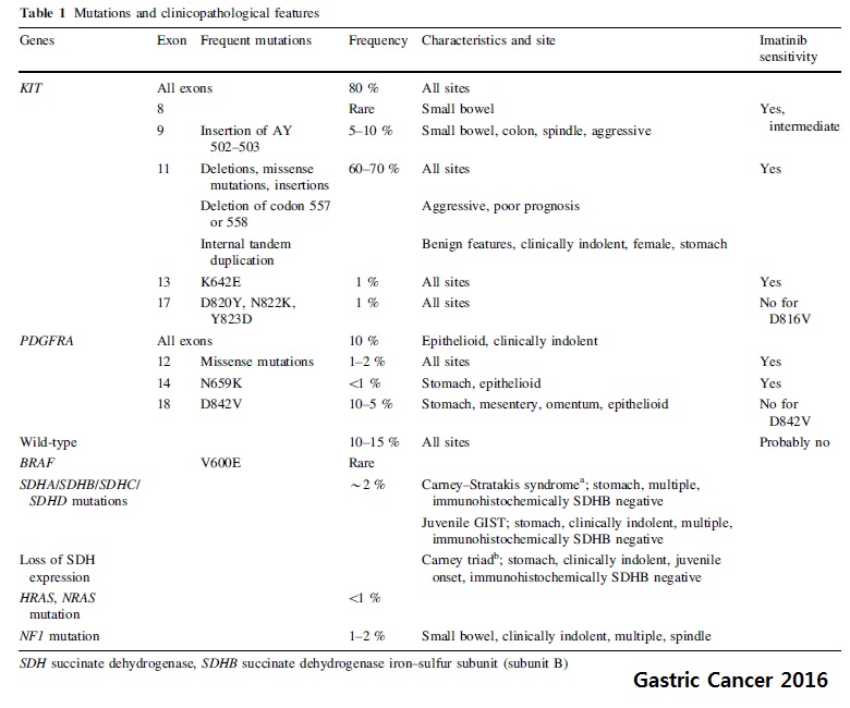
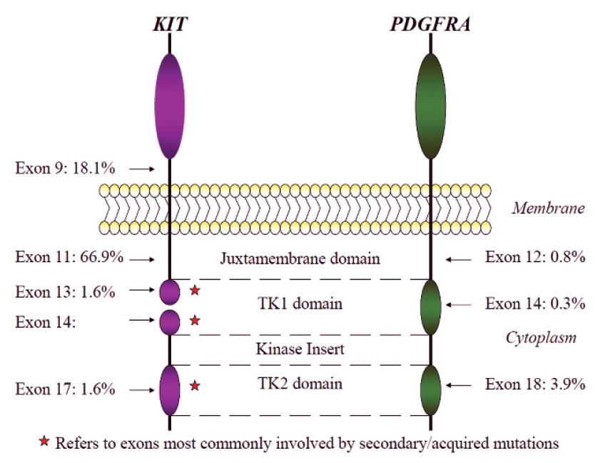
Genetics: KIT 76.0%, SDH deficient 13.9% (SDHA 5.4%, SDHB 2.5%, SDHC 3.9%, SDHC 1.9%, SDHD 0.2%), PDFGRA 10.0%, etc Schaefer. ASCO educational book 2022
GIST immunohistochemistry: c-kit (CD117): 95%, DOG1 (discovered on GIST): 98% positivity
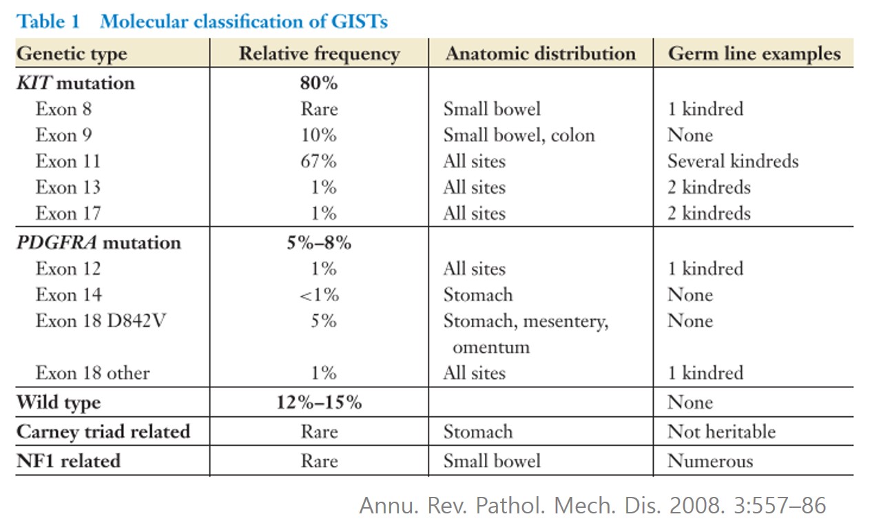
[PDGFRA D842V variant]

Neoadjuvant Glivec 사용하였으나 반응이 좋지 않아서 수술을 시행하였고 정밀검사 결과 PDGFRA D842V로 확인되었음.
[SDH-deficient GIST]
Only arise in the stomach
60% harbor inactivating mutations (nearly always germline), 40% harbor SDHC promoter methylation
Multinodular/plexiform architecture, Epitheloid > mixed morphology (C-kit, DOG1)
Not that rare: 8% of gastric GISTs
Lymph node metastases common
Often clinically indolent in the metastatic setting
Risk assessment criteria do not predict behavior
Imatinib not effective
위 점막하종양의 내시경 조직검사에서 GIST로 확인된 예는 많지 않습니다. 드물게 뚜렷한 함몰부가 있는 경우는 내시경 조직검사로 확인되기도 합니다.
Stomach, wedge resection: Gastrointestinal stromal tumor of low risk of malignant potential by proposed modification for adjuvant therapy (2008) (see note);
1) tumor size: 5.5x4 cm
2) mitosis: 4/50 HPF (high powered fields)
3) histological type: spindle
4) necrosis: absent
5) cellularity: high
6) cellular atypia: mild
7) invasion into mucosa: absent
8) resection margin involvement: absent
Note: low risk by NIH (2002) and low risk (3.6%) of progressive disease by Miettinen (2006).
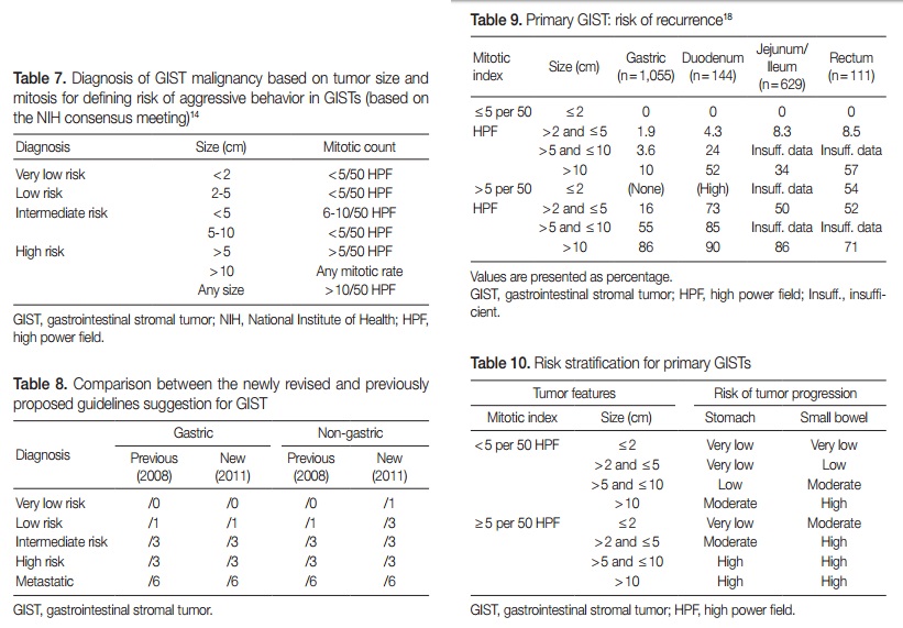
[GIST의 malignant risk 분류]
GIST의 malignant risk에 대한 분류법이 많아서 한 환자가 서로 다른 risk로 분류되기도 합니다. 헷갈리기 그지 없습니다. 게다가 계속 바뀝니다.
조직학적으로 mitosis는 매우 중요한 risk factor입니다.
Mitosis in GIST
Stomach, body, wedge resection: Gastrointestinal stromal tumor of intermediate risk of malignant potential by proposed modification for adjuvant therapy (2008) (see note);
1) tumor size: 2.8x2.5 cm
2) mitosis: 7/50 HPF (high powered fields)
3) histological type: spindle
4) necrosis: absent
5) cellularity: intermediate
6) cellular atypia: moderate
7) invasion into mucosa: absent
8) resection margin involvement: absent
Note: Intermediate risk by NIH (2002) and moderate risk (16%) of progressive disease by Miettinen (2006).
[2014년 새로운 진단 기준]
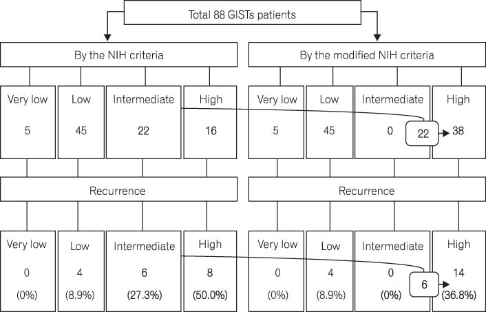
2014년 7월호 Intestinal Research에 실린 서울대 임종필 교수님 팀의 논문 Prediction of Tumor Recurrence in Patients with Non-Gastric Gastrointestinal Stromal Tumors Following Resection according to the Modified National Institutes of Health Criteria을 흥미롭게 읽었습니다. GIST의 병리기준을 과거의 것과 새로운 것(modified NIH criteria)을 비교한 내용이었습니다. 새로운 기준을 사용하였을 때 recur를 더 잘 예측할 수 있었다고 합니다.
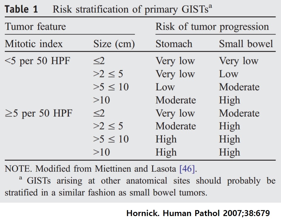
Risk Stratification of Primary Gastrointestinal Stromal Tumors (GISTs) under the National Institutes of Health (NIH) Criteria and Modified NIH Criteria
Risk stratification according to the National Institutes of Health (NIH) and modified NIH criteria. Twenty-two patients with intermediate risk under the original NIH criteria were reclassified into the high-risk category by the modified NIH criteria. Among the 22 reclassified patients, 6 patients experienced tumor recurrence. GISTs, gastrointestinal stromal tumors.
이 논문에 대한 삼성서울병원 장동경 교수님의 editorial도 좋았습니다. Modified NIH criteria에 따라 high risk로 분류된 환자에게 adjuvant imatinib이 필요하다는 내용이었습니다. 과거 기준에 따라 intermediate risk로 분류되었으나 신기준에 따라 high risk로 바뀐 환자에게 도움이 될 것 같습니다.
![]() 3. Mini-GIST 혹은 mini-SMT
3. Mini-GIST 혹은 mini-SMT
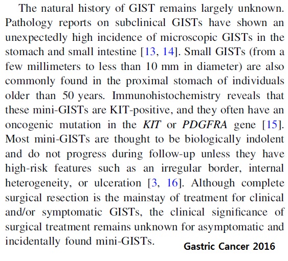
작은 undiagnosed SMT 중 가장 나쁜 종류가 mini-GIST일 것입니다. 그러나 mini-GIST 또한 매우 흔하고 대부분 indolent하고 꼭 치료해야 하는지 명확하지 않습니다.
다만 많은 저자들이 mini-SMT 혹은 mini-GIST를 논하면서 high-risk features에 irregular border나 ulceration과 함께 EUS를 해야만 알 수 있는 internal heterogeneity를 언급하고 있기 때문에 EUS가 과잉 처방되고 있을 뿐입니다. EUS에서 관찰되는 internal heterogeneity는 큰 GIST에서는 나름 의미가 있지만, mini-GIST 혹은 mini-SMT에서 관찰되는 internal heterogeneity의 임상적 의의는 명확하지 않다고 보는 것이 맞습니다.
Undiagnosed SMT에 대한 별도의 가이드라인은 없습니다. GIST 가이드라인의 일부로서 언급되고 있을 뿐입니다. 내시경 전문가의 참여가 부족한 상태로 씌여졌다는 말씀입니다. EGD와 EUS를 혼동하거나 명확히 구분하지 않고 씌여진 가이드라인이 많은 것은 그 때문입니다. 주의가 필요합니다. 예를 들면, 2016년 Gastric Cancer 종설에 실린 다음 문장은 Table의 내용과 일치하지 않습니다. Misleading할 가능성이 높은 틀린 문장입니다. "The standard diagnosis, treatment, and follow-up of GIST When small esophageal or gastric nodules (SMTs smaller than 2 cm) having no high-risk features are detected, they can usually be followed by periodic endoscopic ultrasonography (EUS) until the tumors increase in size or become symptomatic." 마치 5 mm SMT에 대해서도 EUS를 해야 하는 것처럼 읽히지 않습니까? EGD와 EUS를 구분하지 않았다니 정말 황당한 일입니다. 2 cm 미만이고 고위험 인자가 없는 식도/위 SMT는 (EUS가 아니라) EGD로 추적관찰 할 수 있습니다. EUS는 필수가 아니라 선택입니다. 향후 GIST 가이드라인 작업에는 내시경 전문가가 참여해야 합니다.
경과관찰 중 뚜렷한 모양 변화가 있을 때에는 수술을 권하는 것이 좋습니다.
![]() 4. GIST 치료
4. GIST 치료
2 cm 이상의 GIST의 표준 치료는 수술적 절제입니다. 내시경 절제술도 일부 시도되고 있으나 외과의사들은 아직 positive margins이나 tumor spillage를 걱정하고 있습니다. 저는 비교적 3-4 cm 정도의 작은 GIST는 조심스럽게 내시경치료를 해 볼 수 있겠다고 생각합니다.
2 cm 미만의 GIST는 절제술 혹은 경과관찰을 선택할 수 있습니다. 사실 2 cm 미만의 undiagnosed SMT에서는 조직학적 진단이 꼭 필요한 것은 아닙니다. 2 cm 미만에서 조직진단을 하지 않는 철학을 가진 의사는 2 cm 미만 GIST를 만날 일조차 없습니다.
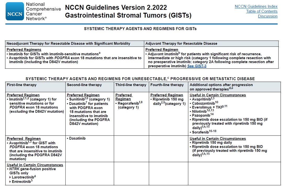
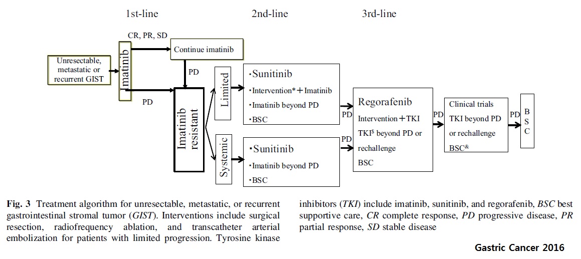
아래는 unresectable, metastatic 혹은 recurrent GIST에 대한 치료 algorithm입니다.
너무 큰 GIST는 neoadjuvant imatinib을 쓰기도 합니다. (Kong Gastric Cancer 2023)
수술 후 high risk이면 aduvant imatinib을 3년간 사용합니다. 5년사용하면 도움이 되는가는 명확치 않아서 현재 임상연구가 진행 중입니다 (NCT02413736)
새로운 약들이 나오고 있습니다.
Tagetted treatment는 매우 많이 개발되고 있습니다.
2025-12-6. 연건동 위암 forum. 구홍회 교수 강의 중
![]() 5. 상부위장관 출혈로 내원한 GIST
5. 상부위장관 출혈로 내원한 GIST
Melena
Stomach, subtotal gastrectomy:
Gastrointestinal stromal tumor of high risk of malignant potential by NIH criteria:
1) tumor size: 6.5x6.0 cm
2) mitosis: 6 /50 HPF (high powered fields)
3) histological type: spindle
4) necrosis: absent
5) cellularity: intermediate
6) cellular atypia: mild
7) invasion into mucosa: present
8) myxoid change: present
9) resection margin involvement: absent
10) no metastasis in 4 regional lymph nodes
Melena
Stomach, cardia, wedge resection:
Gastrointestinal stromal tumor of high risk of malignant potential by NIH (2002) and moderate risk(16 %) of progressive disease by Miettinen(2006) ;
1) tumor size: 3.5x2.5x2.4 cm
2) mitosis: 88/50 HPF (high powered fields)
3) histological type: mixed spindle and epithelioid
4) necrosis: present
5) cellularity: high
6) cellular atypia: moderate
7) invasion into mucosa: present
8) resection margin involvement: absent
Melena로 오셨는데 SMT 같은데 출혈 부위가 없다고 의뢰되었음. 자세히 보니 궤양이 있었음.
Stomach, body, wedge resection:
Gastrointestinal stromal tumor of high risk of malignant potential by proposed modification for adjuvant therapy (2008) (see note);
1) tumor size: 5.5x3.5 cm
2) mitosis: 40/50 HPF (high powered fields)
3) histological type: spindle
4) necrosis: absent
5) cellularity: high
6) cellular atypia: moderate
7) invasion into mucosa: absent
8) resection margin involvement: absent
Note: high risk by NIH (2002) and high risk (55%) of progressive disease by Miettinen (2006).
[Addendum]
Ki-67 : Positive in 10% of tumor cells
DOG-1 : Positive in tumor cells
C-KIT (CD 117) : Positive in tumor cells
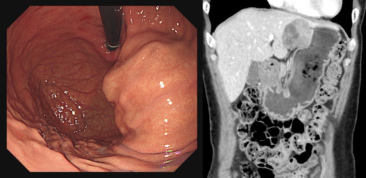
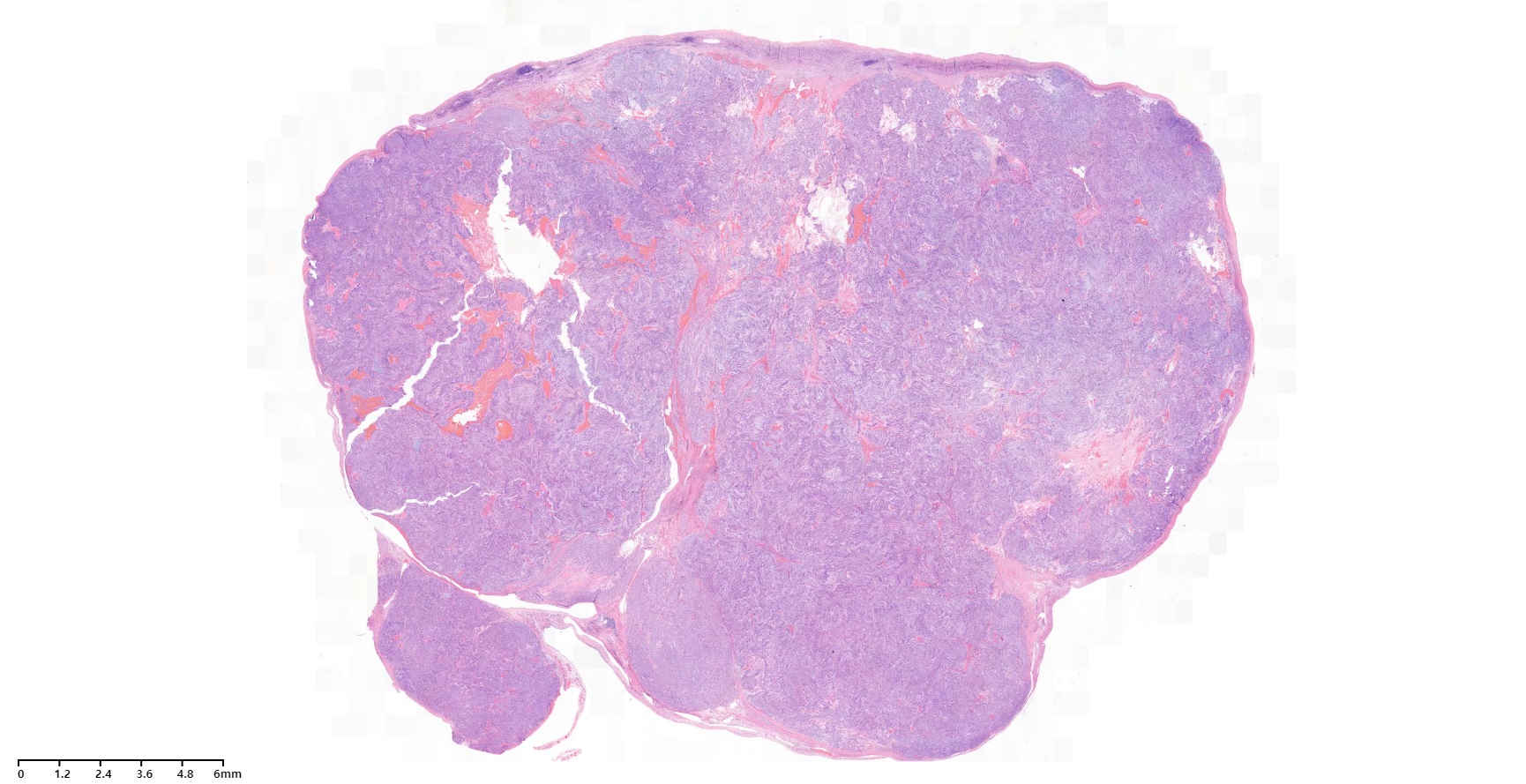
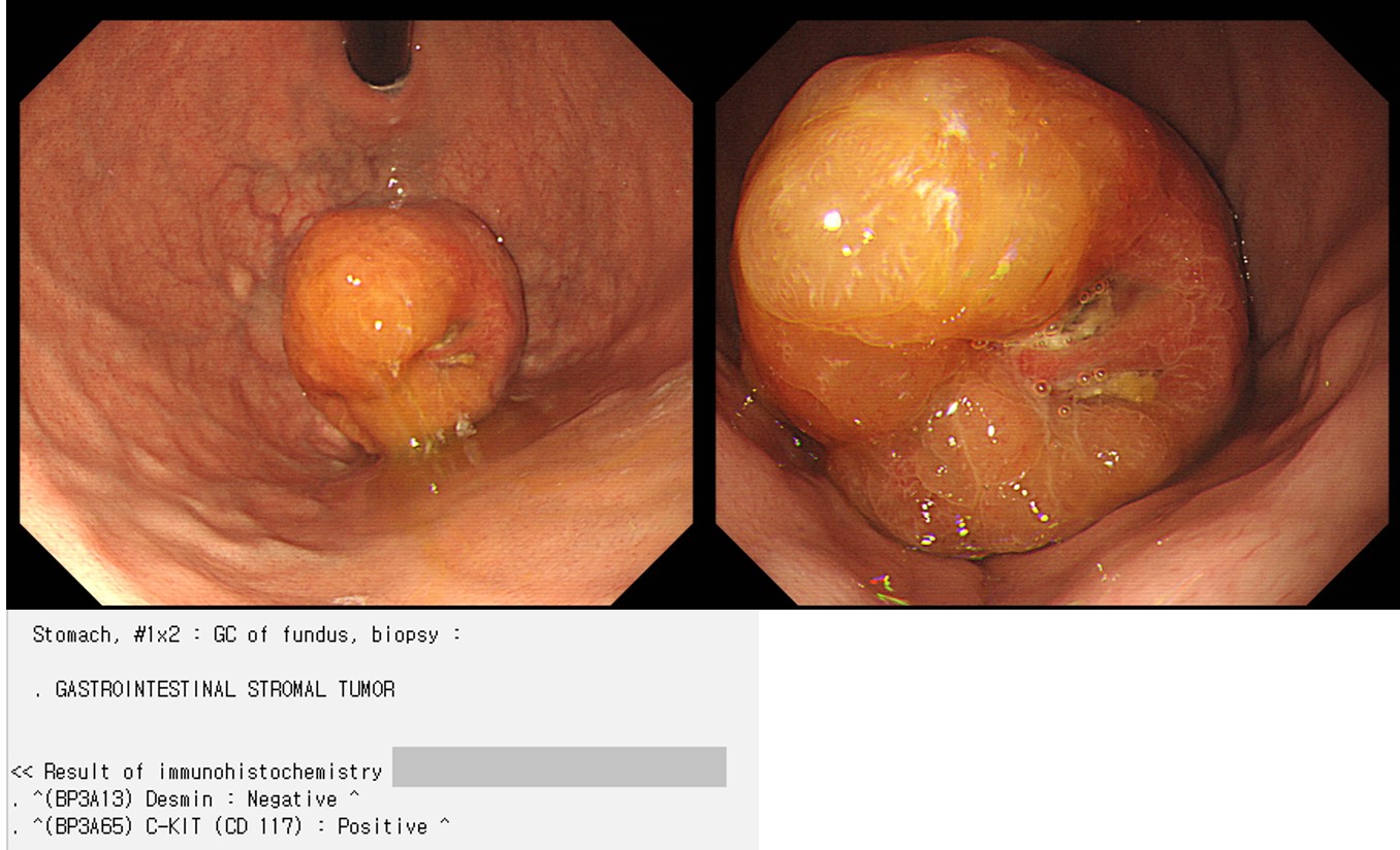
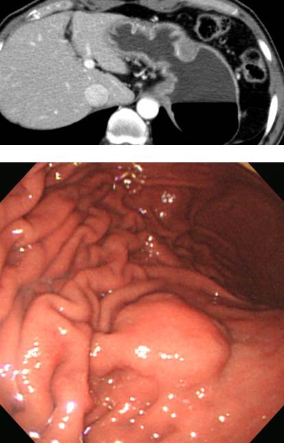
![]() 6. 첫 진단시 전이가 있었던 GIST
6. 첫 진단시 전이가 있었던 GIST
GIST with hepatic metastasis. Fundus의 불규칙한 궤양 부분(두번째, 세번째 사진)만으로 판단하면 AGC, lymphoma, GIST 등을 모두 고려할 수 있겠으나, 불규칙한 궤양부 주변의 넓은 SMT 부분(첫번째 사진)을 고려하면 AGC의 가능성은 매우 떨어지는 경우임을 알 수 있습니다. C-kit (CD117) 양성 GIST였습니다.
Hepatic metastasis
Hepatic metastasis
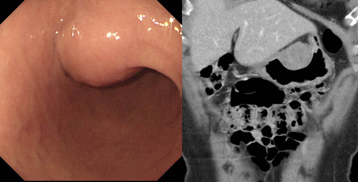
내시경에서 여러 SMT가 있는 것처럼 보였으나 CT에서는 proximal stomach 거의 전체를 감싸는 GIST 였으며, MRI에서는 몇 개의 metastastasis가 있었습니다.
조직검사: Gastrointestinal stromal tumor, high risk of malignant behavior;
1) mitosis: 11/16 HPF
2) histological type: mixed
3) necrosis: absent
4) cellularity: high
5) myxoid change: present
처음부터 간 전이가 있어서 imatinib을 투여하였습니다.
Liver biopsy: Gastrointestinal stromal tumor
C-KIT (CD 117) : Diffusely positive in tumor cells
Ki-67 : Positive in up to 8% of tumor cells
DOG-1 : Focally positive in tumor cells
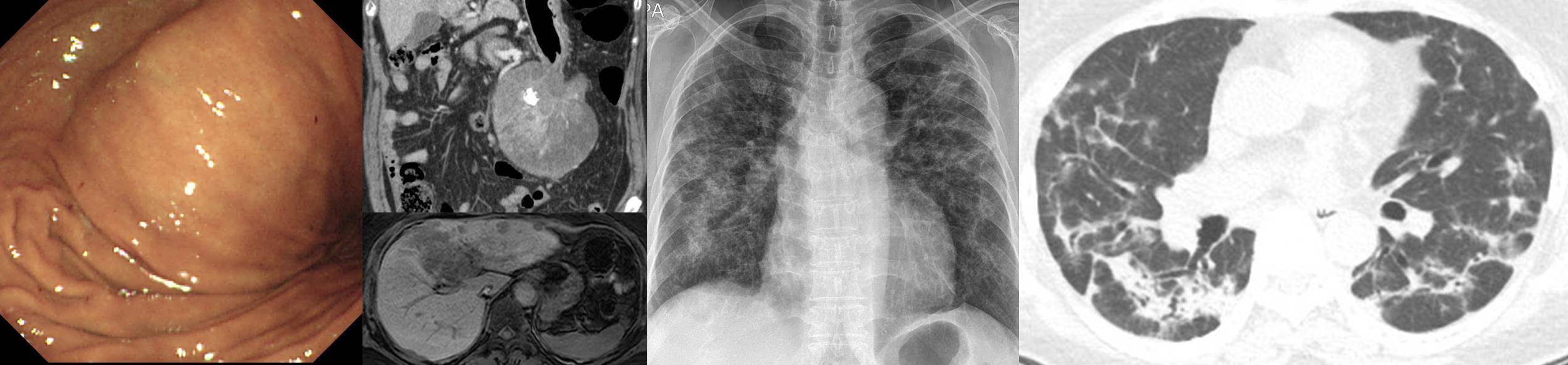
Gleevec 투여 후 호흡곤란 발생. CT에서 drug-induced BOOP 소견으로 일단 Gleevec 중단하였습니다. Gleevec의 드문 합병증이라고 합니다.
(2017, 49세 여성) 우연한 초음파에서 간종괴가 발견되었습니다. 내시경에서는 특이소견이 없었습니다. CT에서 이미 복강에 여러 metastatic nodule 들이 관찰되었습니다. EUS-guided biopsy를 하였고 GIST로 확인되었습니다. Gleevec 치료를 하였습니다. Exophytic growth를 보인 gastric GIST로 첫 진단시 간 침윤과 복강 전이가 있었던 증례로 결론지었습니다.
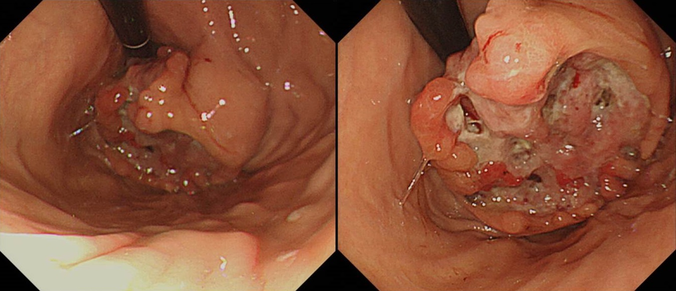
내시경 시행 의사가 위에 clot들이 있는데 조직검사를 해도 좋을지 realtime으로 문의가 와서 조직검사를 하도록 권하였고 c-kit positive GIST로 확인되었음.
![]() 7. 경과관찰 중 궤양이 발생하여 수술한 증례
7. 경과관찰 중 궤양이 발생하여 수술한 증례
3년 경과관찰 중 크기가 증가하였고 작고 얕은 궤양이 발생하여 수술
Stomach, fundus, wedge resection: Gastrointestinal stromal tumor of intermediate risk of malignant potential by proposed modification for adjuvant therapy (2008) (see note);
1) tumor size: 3.8x3.0 cm
2) mitosis: 9/50 HPF (high powered fields)
3) histological type: spindle
4) necrosis: absent
5) cellularity: intermediate
6) cellular atypia: moderate
7) invasion into mucosa: absent
8) resection margin involvement: absent
Note: intermediate risk by NIH (2002) and moderate risk (16%) of progressive disease by Miettinen (2006).
경과관찰 중 궤양
Gastrointestinal tumor of high risk of aggresive behavior:
1) tumor size : 2.7x2x1.5 cm
2) mitosis: 26/50 HPF
3) histologic type : spindle
4) necrosis : present
5) cellularity : high
6) invasion into mucosa : present
7) resection margin involvement : absent
8) histologic subtype: cellular spindle cell type
경과관찰 중 궤양
Stomach, mid body, posterior, wedge resection:
Gastrointestinal stromal tumor of low risk of malignant potential by NIH (2002) and very low risk (1.9 %) of progressive disease by Miettinen (2006):
1) tumor size: 3.5x3x2 cm
2) mitosis: 2/50 HPF (high powered fields)
3) histological type: spindle
4) necrosis: absent
5) cellularity: high
6) cellular atypia: mild
7) invasion into mucosa: absent
8) resection margin involvement: absent
처음 내시경 당시에는 출혈이 없었는데 수술 기다리는 며칠 사이에 melena를 보여 서둘러 수술함
Stomach, body, posterior wall, wedge resection:
Gastrointestinal stromal tumor of high risk of malignant potential:
1) tumor size: 4.2x3.8x3.3 cm
2) mitosis: 10/50 HPF (high powered fields)
3) histological type: spindle
4) necrosis: absent
5) cellularity: intermediate
6) cellular atypia: mild
7) invasion into mucosa: absent
8) resection margin involvement: absent
![]() 8. 경과관찰 중 출혈로 수술한 GIST
8. 경과관찰 중 출혈로 수술한 GIST
추적관찰 중 melena
Stomach, high body, wedge resection:
Gastrointestinal stromal tumor of low risk of malignant potential by proposed modification for adjuvant therapy (2008), low by NIH and very low risk (1.9 %) of progressive disease by Miettinen (2006) :
1) tumor size: 2.2x2 cm
2) mitosis: 4 /50 HPF (high powered fields)
3) histological type: spindle
4) necrosis: absent
5) cellularity: intermediate
6) cellular atypia: moderate
7) invasion into mucosa: present
8) resection margin involvement: absent
추적관찰 중 melena
Stomach, fundus, wedge resection:
Gastrointestinal stromal tumor of low risk of malignant potential by NIH (2002) and very low risk (1.9 %) of progressive disease by Miettinen (2006):
1) tumor size: 2.1x1.9x1.7 cm
2) mitosis: 1/50 HPF (high powered fields)
3) histological type: spindle
4) necrosis: absent
5) cellularity: low
6) cellular atypia: mild
7) invasion into mucosa: absent
8) resection margin involvement: absent
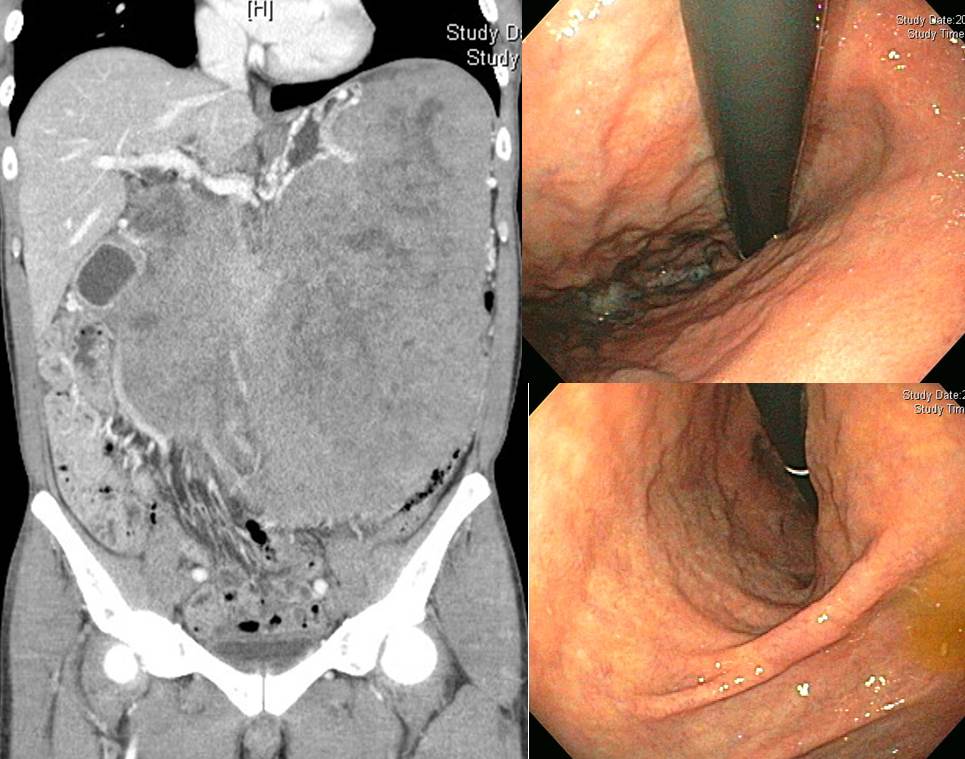
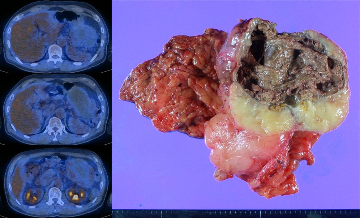
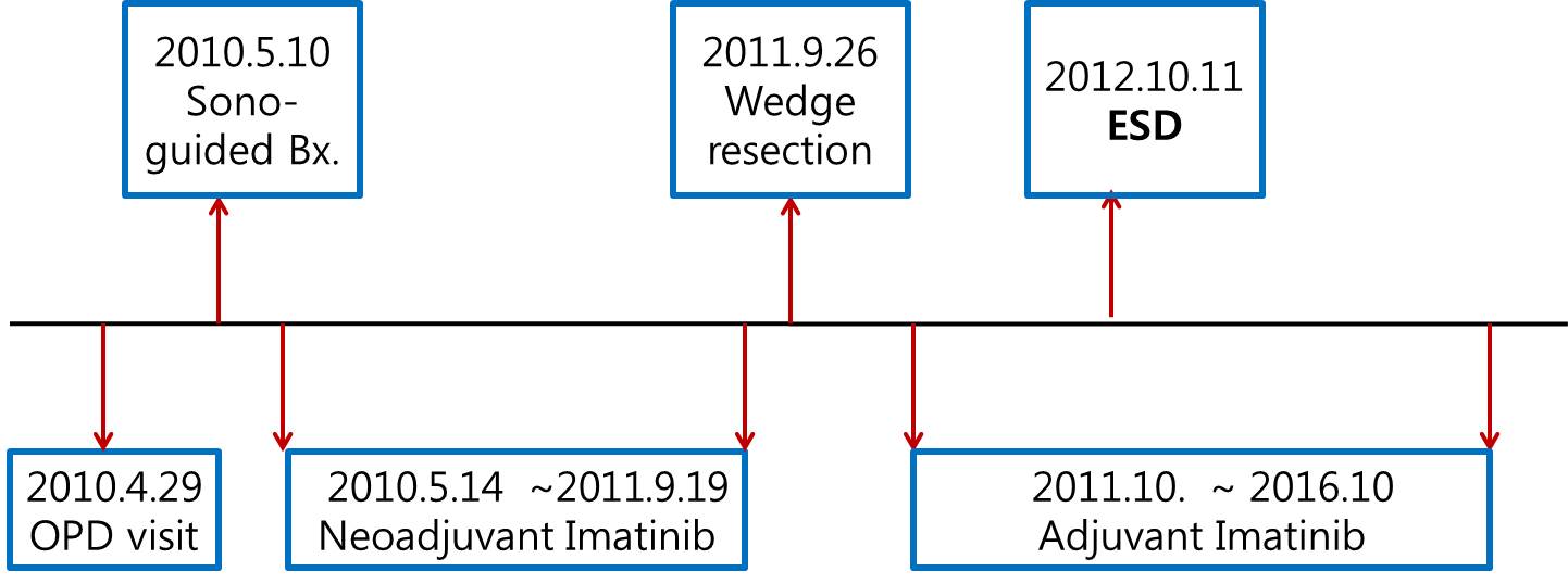
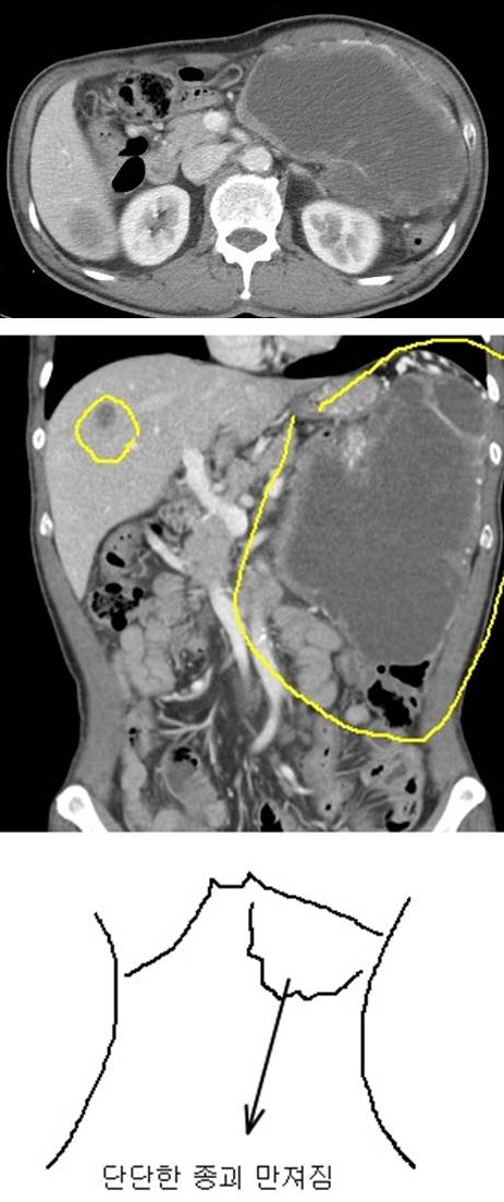
![]() 9. 필자가 경험한 가장 큰 GIST
9. 필자가 경험한 가장 큰 GIST
2달 전부터 시작된 복부 팽만으로 내원하였습니다. 복부 CT에서는 30x24cm로 측정되었지만 사실 더 커보였습니다.
정확한 진단을 위하여 sono-guided biopsy를 하였습니다. GIST (high risk of malignant potential)로 나왔고 당시 mitosis는 4/17 HPFs, 조직형은 mixed spindle and epitheloid였습니다.
Gleevec으로 16개월 치료하였고 병소가 현저히 작아졌습니다.
Surgical wedge resection을 하였습니다. 종양의 크기는 15x9x9cm였고, neat total necrosis로 인하여 mitosis는 측정할 수 없었습니다.
약 1년 후 추적내시경에서 위암 1개, 선종 1개가 발견되어 ESD를 하였습니다.
Surgical wedge resection 후 5년간 adjuvant imatinib 사용하였습니다. 이후 경과관찰 중 결국 재발하셨습니다.
위 환자 정도로 크지 않아도 locally advanced disease는 바로 surgical resection이 어렵기 때문에 일단 Gleevec 치료부터 시작합니다. 반응이 좋으면 수술을 하기도 합니다.
![]() 10. 수술을 권유받았으나 거부하고 지내다가 간전이 상태로 의뢰된 환자
10. 수술을 권유받았으나 거부하고 지내다가 간전이 상태로 의뢰된 환자
여러 병원에서 수술을 권유받고 본 병원 외과를 찾았다가, 환자가 수술하지 않는 것으로 마음을 바꾸어 소화기내과를 찾은 분입니다. 당연히 수술을 권했으나 이후 follow-up loss 되었습니다.
3년 후 전이된 상태로 다시 내원하셨습니다. 안타까운 일입니다.
EUS guided biopsy로 c-kit positive malignant GIST를 진단하였습니다.
![]() 11. More cases of gastric GIST
11. More cases of gastric GIST
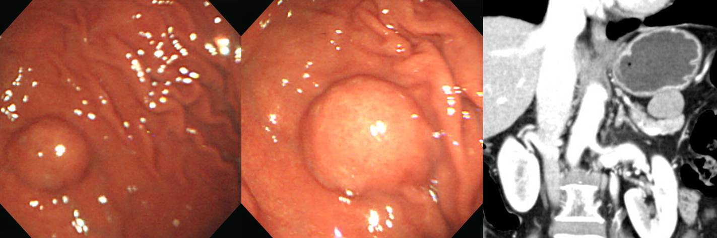
우연히 발견된 작은 gastric SMT는 대부분은 커지지 않지만 방심은 금물입니다. 분명 커지는 경우도 있기 때문입니다. 아래 증례는 대장암 환자에서 우연히 발견된 위 SMT였습니다. 추적관찰을 권하였으나 follow up loss 되었습니다. 6년만에 오셨는데 제법 커진 상태였습니다. CT 검사 후 수술을 하였습니다.
(2012. F/61)
Stomach, wedge resection: Gastrointestinal stromal tumor of high risk of malignant potential by proposed modification for adjuvant therapy (2008) (see note);
1) tumor size: 6.5x4 cm
2) mitosis: 6/50 HPF (high powered fields)
3) histological type: mixed spindle and epithelioid
4) necrosis: absent
5) cellularity: intermediate
6) cellular atypia: moderate
7) invasion into mucosa: absent
8) resection margin involvement: absent
Note: High risk by NIH (2002) and High risk (55%) of progressive disease by Miettinen (2006).
. DOG-1 : Positive in tumor cells
. Ki-67 : Positive in about 10% of tumor cells
. C-KIT (CD 117) : Positive in tumor cells
다음은 10년 사이에 크기 증가가 있어 수술하였던 환자입니다. 의뢰 전 특별한 병력을 가지고 있었습니다. 내시경 조직검사 후 집에서 이틀 동안 검은 변이 있었는데 환자께서는 출혈인 줄 모르고 어지럽다는 이유로 인근 대학병원 신경과를 방문하여 머리 MRI를 찍고 이상이 없다는 이야기를 들었다고 합니다. 119에 두 번 실려갔는데 아무도 내시경 검사 했는지, 혈변은 없었는지 물어보지 않았다고 합니다. 신경과 선생님들께 부탁합니다. 병력 청취를 잘 해주시기 바랍니다.
Stomach, wedge resection: Gastrointestinal stromal tumor of intermediate risk of malignant potential by proposed modification for adjuvant therapy (2008) (see note);
1) tumor size: 2.5x1.5 cm
2) mitosis: 7/50 HPF (high powered fields)
3) histological type: spindle
4) necrosis: absent
5) cellularity: high
6) cellular atypia: moderate
7) invasion into mucosa: absent
8) resection margin involvement: absent
Ki-67 : Positive in up to about 3% of tumor cells
C-KIT (CD 117) : Positive in tumor cells
DOG-1 : Positive in tumor cells
* Note: Intermediate risk by NIH (2002) and moderate risk (16%) of progressive disease by Miettinen (2006).
다양한 증례입니다.
Stomach, wedge resection: Gastrointestinal stromal tumor of low risk of malignant potential by NIH (2002) and very low risk (1.9 %) of progressive disease by Miettinen (2006):
1) tumor size: 3.5x2.5x2 cm
2) mitosis: 5/50 HPF (high powered fields)
3) histological type: spindle
4) necrosis: absent
5) cellularity: high
6) cellular atypia: mild
7) invasion into mucosa: absent
8) resection margin involvement: absent
뚜렷한 bridging fold와 깊은 궤양을 보였던 GIST
Gastrointestinal stromal tumor of intermediate risk of malignant potential;
1) tumor size: 3.5x3.5x2 cm
2) mitosis: 10 / 50 HPF (high powered fields)
3) histological type: spindle (hypercellular spindle type)
4) necrosis: absent
5) cellularity: high
6) cellular atypia: mild
7) invasion into mucosa: absent
8) resection margin involvement: absent
C-kit: Positive, DOG-1: Positive, CD34 : Positive, PDGFRA: Positive, Ki-67: Positive in 11% of tumor cells
Forceps biopsy로 진단되었던 GIST
Gastrointestinal stromal tumor of low risk of aggressive behavior, stomach:
1) tumor size: 6.5x6 cm
2) mitosis: 3/50 HPF
3) histological type: mixed spindle and epithelioid
4) necrosis: absent
5) cellularity: high
6) invasion into mucosa: present
7) resection margin involvement: absent
C-kit: Positive, PKC-Θ: Strong positive, CD34: Strong positive, PDGFRA: Positive with dot-like accentuation, Ki-67: Positive (3%)
우연히 시행한 복부초음파에서 발견된 GIST. 동시에 시행한 내시경에서는 SMT가 의심되지 않았다고 합니다.
Gastrointestinal stromal tumor of moderate risk of malignant potential:
1) size: 11x9x8 cm
2) mitosis : 2/50 HPFs
3) histologic type: epithelioid
4) necrosis: present
5) cellularity: high
6) cellular atypia: moderate
7) invasion to mucosa: absent
8) resection margin involvement: absent
DOG-1 : Positive, C-kit : Negative, PKC-Q : Weak positive, CD34 : Focal positive, PDGFRA: Positive (dot-like), Ki-67 : Positive in 20% of tumor cells
22G Precore 사용하여 EUS-guided FNAB 시행함. GIST로 나와 수술을 시행하였습니다.
Stomach, mid body, wedge resection:
Gastrointestinal stromal tumor of very low by NIH and none risk (0%) of progressive disease by Miettinen (2006) :
1) tumor size: 1.9x1.8x1 cm
2) mitosis: 1/50 HPF (high powered fields)
3) histological type: spindle
4) necrosis: absent
5) cellularity: intermediate
6) cellular atypia: mild
7) invasion into mucosa: absent
8) resection margin involvement: absent
들문부 GIST
Stomach, esophagogastric junction, mass excision:
Gastrointestinal stromal tumor of high risk of malignant potential:
1) tumor size: 5.1x3.5 cm
2) mitosis: 38/50 HPF (high powered fields)
3) histological type: spindle (palisading-vacuolated)
4) necrosis: absent
5) cellularity: high
6) cellular atypia: moderate
7) invasion into mucosa: absent
8) resection margin involvement: absent
들문부 위암과 구분이 어려웠던 GIST
Stomach and esophagus, proximal subtotal gastrectomy :
Gastrointestinal stromal tumor of high risk of malignant potential :
1) tumor size: 5.5x3.5 cm
2) mitosis: 61/50 HPF (high powered fields)
3) histological type: mixed spindle and epithelioid
4) necrosis: present
5) cellularity: high
6) cellular atypia: marked
7) invasion into mucosa: present
8) resection margin involvement: absent
9) no metastasis in 2 perigastric lymph node (0/2: perigastric LN, 0/2)
Non-healing ulcer로 의뢰되었던 큰 GIST입니다.
Stomach, subtotal gastrectomy:
Gastrointestinal stromal tumor of high risk of malignant potential by NIH consensus guideline:
1) tumor size: 12x11 cm
2) mitosis: 1/50 HPF (high powered fields)
3) histological type: spindle
4) necrosis: present
5) cellularity: intermediate
6) cellular atypia: moderate
7) invasion into mucosa: absent
8) resection margin involvement: absent
9) No metastasis in 42 regional lymph nodes
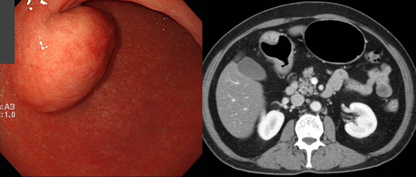
크기가 증가하는 점막하종양으로 의뢰됨. CT에서 위 병소인지, 췌장 병소인지, lesser omentum의 병소인지 애매하였음. EUS-guided biopsy를 할지 말지에 대하여 고민하고 환자와 상의하여 결국 바로 laparoscopic wedge resection을 하였고 GIST로 확인됨.
Gastrointestinal stromal tumor
High risk of malignant potential by NIH (2002) and moderate risk (16 %) of progressive disease by Miettinen (2006) :
1) tumor size: 3.5x2.7 cm
2) mitosis: 14/50 HPF (high powered fields)
3) histological type: spindle
4) necrosis: absent
5) cellularity: intermediate
6) cellular atypia: mild
7) invasion into mucosa: absent
8) resection margin involvement: absent
빙산의 일각 양상의 SMT --> fundus, wedge resection
Gastrointestinal stromal tumor of low risk of malignant potential by proposed modification for adjuvant therapy (2008) and very low risk (1.9 %) of progressive disease by Miettinen (2006):
1) tumor size: 4x3x3 cm
2) mitosis: 3/50 HPF (high powered fields)
3) histological type: spindle
4) necrosis: absent
5) cellularity: intermediate
6) cellular atypia: mild
7) invasion into mucosa: absent
8) resection margin involvement: absent
Stomach, fundus, wedge resection:
Gastrointestinal stromal tumor of low by NIH and very low risk (1.9%) of progressive disease by Miettinen (2006) :
1) tumor size: 3.3x2.5 cm
2) mitosis: 2/50 HPF (high powered fields)
3) histological type: spindle
4) necrosis: absent
5) cellularity: intermediate
6) cellular atypia: mild
7) invasion into mucosa: absent
8) resection margin involvement: absent
Stomach, lesser curvature of lower body, wedge resection:
Gastrointestinal stromal tumor of low by NIH and very low risk (1.9 %) of progressive disease by Miettinen (2006) :
1) tumor size: 2.6x2.3 cm
2) mitosis: 1/50 HPF (high powered fields)
3) histological type: spindle
4) necrosis: absent
5) cellularity: intermediate
6) cellular atypia: mild
7) invasion into mucosa: absent
8) resection margin involvement: absent
Gastrointestinal stromal tumor of high risk by NIH and high risk (55 %) of progressive disease by Miettinen (2006) :
1) tumor size: 6.8x6.6x6.2 cm
2) location: high body, posterior wall of stomach
3) mitosis: 11/50 HPF (high powered fields)
4) histological type: spindle
5) necrosis: present
6) cellularity: high
7) cellular atypia: moderate
8) invasion into mucosa: absent
9) resection margin involvement: absent
10) tumor adhesion but no invasion into pancreas
11) 7 reactive lymph nodes
Stomach, low body, wedge resection:
Gastrointestinal stromal tumor of low risk of malignant potential by proposed modification for adjuvant therapy (2008), low by NIH and very low risk (1.9 %) of progressive disease by Miettinen (2006) :
1) tumor size: 2.2x1.6x1.4 cm
2) mitosis: up to 1/50 HPF (high powered fields)
3) histological type: epithelioid
4) necrosis: absent
5) cellularity: intermediate
6) cellular atypia: moderate
7) invasion into mucosa: absent
8) resection margin involvement: absent
Exophytic growth를 보인 GIST는 내시경 진단이 어렵습니다.
Stomach, lower body, wedge resection:
Gastrointestinal stromal tumor of low risk of malignant potential by proposed modification for adjuvant therapy (2008) (see note);
1) tumor size: 3.5x3 cm
2) mitosis: up to 2/50 HPF (high powered fields)
3) histological type: epithelioid
4) necrosis: absent
5) cellularity: intermediate
6) cellular atypia: moderate
7) invasion into mucosa: absent
8) resection margin involvement: present
Note: Low risk by NIH (2002) and very low risk (1.9 %) of progressive disease by Miettinen (2006).
Exophytic growth를 보인 GIST는 내시경 진단이 어렵습니다.
Stomach, wedge resection: Gastrointestinal stromal tumor of low risk of malignant potential by proposed modification for adjuvant therapy (2008) (see note);
1) tumor size: 2.6x2 cm
2) mitosis: 1/50 HPF (high powered fields)
3) histological type: spindle
4) necrosis: absent
5) cellularity: intermediate
6) cellular atypia: mild
7) invasion into mucosa: absent
8) resection margin involvement: absent
Note: Low risk by NIH (2002) and very low risk (1.9 %) of progressive disease by Miettinen (2006).
Exophytic growth를 보인 GIST는 내시경 진단이 어렵습니다.
Stomach, lower antrum, wedge resection:
. Gastrointestinal stromal tumor of low risk of malignant potential by proposed modification for adjuvant therapy (2008) (see note);
1) tumor size: 2.8x1.9 cm
2) mitosis: 1/50 HPF (high powered fields)
3) histological type: epithelioid
4) necrosis: absent
5) cellularity: intermediate
6) cellular atypia: mild
7) invasion into mucosa: cannot be evaluated
8) resection margin involvement: absent
Note: Low risk by NIH (2002) and very low risk (1.9%) of progressive disease by Miettinen (2006).
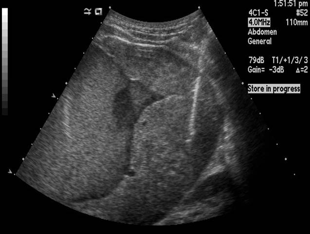
개업가 복부 초음파에서 간 옆의 mass가 발견되었습니다. 내시경과 CT 후 수술이 시행되었고 GIST로 나왔습니다. 복부 초음파로 간과 쓸개만 보는 것이 아닙니다. 이것 저것 볼 것이 많습니다.
Stomach, GIST, wedge resection: Gastrointestinal stromal tumor of low risk of malignant potential by proposed modification for adjuvant therapy (2008) (see note);
1) tumor size: 2.5x2 cm
2) mitosis: 3/50 HPF (high powered fields)
3) histological type: mixed spindle and epithelioid
4) necrosis: absent
5) cellularity: intermediate
6) cellular atypia: mild
7) invasion into mucosa: absent
8) resection margin involvement: absent
위 SMT인데 CT에서 hypervascular 양상이었기 때문에 GIST가 아닐 수도 있겠다고 생각되었습니다. CT 판독도 다음과 같았습니다. Very hypervascular mass abutting stomach. DDx. Gastric subepithelial tumor such as GIST, glomus tumor vs. primary mesenteric origin tumor such as Castleman's disease. 그런데 수술 결과는 역시 GIST로 나왔습니다. GIST는 참으로 다양한 모습을 보일 수 있습니다.
Stomach, posterior wall of low body, wedge resection: Gastrointestinal stromal tumor of low risk of malignant potential
1) tumor size: 2.7x2.4 cm
2) mitosis: 3/50 HPF (high powered fields)
3) histological type: epithelioid
4) necrosis: absent
5) cellularity: intermediate
6) cellular atypia: moderate
7) invasion into mucosa: absent
8) resection margin involvement: absent
Ki-67: positive in 3% of tumor cells
DOG-1: positive in tumor cells
C-kit (CD 117): positive in tumor cells
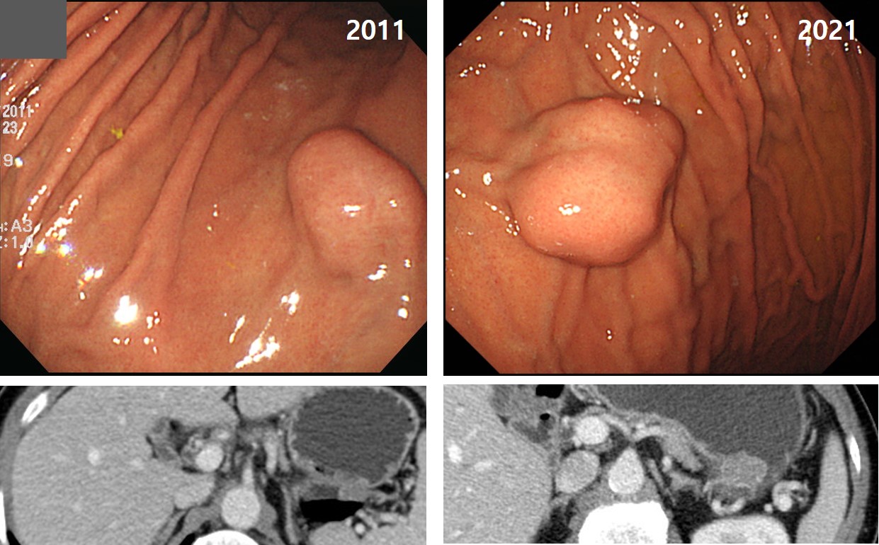
Surgery was recommended for a large gastric SMT 9 years ago, but the patient refused it. Nine years later, the patient visited my clinic again for the surgical treatment. In CT images, the diameter was 4.8cm in 9 years ago, and 5.1cm in recent images. Surgery was done.
9년 전 수술을 권했는데 환자께서 수술을 받지 않고 지내시다가 9년 만에 외부 병원에서 내시경 검사를 받은 후 갑자기 수술을 받겠다고 찾아오셨습니다.
CT를 다시 찍었는데 다행스럽게 아주 조금 자란 것으로 나왔습니다. " Gastric fundus와 맞붙어 있는 submucosal tumor로 판단되는 병변은 2011년 사진에서는 4.8 cm으로 측정되고 현재는 5.1 cm으로 약간 크게 측정됨. 전반적인 양상은 큰 변화 없으며 ascites나 peritoneal nodule 보이지 않음." 수술을 하였습니다.
Stomach, laparoscopic wedge resection: Gastrointestinal stromal tumor of intermediate risk of malignant potential by proposed modification for adjuvant therapy (2008) (see note);
1) tumor size: 5.5x4.0 cm
2) mitosis: 3/50 HPF (high powered fields)
3) histological type: spindle
4) necrosis: absent
5) cellularity: intermediate
6) cellular atypia: mild
7) invasion into mucosa: absent
8) resection margin involvement: absent
Note: intermediate risk by NIH (2002) and low risk (3.6 %) of progressive disease by Miettinen (2006).
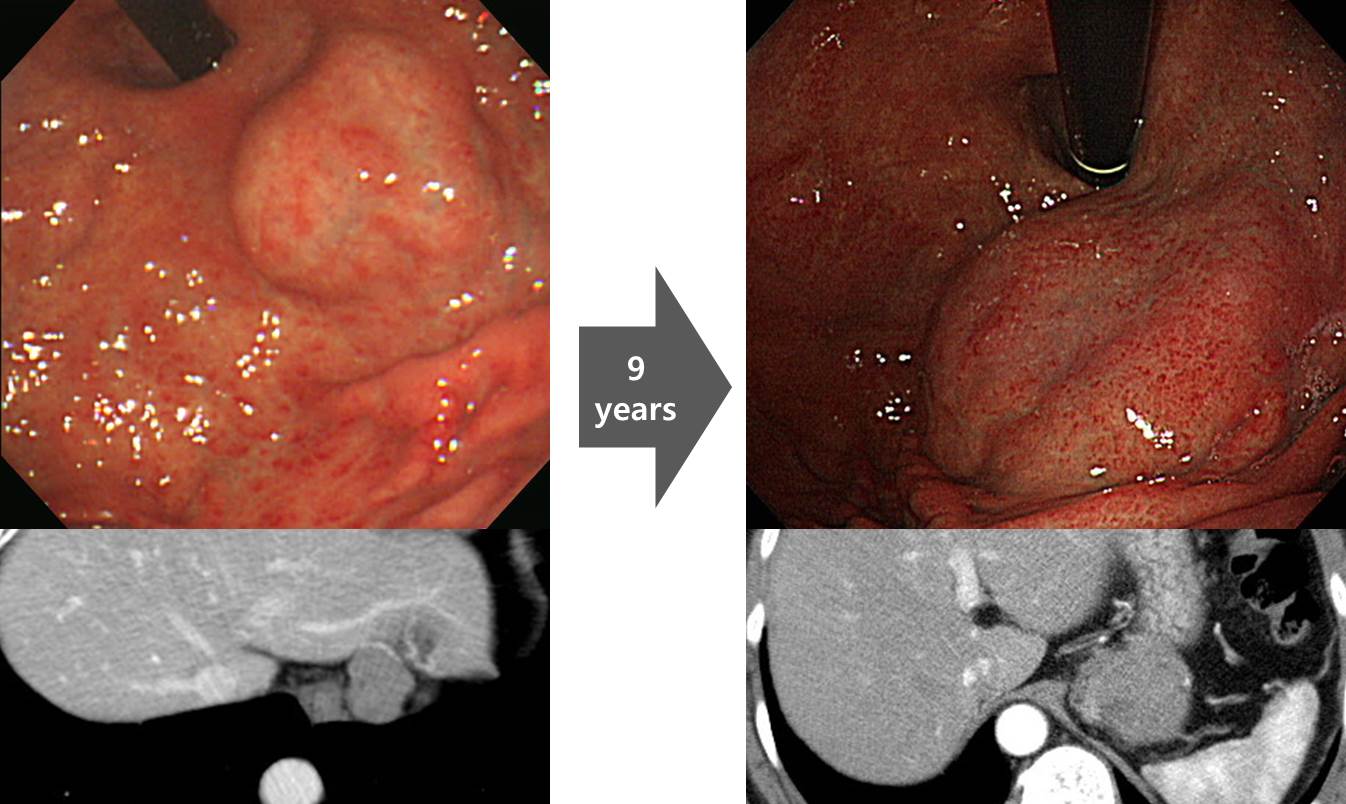
빨리 자라는 GIST도 있고 천천히 자라는 GIST도 있습니다. 아직 정확한 예측이 불가능한 상태입니다. 운명이라고밖에 할 수 없습니다.
M/50 (2017)
![]() 12. 소장 GIST
12. 소장 GIST
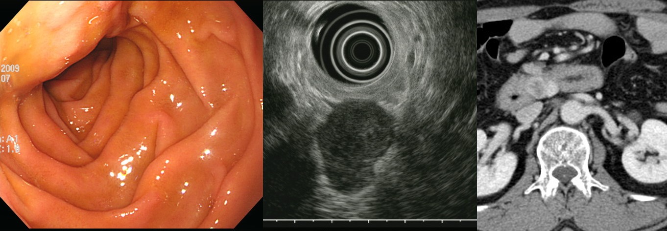
Duodenal GIST. 3.2x1.8cm
십이지장 GIST가 necrosis가 심하면 조직검사에서 나오지 않을 수 있습니다. Melena로 내원하신 분으로 두 번의 EGD 조직검사에서 조직학적 확진이 되지 않아 위에서 EUS-guided biopsy로 조직학적 확진을 내릴 수 있었던 duodenal GIST with hepatic metastais 증례입니다. (F/60, 2018)
Duodenum, common bile duct and pancreas, PPPD: Gastrointestinal stromal tumor of low risk of malignant potential, duodenum;
1) tumor size: 2.7x2.3 cm
2) mitosis: 1/50 HPF (high powered fields)
3) histological type: spindle
4) necrosis: absent
5) cellularity: intermediate
6) cellular atypia: mild
7) invasion into mucosa: absent
8) resection margin involvement: absent
. Chronic cholecystitis with cholelithiasis and cholesterol polyps, gallbladder
빈혈로 내원한 30대 여성의 십이지장에서 SMT가 발견되었고 그 일부가 함몰되어 있었으며 출혈의 원인으로 판단되었고 수술을 시행함 (2017)
Duodenum, 2nd portion, wedge resection: Gastrointestinal stromal tumor of low risk of malignant potential by proposed modification for adjuvant therapy (2008)
1) tumor size: 4.2x4x2.5 cm
2) mitosis: 3/50 HPF (high powered fields)
3) histological type: spindle
4) necrosis: absent
5) cellularity: intermediate
6) cellular atypia: mild
7) invasion into mucosa: present
8) resection margin involvement: absent
9) no metastasis in 2 lymph nodes
Pancreas uncinate process와 duodenum 사이의 enhancing mass가 9년 사이에 조금씩 커져서 수술 후 duodenal GIST로 확진 (2018, F/62)
Excision: Gastrointestinal stromal tumor of low risk of malignant potential by proposed modification for adjuvant therapy (2008) (see note);
1) tumor size: 3x2 cm
2) mitosis: 2/50 HPF (high powered fields)
3) histological type: spindle
4) necrosis: absent
5) cellularity: intermediate
6) cellular atypia: moderate
7) invasion into mucosa: absent
8) resection margin involvement: absent
건진 초음파에서 소장 혹은 췌장 종양이 발견되었습니다 ("R/O Low echoic mass lesion or collapsed bowel, inferior aspect of pancreatic tail"). CT 후 jejunum GIST로 판단되어 수술을 받으셨습니다. (2015)
Small intestine, wedge resection: Gastrointestinal stromal tumor of low risk of malignant potential:
1) tumor size: 4.5x3 cm
2) mitosis: 0/50 HPF (high powered fields)
3) histological type: spindle
4) necrosis: absent
5) cellularity: intermediate
6) cellular atypia: mild
7) invasion into mucosa: absent
8) resection margin involvement: absent
C-kit, PKC-e, DOG-1, CD34: all positive
Ki-67: positive in less than 2% of tumor7년 후 복부 증상으로 검사를 받아 간 종양으로 의뢰되었습니다. 간 조직검사를 하였습니다.
Liver, S8/7, biopsy : Gastrointestinal stromal tumor
C-KIT (CD 117) : Diffusely positive in tumor cells
DOG-1 : Diffusely positive in tumor cells
Cytokeratin (AE1/AE3) : Negative in tumor cells
Ki-67 : Positive in about 3% of tumor cells
Actin (Smooth muscle) : Focally positive in tumor cells다른 곳의 병소가 없었으므로 과거 jejunal GIST의 간전이로 판단하였습니다. 글리백 치료를 위하여 종양내과로 의뢰되었습니다. 소장 GIST는 무섭습니다. "Low risk of malignant potential"로 나왔던 환자에서도 간전이를 보이는 경우가 있으니까요. Low는 No가 아닙니다.
[More cases]
Gastrointestinal stromal tumor of intermediate risk of malignant potential by NIH (2002) and moderate risk (24 %) of progressive disease by Miettinen (2006) :
1) tumor size: 5.5x5.5x5 cm
2) mitosis: 0/50 HPF (high powered fields)
3) histological type: spindle
4) necrosis: absent
5) cellularity: intermediate
6) cellular atypia: mild
7) invasion into mucosa: present
8) resection margin involvement: absent
9) no metastasis in 3 regional lymph node (0/3 : "mesenteric LN", 0/3)
Jejunal GIST
Gastrointestinal stromal tumor of high risk of malignant potential by NIH (2002) and high risk (85%) of progressive disease by Miettinen (2006) :
1) tumor size: 7.8x7x5 cm
2) mitosis: 22/50 HPF (high powered fields)
3) histological type: spindle
4) necrosis: present
5) cellularity: intermediate
6) cellular atypia: mild
7) invasion into mucosa: present
8) myxoid change: present
9) resection margin involvement: absent
Jejunal GIST
small bowel malignant GIST with peritoneal seeding (exon 11 mutation +)
Ileal GIST with liver metastasis
다발성 간전이를 동반한 GIST였습니다. EGD에서 위와 십이지장의 mucosal lesion은 없었으나 CT 소견을 근거로 gastric GIST with liver metastasis로 잠정 진단하고 imatinib을 사용하였습니다. 간 병소는 다 없어지고 원발병소만 남아서 수술을 하였는데 병소의 위치가 위가 아니라 십이지장이었습니다. 처음부터 duodenal GIST with liver metastasis였을 것으로 판단하였습니다.
난소암 의심하여 개복술 시행하였으나 소장 GIST로 나왔습니다.
명치부 불편감으로 시행한 CT에서 우연히 발견되 duodenal 3rd portion hypervascular mass였으며 소아 대장경을 이용한 push enteroscopy 후 수술 시행
Duodenal wedge resection: Gastrointestinal stromal tumor of very low risk of malignant potential by proposed modification for adjuvant therapy (2008);
1) tumor size: 1.8x1.8 cm
2) mitosis: 0/50 HPF (high powered fields)
3) histological type: spindle
4) necrosis: absent
5) cellularity: intermediate
6) cellular atypia: moderate
7) invasion into mucosa: absent
8) resection margin involvement: absent
Note: Very low risk by NIH (2002) and none risk (0%) of progressive disease by Miettinen (2006).
. Ki-67 : Positive in up to about 2% of tumor cells
. C-KIT (CD 117) : Positive in tumor cells
. DOG-1 : Positive in tumor cells
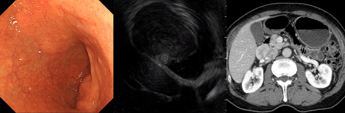
UGI bleeding으로 몇 번 내시경하여 어렵게 진단된 십이지장 GIST (남자 55세, 2021년) GIST high risk
![]() 13. 대장 GIST 증례
13. 대장 GIST 증례

Rectum, trans-anal endoscopic microsurgery:
. Gastrointestinal stromal tumor of very low risk of malignant potential by proposed modification for adjuvant therapy (2008), none risk (0 %) of progressive disease by Miettinen (2006) :
1) tumor size: 1.4x1.2x0.4 cm
2) mitosis: 3/50 HPF (high powered fields)
3) histological type: spindle
4) necrosis: absent
5) cellularity: intermediate
6) cellular atypia: mild
7) invasion into mucosa: absent
8) resection margin involvement: absent
. Pho-H-H3: Positive in less than 5% of tumor cells
. C-kit : Positive
. DOG1 : Positive
. Ki-67: Negative
. PKC-Θ: Weakly positive제 전공은 아니지만... 이렇게 작은 GIST는 follow up 하면 어떤가 생각해 보았습니다.
![]() 14. 기타 장기 GIST
14. 기타 장기 GIST
식도 GIST. 모두 수술로 절제한 후 식도 GIST로 나온 증례입니다. 식도 GIST는 leiomyoma보다 매우 rare합니다. 하부식도에 위치하고 lobulated 되는 경향이 있고 leiomyoma보다 soft 합니다. Leiomyoma는 보통 매우 단단합니다.
췌장 GIST
Small intestine, pancreas, CBD and gallbladder, PPPD : Gastrointestinal stromal tumor of low risk of malignant potential by proposed modification for adjuvant therapy (2008), low risk (8.3%) of progressive disease by Miettinen (2006)
1) tumor size: 2.3x2x1.8cm
2) mitosis: up to 1/50HPF (high powered fields)
3) histological type: epithelioid
4) necrosis: absent
5) cellularity: low
6) cellular atypia: mild
7) invasion into mucosa: absent
8) resection margin involvement: absent
. 20 reactive lymph nodes
![]() 15. Code
15. Code
수술한 위 GIST의 코드에 대한 논란이 있어 잠정안을 마련하였습니다. (2017/3)
돈자루 및 칼자루를 가지고 있는 정부에서는 기준부터 명료하게 만들어야 할 것입니다. 환자의 질병에 대하여 논의할 시간도 부족한데 코드 가지고 자꾸 시간을 낭비하다니 안타깝습니다. 주지하는 바와 같이 '암등록본부'가 기준은 아닙니다. 아무도 기준을 안 알려주니 여기 저기 찔러볼 뿐입니다. '암등록본부'도 그 중 하나입니다.
![]() [FAQ]
[FAQ]
[2016-11-14. 2016년 발표된 GIST management 종설에 대한 김상균 교수님 편지]
1) 2-5 cm GIST 중 실제 malignant risk는 very low risk에서 intermediate까지 다양합니다. Very low rsk에 해당하는 경우가 많은 상황에서 모든 환자에게 수술을 권유하는 것은 과잉치료가 될 수 있습니다. High risk feature를 갖는 환자에 한해 치료가 이루어지는 것이 바람직할 것으로 보이나, 아직까지 과연 어떤 것이 그런 경우인지를 미리 확인하는 것이 현실적으로 어렵다는 한계가 있습니다만 (크기, 궤양의 여부, 내부 necrosis 여부 등이 실제로 큰 관계가 없는 경우가 많습니다).
2) 2 cm 미만의 GIST는 mitotic index와 관련없이 모두 very low risk에 해당합니다 (Hornick JL. Hum Pathol 2007에 따르면 소장은 아니지만 위 GIST의 경우 크기가 2 cm 이하이면 mitotic index가 5/50 HPF 이하와 이상 모두 very low risk에 해당합니다). 이 경우는 수술이 필요없다고 생각합니다. 단, very low risk는 진단 당시의 시점만을 반영하는 것이므로 시간 경과에 따른 변화 여부는 꼭 확인해야 합니다 (크기 변화는 전체의 5% 미만).
[2016-11-20. 이준행 답변]
좋은 의견 감사합니다.
위 GIST가 2cm 이하면 같은 크기의 소장이나 대장 GIST보다 위험성이 덜합니다. 2 cm 이하의 위 SMT는 비록 조직학적으로 GIST로 확인되더라도 무조건 수술을 권하지 않습니다. 2016년 발표된 GIST management 종설의 algorithm에서 알 수 있듯이 비록 조직학적으로 GIST라고 확인되더라도 high risk feature가 없으면 경과관찰이 가능합니다.
2 cm 이하의 위 SMT 혹은 위 GIST에 대한 과잉 검사와 과잉 치료의 가능성을 지적해 주셔서 감사합니다.
[2017-2-15. 애독자 질문]
교수님께서 보여주신 증례 중에 아래 증례같은 경우 (1-3 cm 사이의 경우) 저는 수술할지 경과 관찰할지 항상 고민입니다. FNAB나 incision을 해서 조직검사하는 것을 적극적으로 권고하시는지요?
[2017-2-15. 이준행 답변]
중요한 질문이라고 생각합니다. 현재 우리나라에서 인정된 '무증상 성인의 위 SMT에 대한 가이드라인'은 없기 때문에, 아래와 같은 일본 가이드라인을 따르는 경우가 많습니다. 물론 공인된 정답은 아닙니다.
일본측 의견에 따르면 2cm가 되지 않는 SMT에서 특별히 나쁜 소견이 없으면 조직을 얻지 말고 경과관찰을 하면 됩니다. 이렇게 권하는 이유 중 하나는 조직을 얻어서 GIST가 나오면 수술을 할 수 밖에 없기 때문입니다. 조직을 모르는 상태에서는 조심스럽게 경과관찰을 할 수 있지만, 일단 GIST인 것을 알면 수술을 하지 않을 수 없습니다. 법적인 이유도 있기 때문입니다. '모르는 게 약이다'라는 말이 있는데 딱 여기에 해당합니다.
통일된 국내 가이드라인이 없기 때문에, 전문가 사이의 의견차가 발생합니다. 제법 생각의 차이가 큽니다. 한 병원 내에서도 의견이 통일되어 있지 못합니다. 질문하신 증례는 제 환자는 아니었습니다. EUS에서 장경 18.9mm로 측정되었고, EUS-guided로 조직이 얻어졌고, GIST로 나왔고, 수술이 진행되었습니다. 타당한 검사와 타당한 수술이었습니다. 다만 모든 의사가 다 그렇게 해야만 하는 상황은 아니었습니다.
우선 CT를 찍어 2cm 미만으로 나오면 내시경 추적 관찰도 가능한 option이었습니다. 더 커졌거나 문제가 있을 때만 수술하자는 전략입니다. 저는 그렇게 했을 것 같습니다.
결국 조금 적극적인 견해도 가능하고 다소 보수적인 견해도 가능한 환자였습니다. 빨리 통일된 국내 가이드라인이 필요합니다. 그러나 매우 매우 매우 매우 어려운 일일 것입니다. 이유는 아시겠지만 ..... (글로는 쓸 수 없습니다. 제 생각을 알고 싶으시면 학회나 집담회때 개인적으로 논의해 봅시다. 술 한잔 사 달라고 찾아오셔도 좋습니다.)
[2023-5-21]
다시보기가 가능한 web seminar의 힘은 대단합니다. 4월 18일 KSGE 세미나 이하예민 교수님 강의를 리뷰하였습니다.
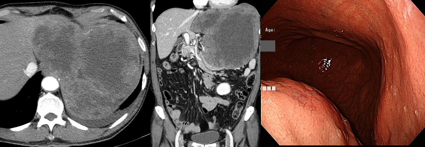
![]() [2025-1-19] Exophytic SET at antrum에 대한 질문에 답합니다.
[2025-1-19] Exophytic SET at antrum에 대한 질문에 답합니다.
![]() [2026-3-27]
[2026-3-27]
![]() [References]
[References]
1) EndoTODAY 점막하종양 submucosal tumor
2) The standard diagnosis, treatment, and follow-up of GIST - 2016년 Gastric Cancer지 1월호 종설
© 일원내시경교실 바른내시경연구소 이준행. EndoTODAY Endoscopy Learning Center. Lee Jun Haeng


