![]() [Description exercise 1 해설] - 終
[Description exercise 1 해설] - 終
DEX quiz 시작을 축하합니다. 많은 분들의 답변 중 비교적 공통적으로 틀리는 부분을 중심으로 해설을 붙였습니다. 다른 분들의 답변과 그에 대한 저의 comment를 천천히 읽어보시기 바랍니다. 가끔 YouTube 동영상도 있을 것입니다 (YouTube 내시경교실).
가장 중요한 소견이 무엇인가 생각하고 답을 작성해 봅시다. 애매한 소견을 모두 기술할 필요는 없습니다. 피부과 의사가 점을 하나하나 기술하지 않는 것처럼...
그러나 주소견에 대한 부소견은 가급적 자세히 써보실 것을 권합니다. 배우는 입장에서는 맞든 틀리든 일단 길게 쓰다보면 뭔가 새롭게 알게 되는 것이 있을 것입니다.
2022년 1월 10일 발간된 아래 책자의 해당 부분을 읽어보시면 크게 도움이 될 것입니다.
![]() Case 1
Case 1
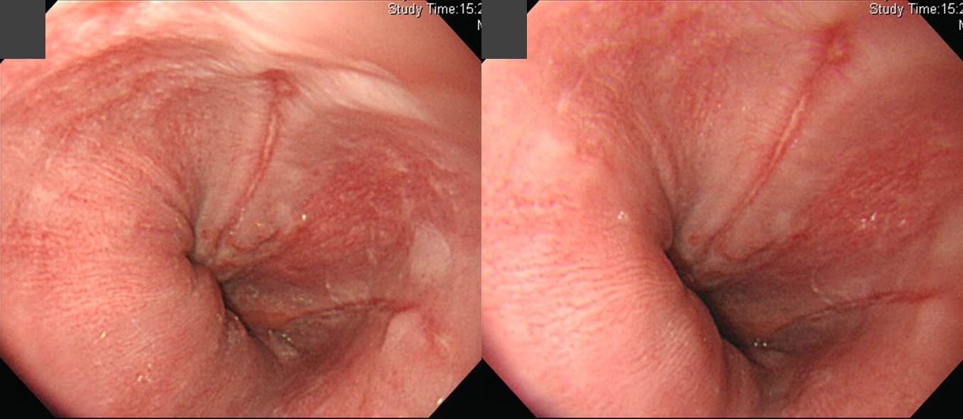
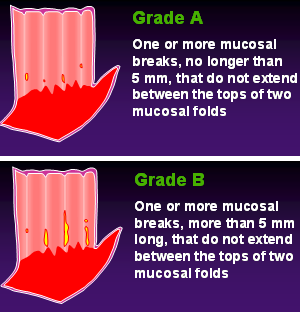
Findings: 위식도접합부에서 시작하여 상방으로 5 mm 이상의 linear mucosal break가 있음 (1시 방향). 3시 방향에는 5 mm 이하의 또 다른 mucosal break가 있음. 주변 점막은 약간 하얗고 두터워보임. 이로 인하여 mucosal break 인접 점막이 불투명해보임. (At far distal esophagus, a linear mucosal break longer than 5 mm is located at 12 o'clock direction. Another short mucosal break less than 5 mm is seen at 3 o'clock direction. Surrounding mucosa shows dirty-white mucosal hypertrophy.)
Impression: Reflux esophagitis, LA-B
[이준행 comment]

가끔 이 증례에 대하여 confluent하다고 보고 LA-C를 주시는 분이 계시는데.... over입니다. Confluent하다고 하려면 아래와 같이 보다 명확해야 합니다.
한 선생님께서 위치 표시를 위하여 LES라는 용어를 사용하셨기에 아래와 같이 comment 해 주었습니다. 내시경은 육안 소견 위주의 검사법입니다. 육안 소견에 충실하게 기술하는 것이 좋습니다.
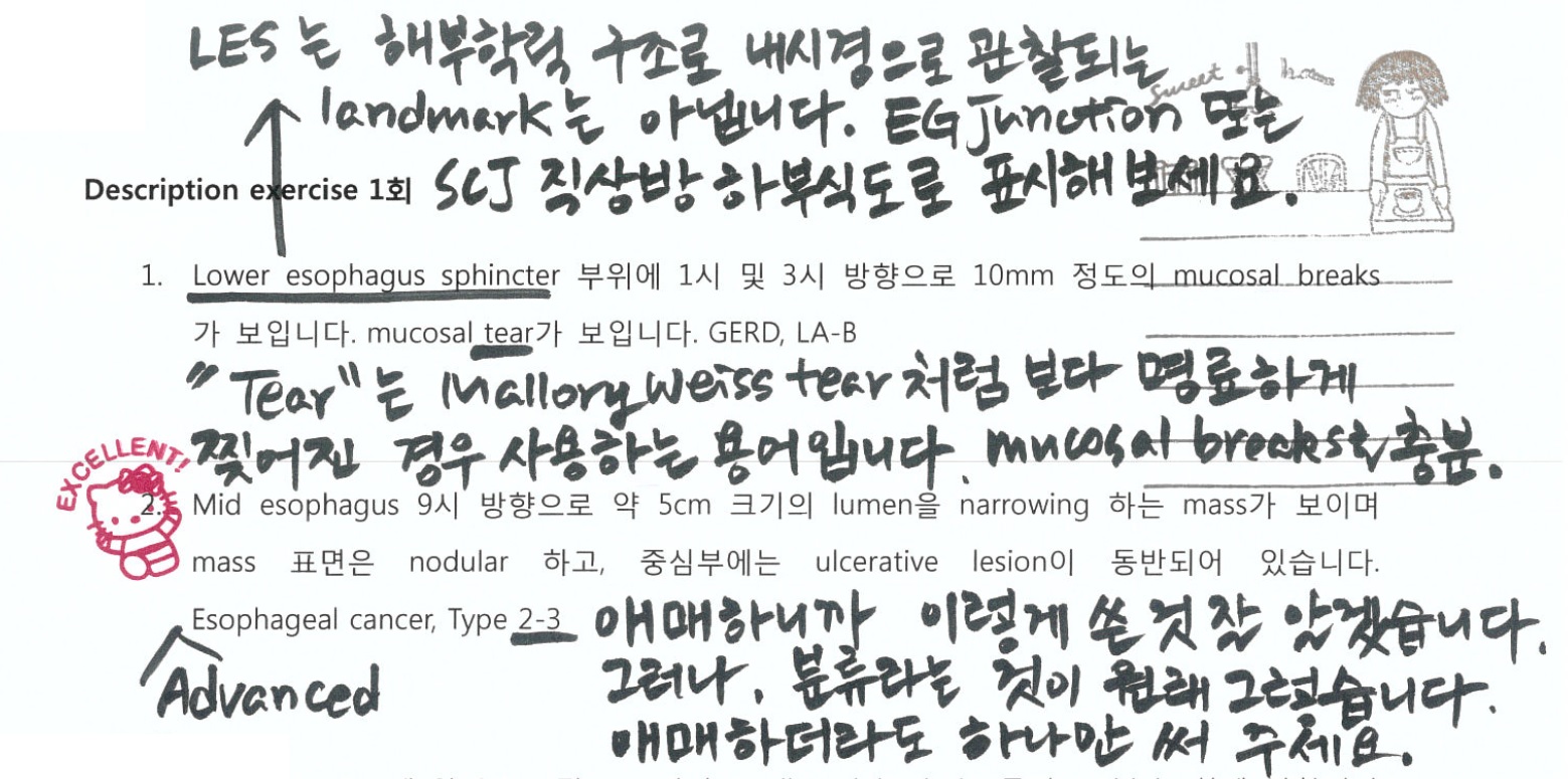
바렛 식도가 의심된다는 의견을 주신 분이 계셔서 아래와 같이 답변드렸습니다.
DEX 문제는 높임말로 답하지 마시고 공문서처럼 쓰기 바랍니다.
Mucosal break 주변 점막이 squamous epithelial hyperplasia로 인하여 하얗고 불투명해 보이는 것이 역류성 식도염의 전형적 소견입니다.
내시경 동아리 성시경 학생 답변입니다. DEX 문제는 높임말로 답하지 마시고 공문서처럼 쓰기 바랍니다.
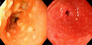
간혹 mucosal break (longitudinal erosion)이 아니라 tear로 기술하신 분이 있습니다. Mallow Weiss tear는 이름 그대로 좀 더 찢어진 느낌입니다. 확연히 다릅니다.
Mallory Weiss tear 증례 사진들
마지막으로 동영상 강의를 소개합니다.


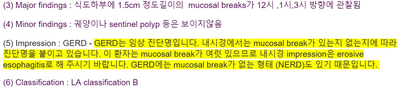
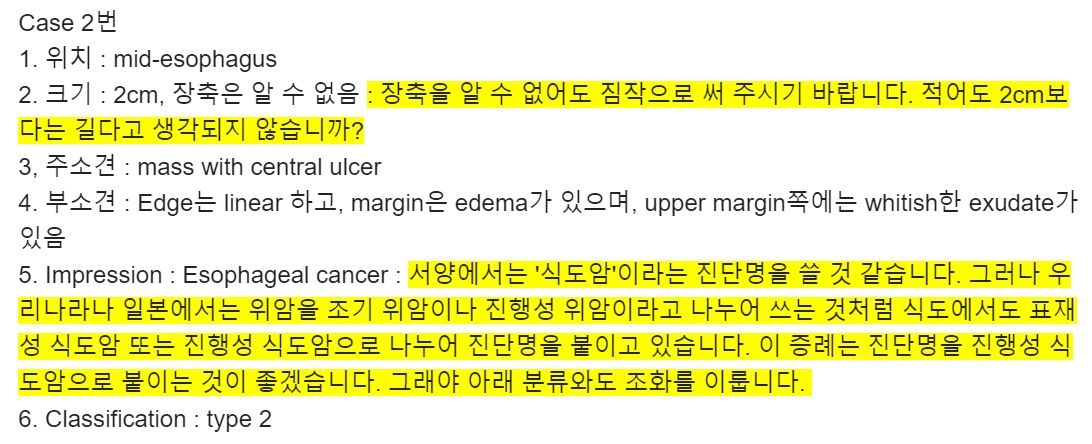
![]() Case 2. Mid-esophagus
Case 2. Mid-esophagus
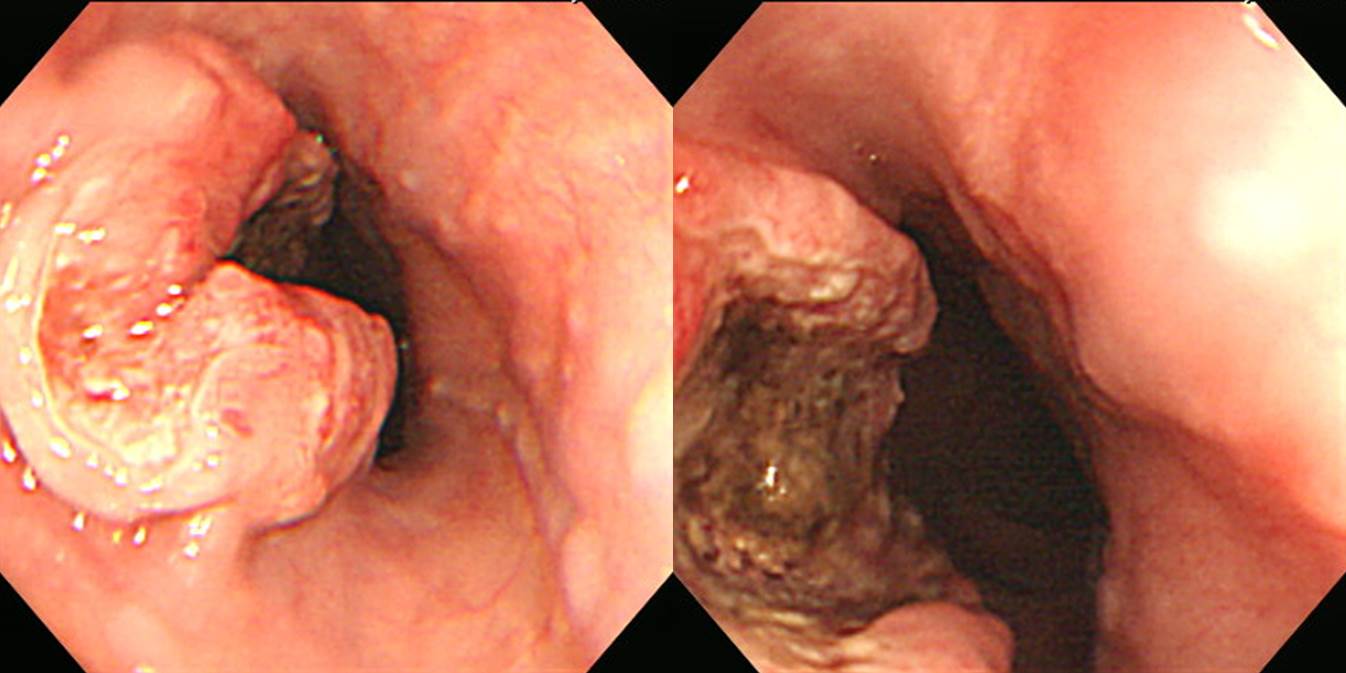
Findings: 중부 식도 내강을 좁히고 있는 4x2 cm의 mass가 있고 중앙부에는 deep ulceration이 있음. Ulcer base는 exudate가 지저분하게 덮여있고 주변 융기부에서는 irregular surface, erythema, mucosal friability 가 보임. (At mid-esophagus, a 4x2 cm sized luminal-narrowing mass with deep central ulceration was seen. The ulcer base was covered with dirty necrotic exudate. Protruded portion showed irregular surface, erythema, and mucosal friability.)
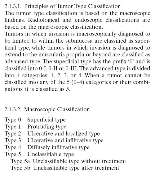
Impression: Advanced esophageal cancer, type 2
[이준행 comment]
식도암도 위암처럼 superficial esophageal cancer 또는 advanced esophageal cancer로 impression을 붙이는 것이 관례입니다. Early esophageal cancer라는 말은 내시경 진단에서 사용하지 않습니다. Early esophageal cancer는 림프절 전이가 없다는 것이 확인된 이후 쓰는 말이므로 내시경 진단에서는 언제나 superficial을 씁니다. (서양에서는 EGC/AGC 개념이 명확하지 않아서 impression을 gastric cancer로 붙이고 classification을 early로 하는 것이 가능합니다. 그러나 우리나라에서는 EGC 혹은 AGC로 진단명을 붙이고 있습니다.^^) 그리고 classification도 위암 비슷하게 해 주시기 바랍니다.
크기 측정에 어려움을 겪는 분들이 많았습니다.

최종 병리결과도 아래와 같았습니다. 역시 4 cm.
Invasive squamous cell carcinoma, moderately differentiated, esophagus:
1) tumor size: 4x2 cm
2) extension to perimuscular adventitia
3) endolymphatic tumor emboli: not identified
4) perineural invasion: present
5) involvement of radial margin
6) negative resection margins (proximal, 3 cm ; distal, 10 cm)
7) metastasis to 3 out of 62 regional lymph nodes (3/62: "LC omentum", 0/1; "RRLN", 0/0; "LRLN", 0/9; "5", 0/6; "7", 2/15; "8u", 0/1; "R9", 0/2; "L10", 0/1; "G1", 0/2; "G2", 0/8; "G3", 1/17)
식도암의 기술과 분류는 다소 애매한 측면이 있습니다만, 이 경우는 ulcerative mass (= type 2) 정도가 좋지 않을까요? 식도는 좁은 tubular organ이므로 위에서 보이는 전형적인 궤양형 (= type 2)은 드문 것 같습니다. (참고: EndoTODAY 분류법)
내시경 결과는 담백하게 쓰는 것이 좋습니다. Keep it simple and smart (KISS)!

![]() Case 3
Case 3
Findings: 위전정부에 multiple white small flat nodule들이 diffuse하게 scattered 되어 있음. (At antrum, multiple white small flat nodules were diffusely scattered.)
Impression: Metaplastic gastritis
[이준행 comment]
어디가 병인지 모르겠다고 분들이 많은 문제입니다. 그렇습니다. 질병은 크게 4가지 종류가 있습니다. Two by two table과 비슷합니다. 한쪽은 focal/diffuse 축이고 다른 쪽은 benign/malignant 축입니다. 이 문제는 'diffuse + benign'의 예입니다. 위염의 감별진단이라는 말씀입니다.
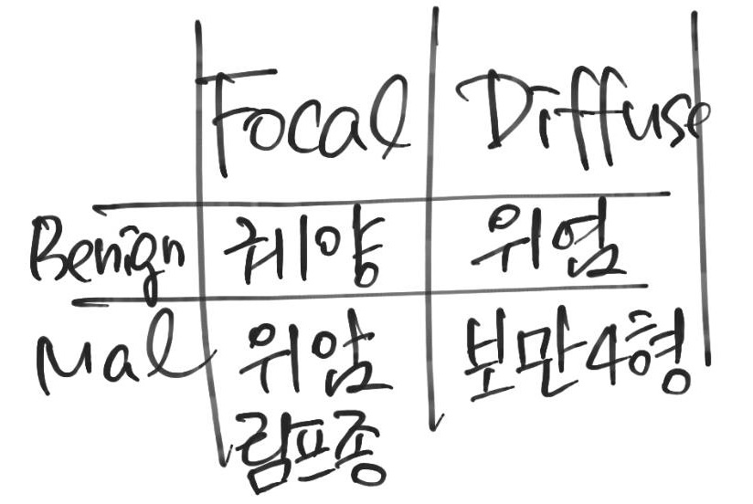
Diffuse/benign 병소에 대하여 크기를 쓸 필요는 없습니다. 반면 Diffuse/malignant인 보만 4형 진행성 위암은 가급적 크기를 써 주기 바랍니다. 암인데 크기를 쓰지 않는 것은 조금 이상하니까.
Impression을 "intestinal metaplasia"라고 쓰신 분이 계십니다. Intestinal metaplasia가 현저하므로 impression (= 진단명)은 metaplastic gastritis 입니다. Chronic이라는 말은 생략하는 것이 관행입니다. Metaplastic gastritis는 모두 chronic이니까.
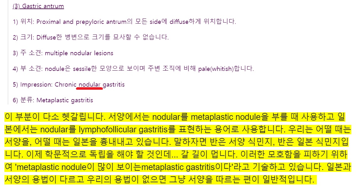
Nodular gastritis로 답한 분도 계셨습니다. Nodular gastritis는 헬리코박터에 의한 lymphofollicular gastritis의 (일본식) 명칭이면서, metaplastic gastritis의 서양식 이름입니다. 무척 헷갈릴 수 밖에 없어서 저는 nodular gastritis라는 이름을 잘 쓰지 않습니다. 이 증례는 전형적인 metaplastic gastritis입니다.

몇 년 전 내과 전문의 시험에서 metaplastic gastritis 사진이 나왔습니다. 소화기내과에서 내시경 교육을 받은 사람들은 쉽게 맞추었는데, 그렇지 못한 대부분의 내과 전공의들은 틀렸다는 후문이었습니다. 직접 내시경을 해 보지 못한 분들에게는 오히려 위염이 더 어려울 수 있습니다. Metaplastic gastritis 사진 몇 장을 아래에 소개합니다.
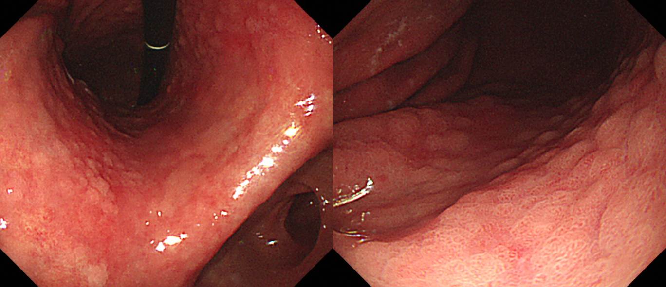

위 거의 전체가 위축성 화생성 변화가 현저하였던 70대 남성
위전정부는 비교적 깨끗한데 위체부 화생성 변화가 현저한 경우
EndoTODAY '위내시경 삽입과 관찰' 150쪽을 참고하십시오.
위염 강의 동영상을 소개합니다.
2017년 4월 15일 금요일 저녁 6시-8시
* 참고: EndoTODAY 위염
![]() Case 4
Case 4
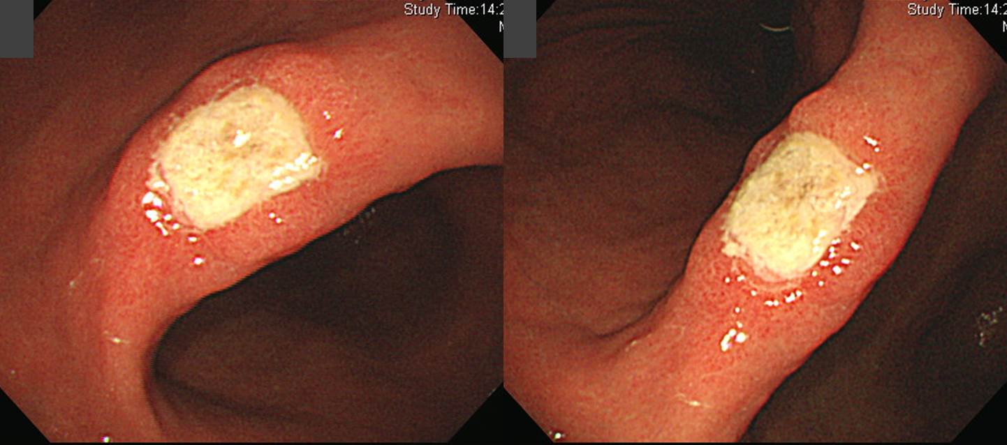
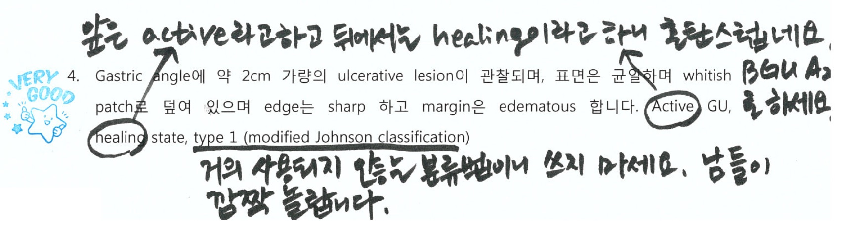
Findings: 위각에 2cm 크기의 궤양이 있음. 궤양 바닥은 두꺼운 exudate로 덮여 있고, edge는 sharp하고, margin은 edematous 함. 주름 변화는 없음. (At the center of the angle, there was a 2 cm ulcer the sharp edge and edematous margin. The ulcer base was covered with thick white-yellow exudate. There was no fold change.)
Impression: Benign gastric ulcer, active 2 stage
[이준행 comment]

Edge와 margin을 구분하여 사용할 것을 권합니다.
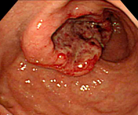
Edge와 margin은 혼동되는 용어입니다. 사전을 찾아보다도 정확한 의미 차이를 알기 어렵습니다. 출판계에서는 edge와 margin을 정확하게 구분하여 사용하고 있습니다. 왼쪽 그림을 보시기 바랍니다. edge는 선이고 margin은 면입니다. 이와 같은 edge와 margin의 의미 차이를 고려하면 오른쪽 사진은 ‘궤양의 edge는 sharp하고 margin은 edematous하다’고 말할 수 있습니다. 이제 ‘조직검사는 edge에서 시행한다’는 것이 어디를 말하는지 정확히 이해하셨을 것입니다.
소화성 궤양의 내시경 진단에 대해서는 아래 강의 동영상을 꼭 보시기 바랍니다.


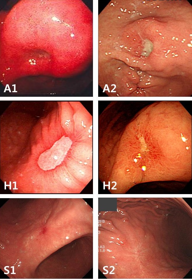
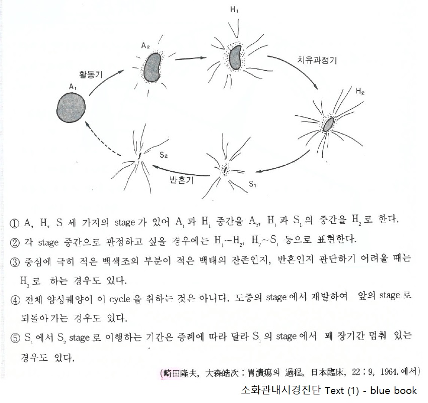
위/십이지장궤양의 병기는 AHS (active-healing-scar) 방식을 사용합니다.
- Active stage 1 (A1): Active and blurred edge. 방금 궤양이 만들어진 경우로 비교적 작고 깊은 ulcer crater가 있고 주변 점막이 심하게 부어 있습니다. 아직 regenerating epithelium이나 fold 변화는 없습니다.
- Active stage 2 (A2): Active and sharp edge. 며칠 정도 지난 궤양입니다. Ulcer crater는 A1 stage보다 넓어지지만 주변 점막 부종이 다소 완화되어 보입니다. Regenerating epithelium과 fold 변화가 아주 살짝 보일 수 있습니다.
- Healing stage 1 (H1): Healing with regenerating epithelium. 주변 점막 부종은 거의 가라앉은 상태이며 regenerating epithelium이 현저히 보입니다. 그러나 아직 ulcer crater가 제법 많이 남아있습니다. Fold 변화도 동반됩니다.
- Healing stage 2 (H2): Almost healed by regeneration. 주변 점막 부종은 모두 가라앉았고, regenerating epithelum이 대부분을 자치하고 ulcer crater는 아주 조금 남아있습니다. Fold 변화가 현저합니다.
- Scar stage 1 (S1): Red scar. Ulcer crater는 없고 regenerating epithelium이 약간 붉은 색조로 보이고 fold만 남아있습니다.
- Scar stage 2 (S2): White scar. Fold만 남아있고 ulcer crater나 regenerating epithelium이 보이지 않습니다. Regenerating epithelium이 오래되어 정상 점막과 구분이 어려운 경우입니다.
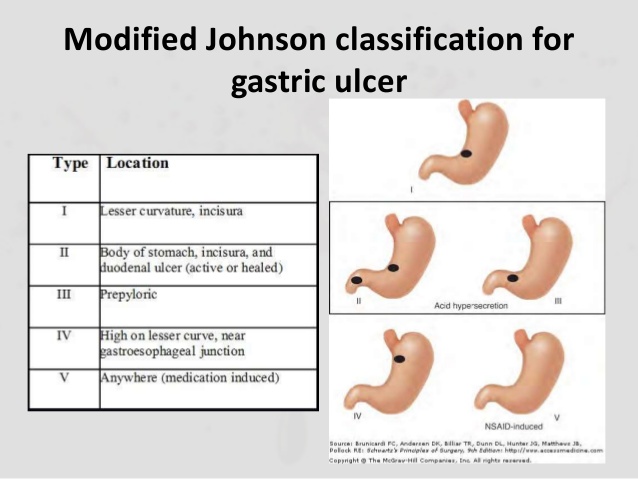
한 선생님께서 위궤양의 분류로 modified Johnson classification을 언급해 주셨습니다. 위치와 pathogenesis를 고려하여 만들어진 분류인데, 유용성에 대한 의문이 있어서 요즘은 거의 사용하지 않고 있습니다. 안 쓰셔도 좋습니다. 아니 쓰지 마세요. 남들이 거의 잘 모르는 분류이니까...
위각 (gastric angle)에 대한 질문이 있어서 답합니다.
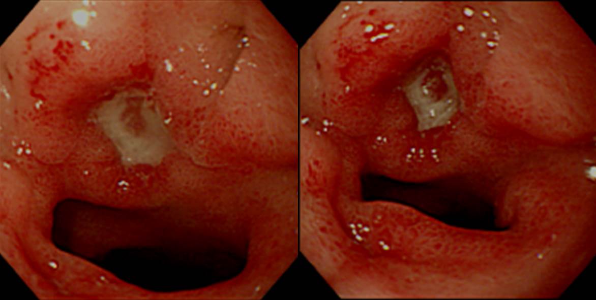
![]() Case 5
Case 5
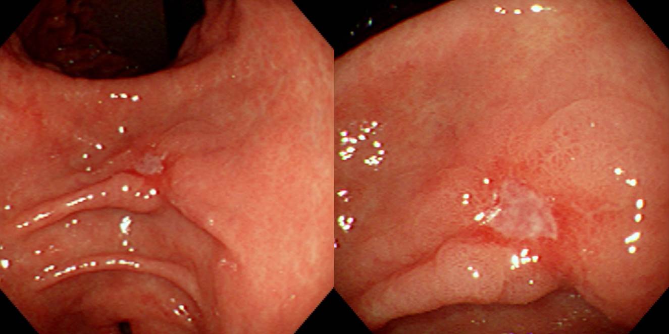
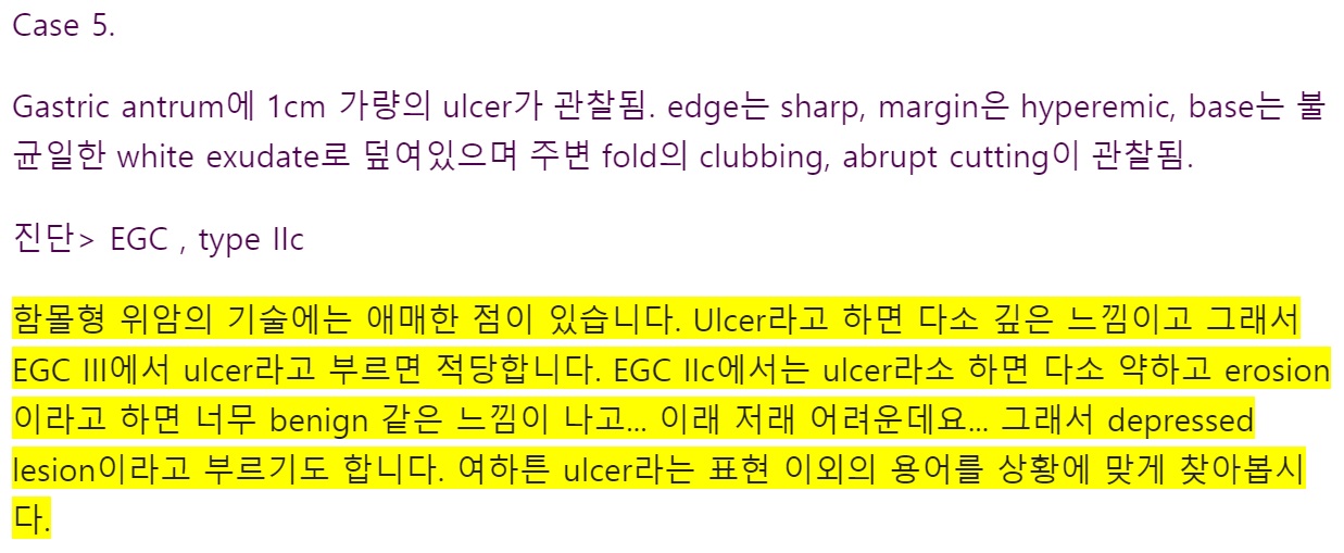
Findings: 전정부 소만에 1cm 가량의 slightly depressed lesion이 있음. Edge는 비교적 sharp하지만 일부 spiculation 되어 있고 (5시와 8시 반 방향) 8시와 5시 반 방향에서 fold가 끌려오고 있으며 그 끝은 함몰부의 edge에서 약간 뭉퉁하고 abrupt하게 끊기고 있음. (At the lesser curvature of the gastric antrum, a 1 cm sized depressed lesion was seen. The edge is relatively sharp, but there were some areas of spiculation. Multiple abnormal converging folds are seen (cutting and clubbing).)
Impression: Early gastric cancer, type IIc
[이준행 comment]
This is tricky. Because the center is depressed and margin is slightly elevated. We call it depressed lesion because marginal elevation is considered as secondary change. Actually, folds are formed only in the depressed lesions. So, the endoscopic diagnosis is EGC IIc (not IIa).
This is an old case, so surgery was done. In the current clinical practice, endoscopic submucosal dissection can be considered.
Stomach, subtotal gastrectomy:
Early gastric carcinoma
1. Location: middle third, center at body and posterior wall
2. Gross type: EGC type IIc+III
3. Histologic type: tubular adenocarcinoma, well differentiated
4. Histologic type of Lauren: intestinal
5. Size: 1.6x1.1 cm
6. Depth of invasion: extension to mucosa (muscularis mucosa) (pT1a)
7. Resection margin: free from carcinoma (safety margin: distal 6 cm, proximal 3.7 cm)
8. Lymph node metastasis: no metastasis in 31 regional lymph nodes (pN0)
9. Lymphatic invasion: not identified
10.Venous invasion: not identified
11.Perineural invasion: not identified
* 참고: EndoTODAY 조기위암 아틀라스
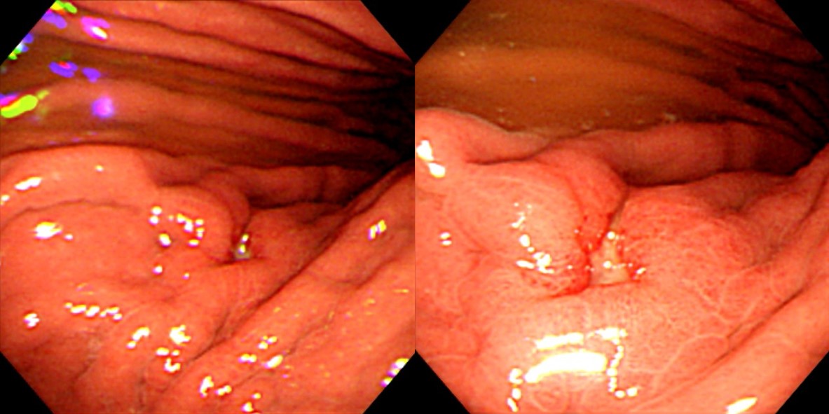
![]() Case 6
Case 6
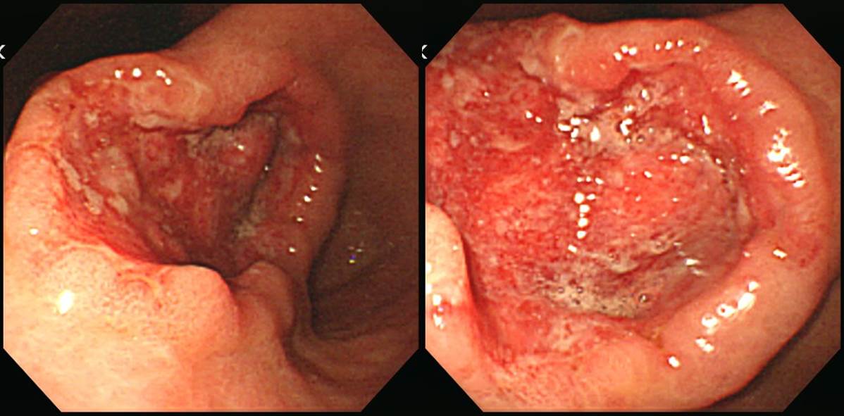
Findings: Proximal antrum, lesser curvature (위각 직하부)에 5 cm 가량의 mass가 있고 그 중앙에 깊고 불규칙한 ulceration이 있음 (ulcerative mass). Ulcer base에서는 erythema, friability, dirty exudate가 있고 깔려있음. (There is a 5 cm sized mass at the lesser curvature side of the proximal antrum, just below the angle. The center was deeply ulcerated. The uneven ulcer base shows erythema, friability, and dirty exudate.)
Impression: Advanced gastric cancer, Borrmann type II
[이준행 comment]
Mass with deep central ulceration (with narrow bank) 이라고 불러도 좋고 ulcerative mass라고 불러도 좋습니다.
Stomach, subtotal gastrectomy:
Advanced gastric carcinoma
1. Location : lower third, Center at antrum and posterior wall
2. Gross type : Borrmann type 3
3. Histologic type : tubular adenocarcinoma, poorly differentiated
4. Histologic type by Lauren : intestinal
5. Size : 5.5x4.5x0.8 cm
6. Depth of invasion : penetrates serosa (pT3)
7. Resection margin: free from carcinoma, safety margin: distal 2.3 cm, proximal 4 cm
8. Lymph node metastasis : no metastasis in 37 regional lymph nodes (pN0)
9. Lymphatic invasion : present
10. Venous invasion : not identified
11. Perineural invasion : present
12. Stage by AJCC : II (T3, N0, MX)아직 처음이라 그런지 어떤 분이 붉은 부분을 보고 bleeding 소견이 있다고 언급하셨습니다. 피가 날 때만 bleeding이라는 용어를 사용합시다.
용어에 대한 comment입니다.
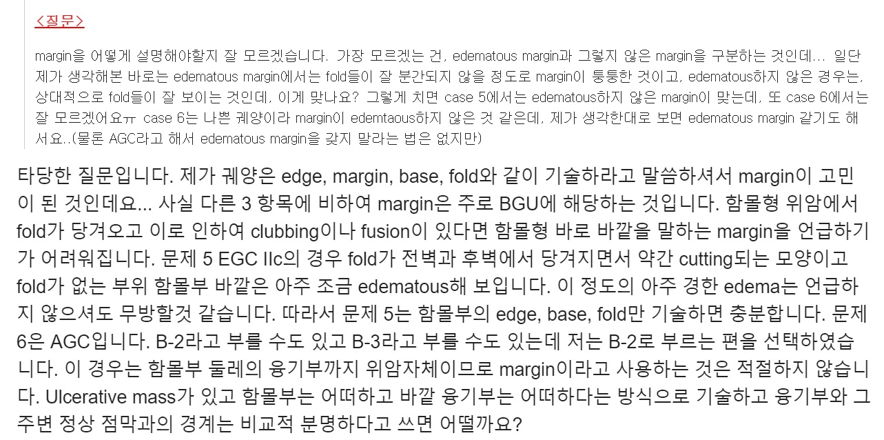
[2020-5-19. 성시경 학생 질문]
교수님의 빠른 답변에 정말 감사드립니다! 자세히 적혀있는 해설을 읽어보니 저의 description에 대한 어휘가 부족함을 느꼈습니다.
교수님의 첨삭과 해설을 읽고 나서 한 가지 질문이 생겼습니다. 제가 edematous하다고 기술한 것이 맞는지 잘 모르겠습니다. Ulcer가 있을 때 margin이 주변보다 융기된 것을 모두 edematous하다고 보면 되는 것인가요? 증례 6과 같은 AGC B-II에서 융기된 margin을 edematous하다고 부르는지 궁금했습니다.
[2020-5-19. 이준행 답변]
매우 좋은 질문입니다.
DEX introduction 강의에서 전형적인 BGU 증례로 minor findings 쓰는 방법을 가르치고 있습니다 (EndoTODAY DEX 해설). 위궤양이나 함몰형 위암에서 (1) edge, (2) margin, (3) base, (4) fold를 관찰하고 기술하라고 말씀드리는데요... 융기형 병소나 함몰형이 아닌 위암에서도 (1) edge, (2) margin, (3) base, (4) fold를 써야 하는 것은 아닙니다. 함몰형은 함몰형답게, 융기형은 융기형답게 기술하십시오.
크기 짐작하는 법에서 설명한 바 있습니다만, 병소의 진단에 따라 크기를 다르게 측정해야 합니다. 양성 위궤양은 ulcer crater의 크기는 작으나 edema가 넓을 수 있습니다. 양성 위궤양에서는 ulcer crater를 병소의 크기로 생각하십시오. 함몰형 위암의 경우나 '함몰 + 융기'형 위암의 경우 암 전체의 크기를 병소의 크기로 보시기 바랍니다. 함몰부 크기가 위암의 크기가 아니기 때문입니다. 예를 들면 이렇습니다.
이 증례를 양성 위궤양으로 생각하면 '5-6mm 크기의 궤양이 있고 주변의 edema가 보인다' 라고 쓸 수 있습니다 (아래 사진 작은 동그라미). '융기 + 함몰'형 위암으로 판단되면 '2cm 크기의 융기형 병소의 중앙에 5-6mm 크기의 함몰된 부위가 있다'라고 쓸 수 있습니다 (아래 사진 큰 동그라미).
EndoTODAY 위내시경 삽입과 관찰 (82쪽)
Description도 마찬가지입니다. 아래 증례를 BGU로 생각하였다면 '크고 깊은 궤양이 있고 주변이 edematous한데 다소 울퉁불퉁하다'고 쓰게 될 것이고, AGC로 생각하였다면 '4cm 크기의 mass with central ulceration (= ulcerative mass)가 있다. Mass와 주변 정상부와의 경계는 명료하다. 함몰부의 지름은 2.5cm 가량인데 함몰부와 융기부의 경계는 일부는 명확하고 일부는 blurred 되어 있다. 함몰부 바닥은 uneven하면서 심한 발적과 일부 clot이 보인다. 융기부는 1cm 두께로 함몰부를 감싸는 형태인데 일부는 매끈하나 일부는 uneven, slightly nodular한 양상이다.'라고 쓰시면 됩니다.
요컨데 AGC 보만 2형이나 3형의 함몰부 주변의 융기부는 edematous한 margin으로 표현하지 마시고 그냥 mass의 일부인 것으로 생각하고 보이는 그대로 쓰시기 바랍니다.
![]() 증례 7 - 출혈로 응급실을 찾은 환자의 십이지장 구부입니다.
증례 7 - 출혈로 응급실을 찾은 환자의 십이지장 구부입니다.
소견: 십이지장 구부에 1cm 가량의 deep ulcer가 있음. Ulcer 중심에 red spot이 보임..
진단: Duodenal ulcer, A1, Forrest classification IIc
[이준행 comment]
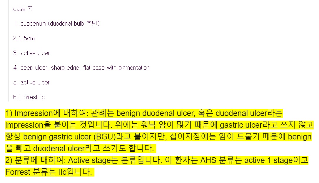
내시경 결과는 임상가에게 의미있는 guide가 되어야 합니다. 결과를 보고 뭔가의 행동을 하거나 판단을 할 수 있도록 도와주어야 합니다. 문제에 melena라고 씌인 점에 주의하십시오 출혈 환자의 내시경에는 두 가지가 중요합니다. (1) 당장 치료 내시경을 할 것인가? (2) 재출혈 위험은 어느 정도인가? 이러한 질문에 답하기 위하여 개발된 것이 Forrest 분류입니다. 출혈로 내시경을 한 경우는 AHS (active, healing, scar) 시스템에 따른 분류뿐만 아니라, Forrest 분류를 꼭 붙여주시기 바랍니다.
Forrest classification
Ulcer 중심에 보이는 것은 red spot (Forrest classification IIc)이라고 생각합니다. 약간 튀어나온 모양이라고 해석하여 exposed vessel이라고 답한 분도 많습니다. Exposed vessel은 노출된 혈관의 aneurysmal dilatation입니다. 어짜피 사진 한 장으로는 확실하지 않지만 저는 red spot으로 보았습니다.
![]() [EndoTODAY Weekly Seminar 다시보기 (2019년)]
[EndoTODAY Weekly Seminar 다시보기 (2019년)]
내시경 증례 퀴즈 2019-19
내시경 증례 퀴즈 2019-18
내시경 증례 퀴즈 2019-17
내시경 증례 퀴즈 2019-16
내시경 증례 퀴즈 2019-15
내시경 증례 퀴즈 2019-14
내시경 증례 퀴즈 2019-10
내시경 증례 퀴즈 2019-9
내시경 증례 퀴즈 2019-8
내시경 증례 퀴즈 2019-7
내시경 증례 퀴즈 2019-6
내시경 증례 퀴즈 2019-5
내시경 증례 퀴즈 2019-4
내시경 증례 퀴즈 2019-3
내시경 증례 퀴즈 2019-2
내시경 증례 퀴즈 2019-1
© 일원내시경교실 바른내시경연구소 이준행. EndoTODAY Endoscopy Learning Center. Lee Jun Haeng.