EndoTODAY 내시경 교실
Beginner | ESA | Schedule | OPD
Seminars | Atlas | Recent | Links
 [조기위암 아틀라스. Early Gastric Cancer. EGC] - 終
3. EGC type I
4. EGC type IIa
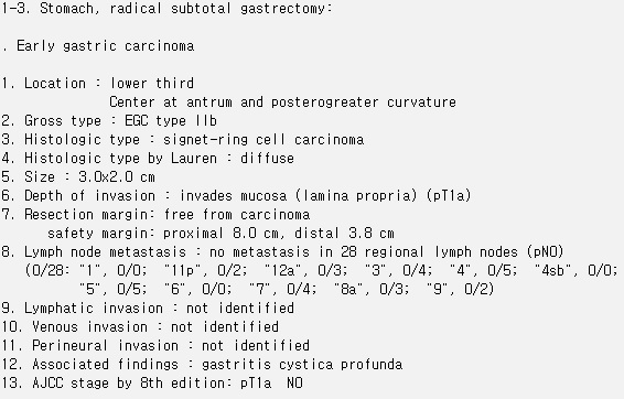
5. EGC type IIb
6. EGC type IIc
7. EGC type III
9. Superficial spreading type EGC
10. AGC-like EGC
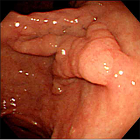
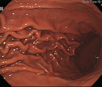
 1. Folds of EGC. 조기위암 fold 변화
EndoTODAY Folds로 옮겼습니다.
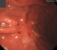
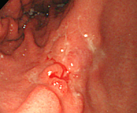
2. Bleeding is important sign
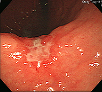
3. EGC type I
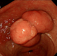
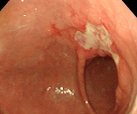
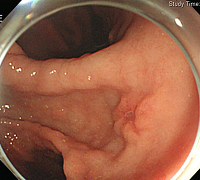
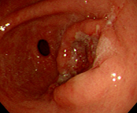
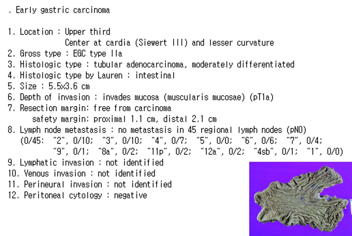
4. EGC type IIa
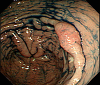
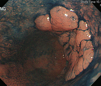
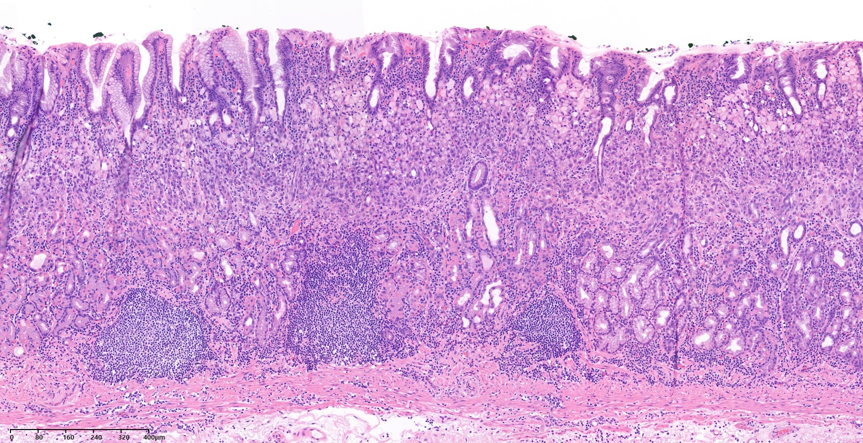
EGC IIa의 ESD를 시행하였습니다. 자세히 관찰한 바 사진만으로 판단한 것에 비하여 넓은 병소였기 때문에 잠시 놀랐지만 시술은 잘 되었습니다.
ESD : Early gastric carcinoma
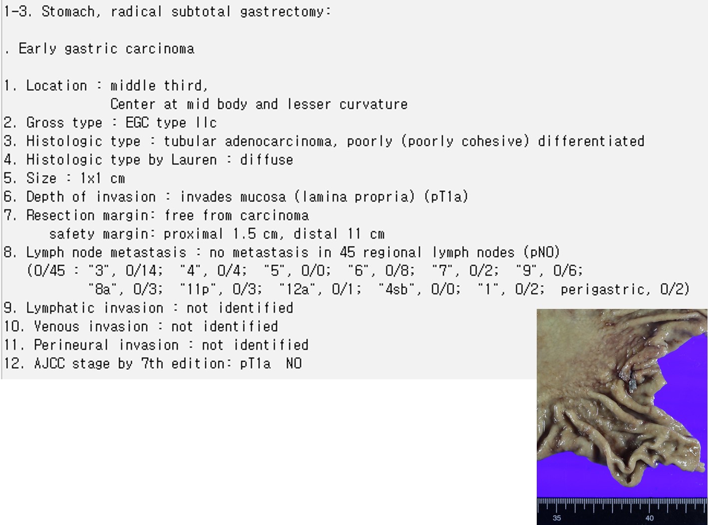
1. Location : angle, lesser curvature
2. Gross type : EGC type IIa+IIb
3. Histologic type : tubular adenocarcinoma, moderately differentiated
4. Histologic type by Lauren : intestinal
5. Size of carcinoma : (1) longest diameter, 58 mm (2) vertical diameter, 24 mm
6. Depth of invasion : invades mucosa (lamina propria) (pT1a)
7. Resection margin : free from carcinoma(N), safety margin : distal 8 mm, proximal 7 mm, anterior 10 mm, posterior 10 mm, deep 1800 ㎛
8. Lymphatic invasion : not identified(N)
9. Venous invasion : not identified(N)
10. Perineural invasion : not identified(N)
11. Microscopic ulcer : absent
12. Histologic heterogeneity: absent

5. EGC type IIb
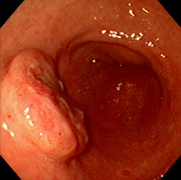
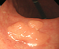
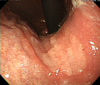
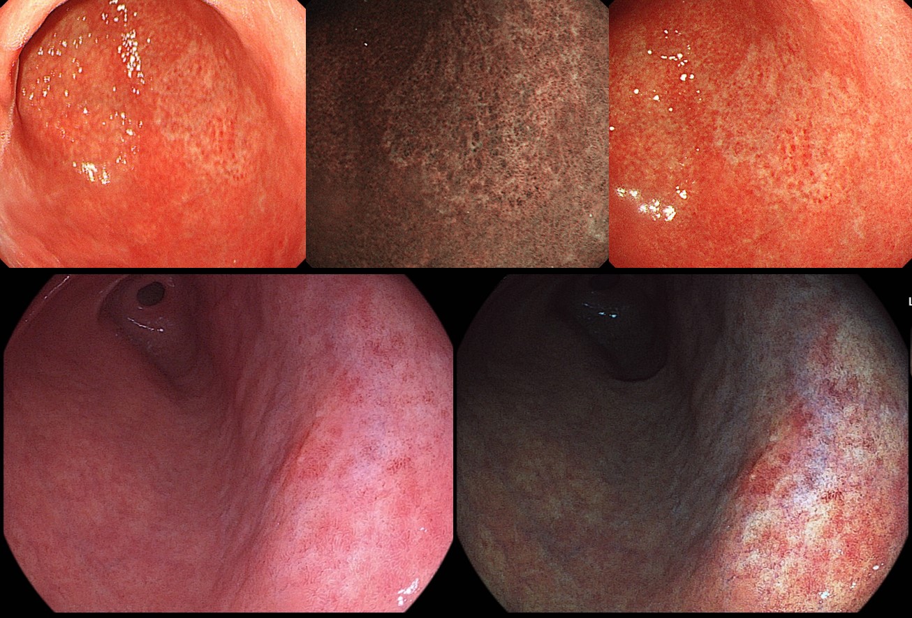
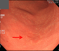
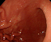
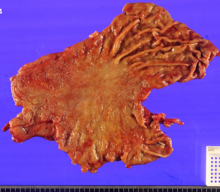
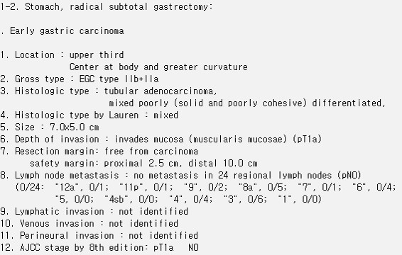
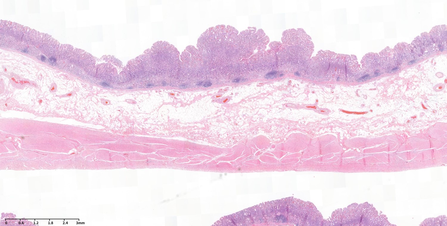
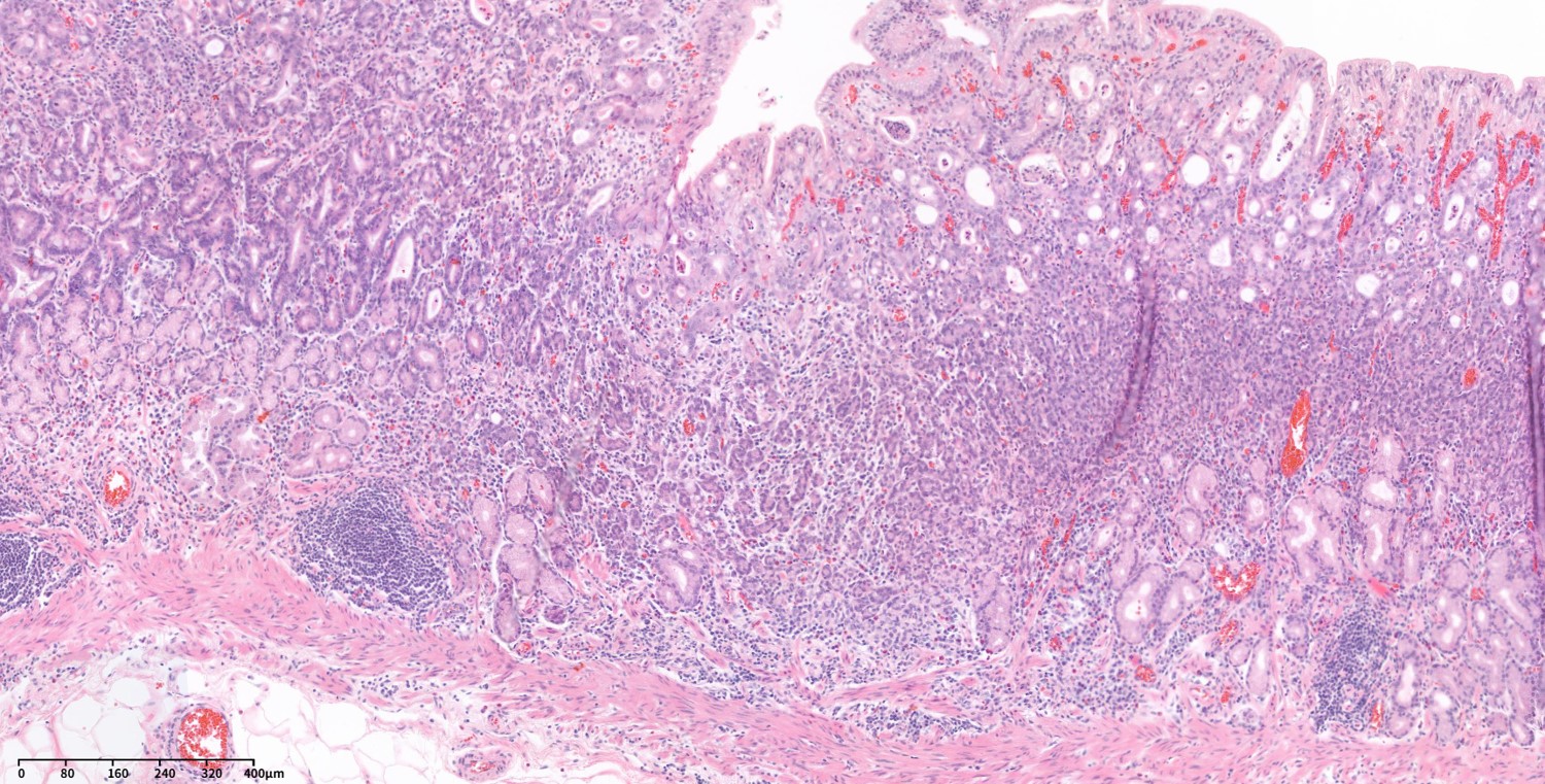
EGC IIb의 중앙에 작은 함몰부나 불규칙한 부분이 발견되는 경우가 있습니다. 검진 내시경 검사에서 두 개의 위궤양으로 추정되었으나 조직검사에서 암이 나와 의뢰된 분입니다. 궤양 주변의 퇴색부가 있었고 넓게 보면 큰 EGC IIb임을 알 수 있습니다. 전체적으로 아주 약간 함몰되어 보이지만 큰 눈으로 보면 평탄한 타입이므로 EGC IIb + IIc로 보는 것이 좋을 것 같습니다. 수술을 하였는데 병리에서는 오히려 융기되어 있어서 EGC IIb + IIa로 병리진단이 되었습니다.
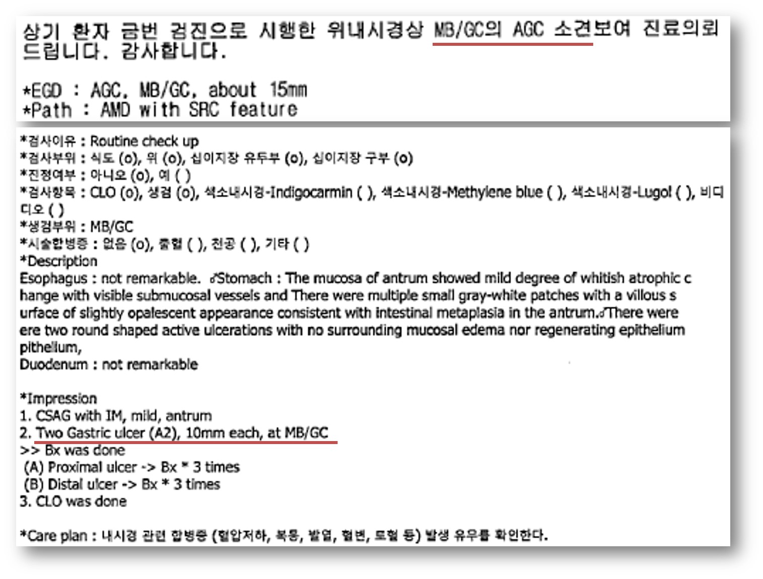
건진 내시경
의뢰 후 재검
6. EGC type IIc
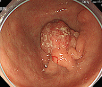
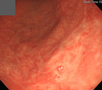
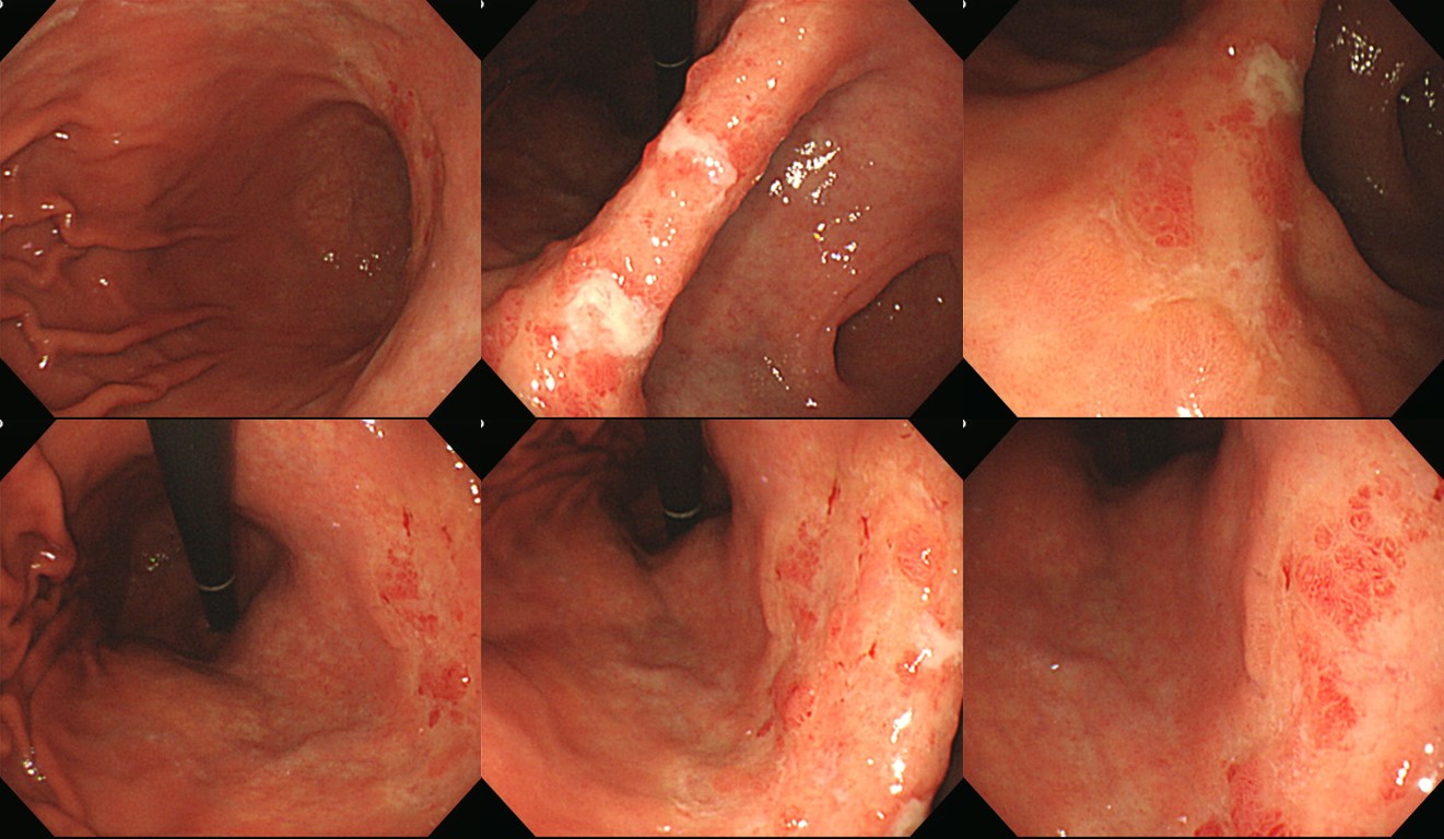
아래와 같은 넓은 EGC는 분류가 쉽지 않습니다. EGC IIc + III라고 할 수는 있겠습니다. 일종의 laterally spreading tumor입니다.
7. EGC type III
전형적인 EGC type III는 많지 않습니다. 뚜렷한 궤양이 있는 환자에서 수술 후 결과가 AGC로 나오는 경우가 많기 때문입니다.
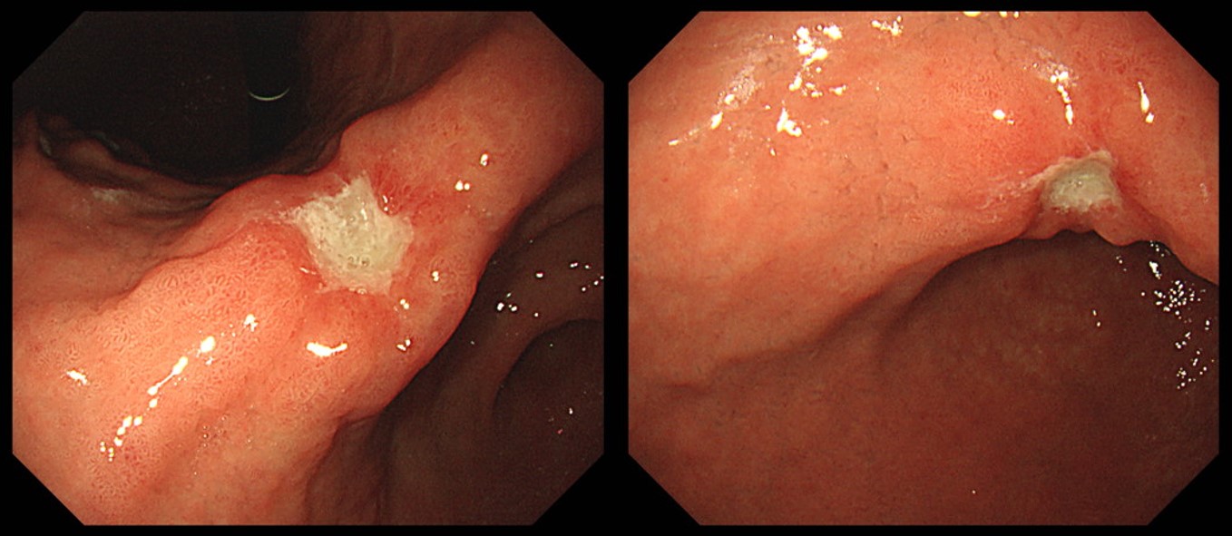
Stomach, radical subtotal gastrectomy: Early gastric carcinoma
1. Location : lower third, Center at body and lesser curvature
2. Gross type : EGC type III
3. Histologic type : tubular adenocarcinoma, moderately differentiated
4. Histologic type by Lauren : intestinal
5. Size : 3.3x1.7 cm
6. Depth of invasion : invades submucosa (sm2) (pT1b)
7. Resection margin: free from carcinoma, safety margin: proximal 2.5 cm, distal 7.3 cm
8. Lymph node metastasis : no metastasis in 32 regional lymph nodes (pN0) (0/32: "1", 0/0; "5", 0/0; "6", 0/0; "7", 0/3; "9", 0/1; "8a", 0/2; "11p", 0/3; "12a", 0/2; "4sb", 0/2; "4", 0/0; 3,5, 0/3; 4,6, 0/4; perigastric LN, 0/12)
9. Lymphatic invasion : not identified
10. Venous invasion : not identified
11. Perineural invasion : not identified
12. Associated findings : ulceration
13. AJCC stage by 8th edition: pT1b N0
8. EGC type IIa + IIc
9. Superficial spreading EGC
10. AGC-like EGC (= EGC, mimicking AGC)
© 일원내시경교실 바른내시경연구소 이준행. EndoTODAY Endoscopy Learning Center. Jun Haeng Lee (since 1999-8-23)
 home
home