EndoTODAY 내시경 교실
Beginner | ESA | Schedule | OPD
Seminars | Atlas | Recent | Links
 [일원내시경교실 목요점심집담회 2015-10-29]
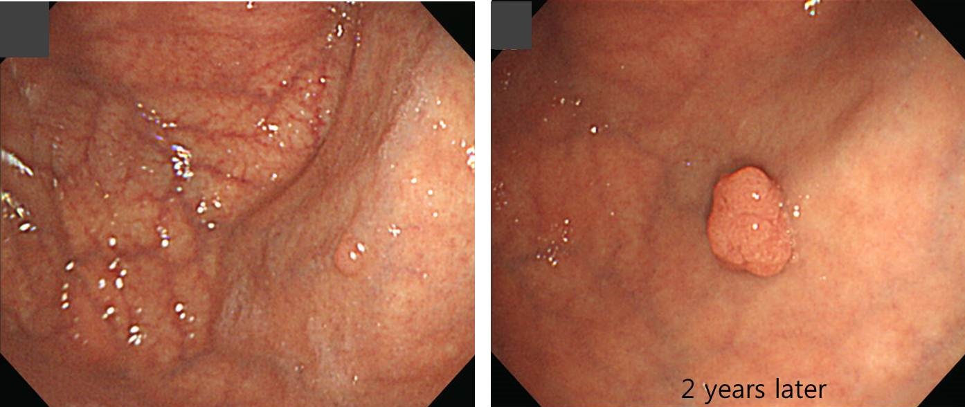
 1. Fundus EGC type I
2년 만에 크기가 증가하였고 조직검사에서 adenoma가 나와 EMR하였는데 암으로 결과가 나왔습니다.
Early gastric carcinoma
1. Location : fundus, anterior wall
2. Gross type : EGC type I
3. Histologic type : tubular adenocarcinoma, well differentiated (foveolar type), in adenoma (foveolar type) with high grade dysplasia
4. Histologic type by Lauren : intestinal
5. Size of carcinoma : (1) longest diameter, 8 mm (2) vertical diameter, 6 mm
6. Depth of invasion : invades mucosa (lamina propria) (pT1a)
7. Resection margin : free from carcinoma(N)
8. Lymphatic invasion : not identified(N)
9. Venous invasion : not identified(N)
10. Perineural invasion : not identified(N)
11. Microscopic ulcer : absent
12. Histologic heterogeneity: absent
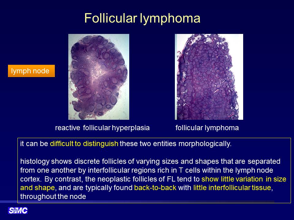
2. Follicular lymphoma of duodenum
Duodenum, 2nd portion, biopsy: follicular lymphoma, grade 1
- Bcl-2: Positive in tumor cells
- CD10: Positive in tumor cells
RT 후 호전되었습니다.
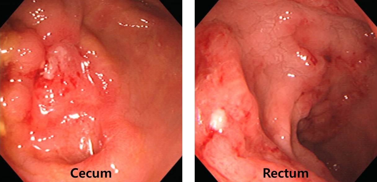
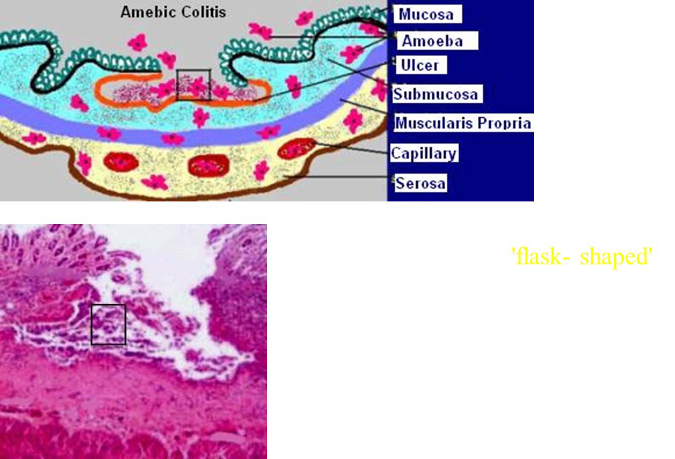
3. Amebic colitis
혈변으로 내원하였고 cecum과 rectum에 병변이 있었습니다.
@ 참고자료: EndoTODAY 아메바 장염
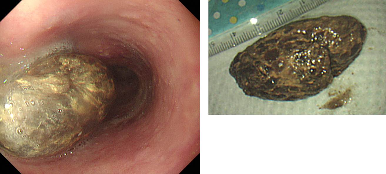
4. 식도이물 = 고기 + 쌈
특별한 organic disease가 없는 분인데 식도의 second physiologic narrowing에 음식물 덩어리가 걸려있어서 net로 제거하였습니다.
@ 참고자료: EndoTODAY 식도 이물
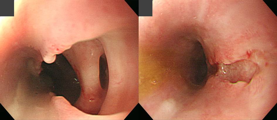
5. 생선가시
식도에 걸린 생선가지 제거 후 mediastinitis가 합병되어 식도 천공이 발생한 사례입니다.
@ 참고자료: EndoTODAY 식도 이물
© 일원내시경교실 바른내시경연구소 이준행. EndoTODAY Endoscopy Learning Center. Lee Jun Haeng.