 EndoTODAY 내시경 교실
Beginner | ESA | Schedule | OPD
Seminars | Atlas | Recent | Links
![]() [일원내시경교실 목요점심집담회 2015-12-3]
[일원내시경교실 목요점심집담회 2015-12-3]
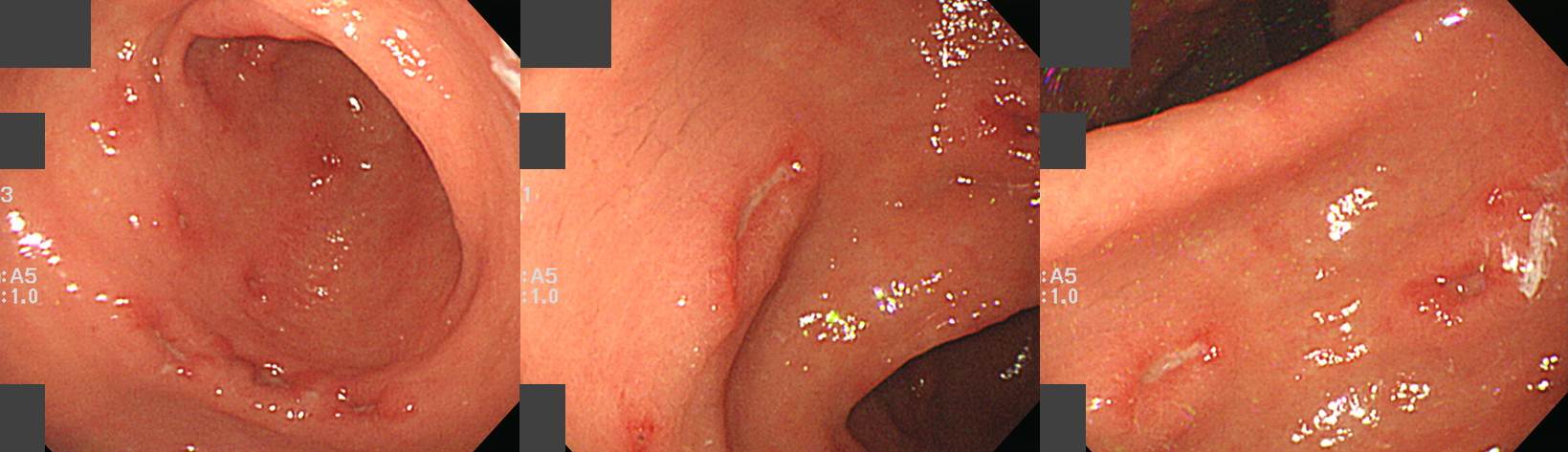
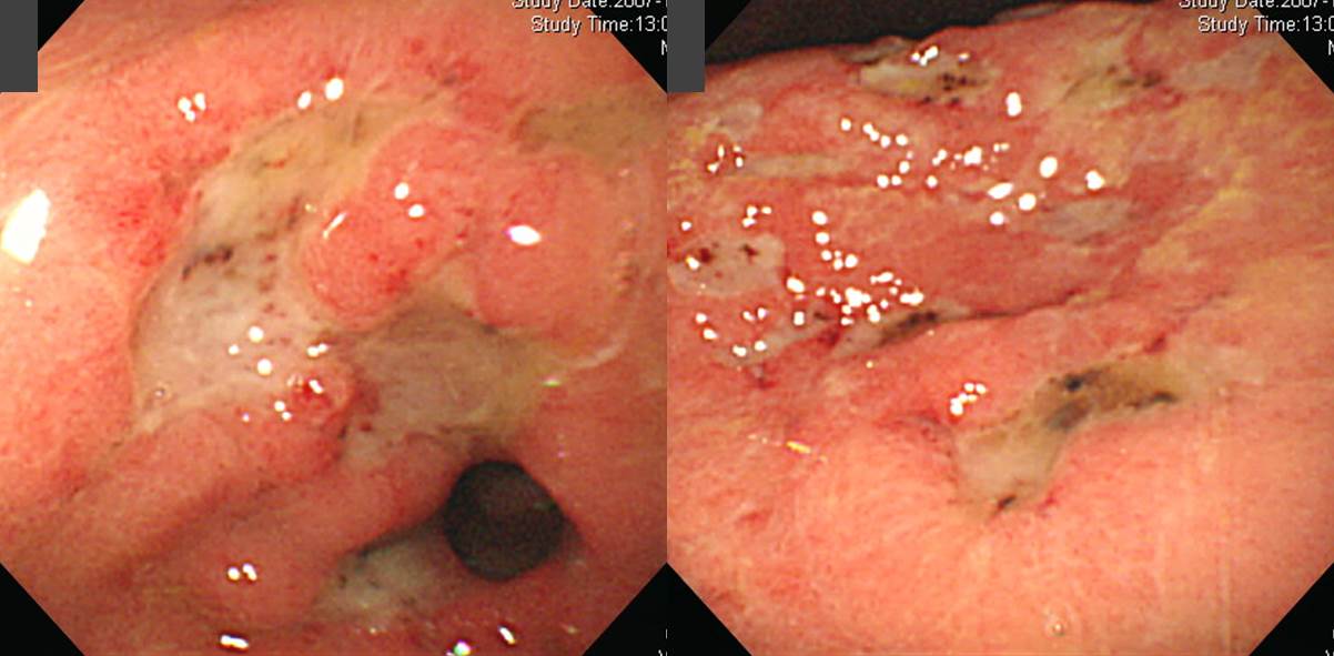
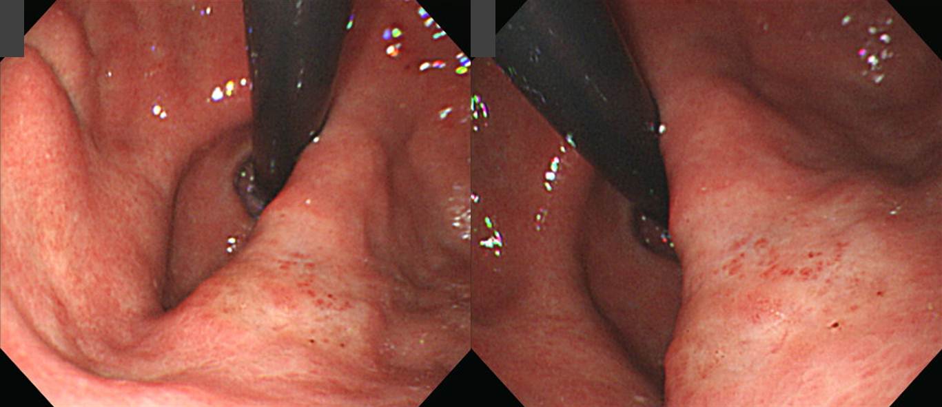
![]() 1. MALT 림프종
1. MALT 림프종
불규칙한 edge의 여러 궤양이 있고 주변 점막이 매우 uneven 하였습니다. MALT 림프종 아니면 undifferentiated type의 EGC 둘 중 하나로 추정하였는데 조직검사는 MALT 림프종으로 확인되었습니다.

* 참고: EndoTODAY MALT 림프종
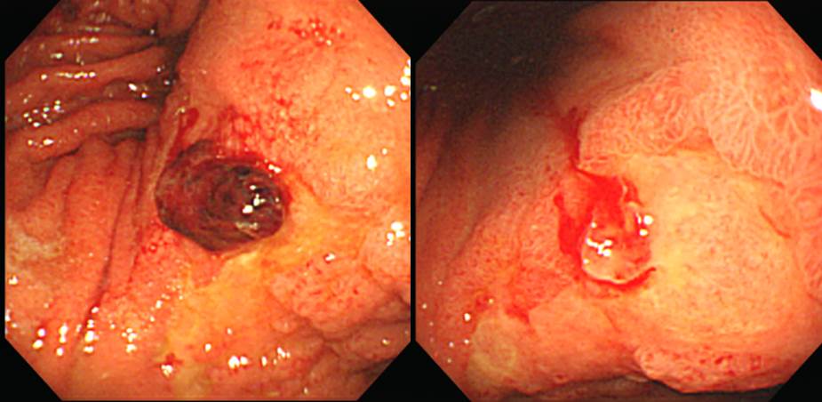
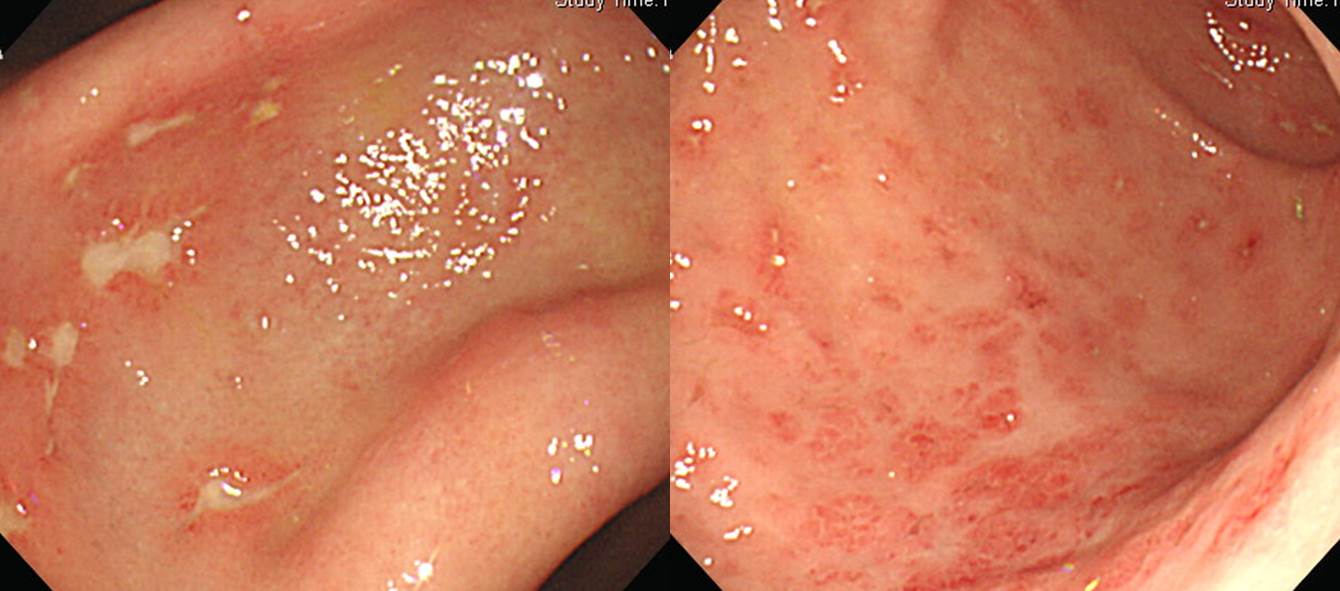
![]() 2. CMV 위염
2. CMV 위염
심장 (heart) 이식 수 개월 후 복통. 다발성 궤양이 발견되었고 조직검사로 CMV 위염을 확인 후 ganciclovir 사용하고 호전되었습니다.
이식 환자에서 발생한 또 다른 CMV 위염 증례들을 소개합니다. 이식 환자에서 발생한 CMV gastritis
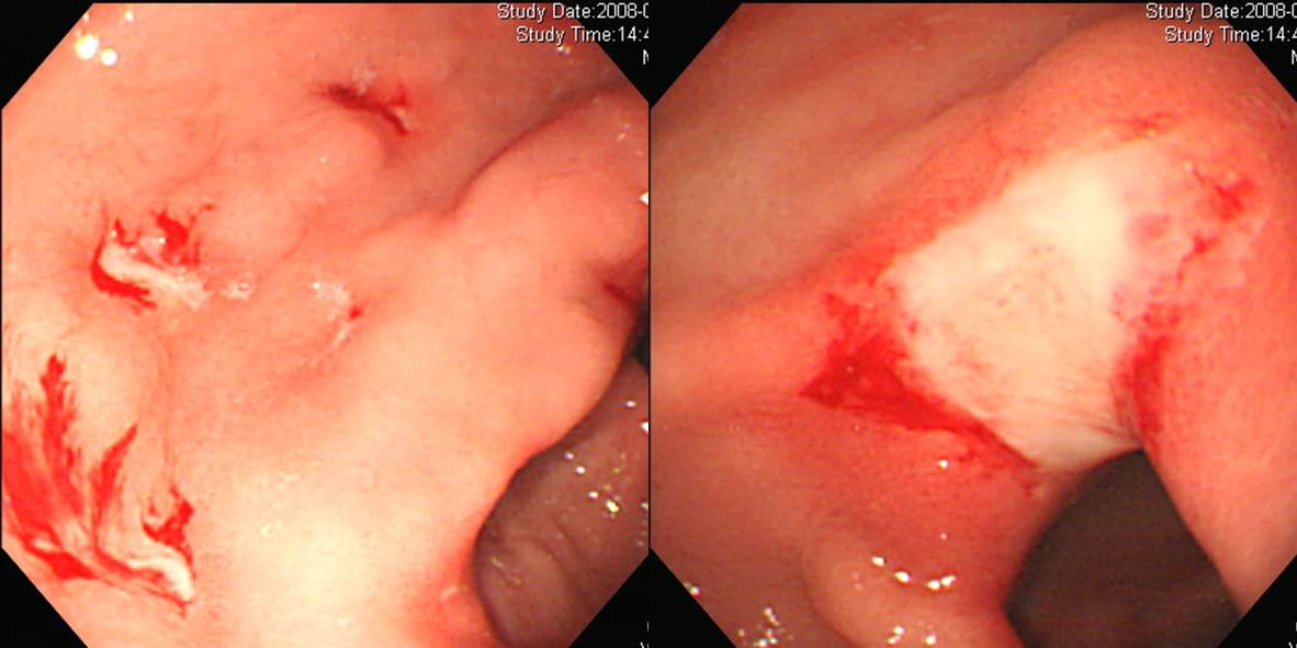
 간 이식. 출혈
 간 이식
간 이식
 간 이식
간 이식
 심장 이식
심장 이식
 심장 이식
심장 이식
 신장 이식
신장 이식
 골수 이식
골수 이식
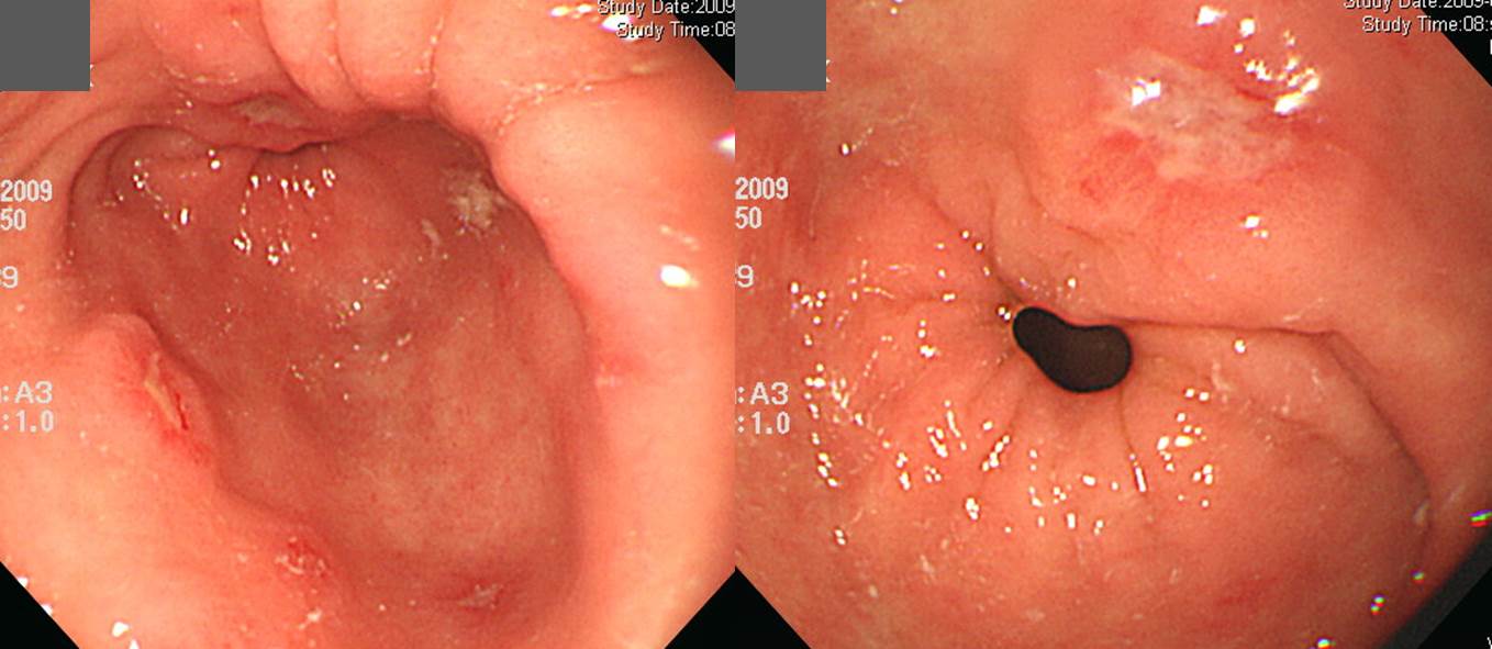
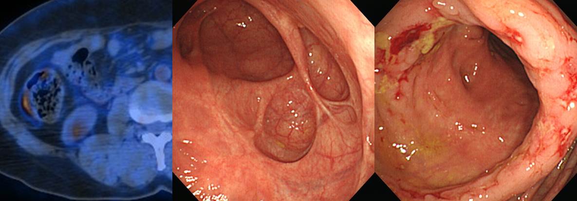
![]() 3. 진행성위암 수술 6년 후 잔위암 (들문분)
3. 진행성위암 수술 6년 후 잔위암 (들문분)
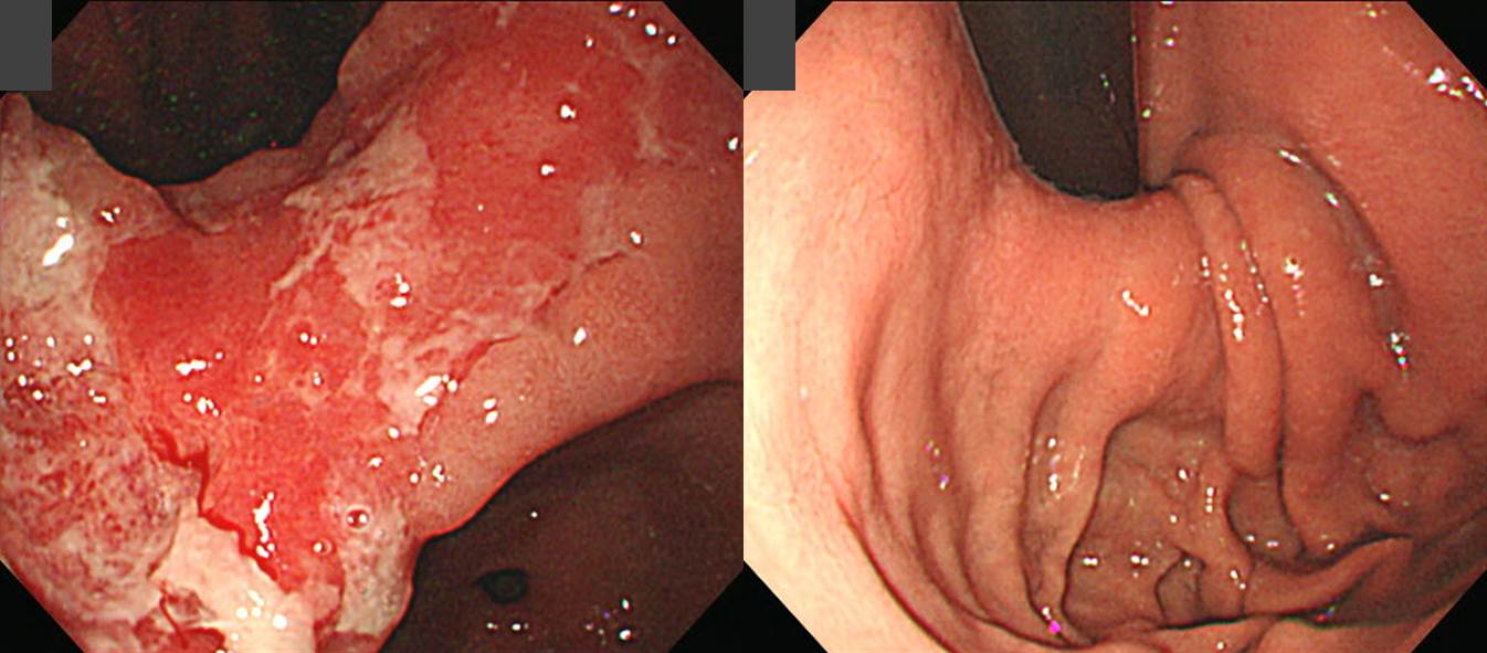
Advanced gastric carcinoma
1. Location : lower third Center at antrum and lesser curvature
2. Gross type : Borrmann type 3
3. Histologic type : tubular adenocarcinoma, poorly differentiated
4. Histologic type by Lauren : intestinal
5. Size : 4x3x0.6 cm
6. Depth of invasion : extension to subserosa (pT2b)
7. Resection margin: free from carcinoma
8. Lymph node metastasis : metastasis to 6 out of 38 regional lymph nodes (pN1)
9. Lymphatic invasion : present
10. Venous invasion : not identified
11. Perineural invasion : not identified
Status post subtotal gastrectomy
Early gastric carcinoma
1. Location : upper third Center at cardia and posterior wall
2. Gross type : EGC type IIb
3. Histologic type : signet-ring cell carcinoma
4. Histologic type by Lauren : diffuse
5. Size : 0.9x0.8 cm
6. Depth of invasion : invades mucosa (lamina propria) (pT1a)
7. Resection margin: free from carcinoma
8. Lymph node metastasis : no metastasis in 6 regional lymph nodes (pN0)
9. Lymphatic invasion : not identified
10. Venous invasion : not identified
11. Perineural invasion : not identified
12. Peritoneal cytology : negative
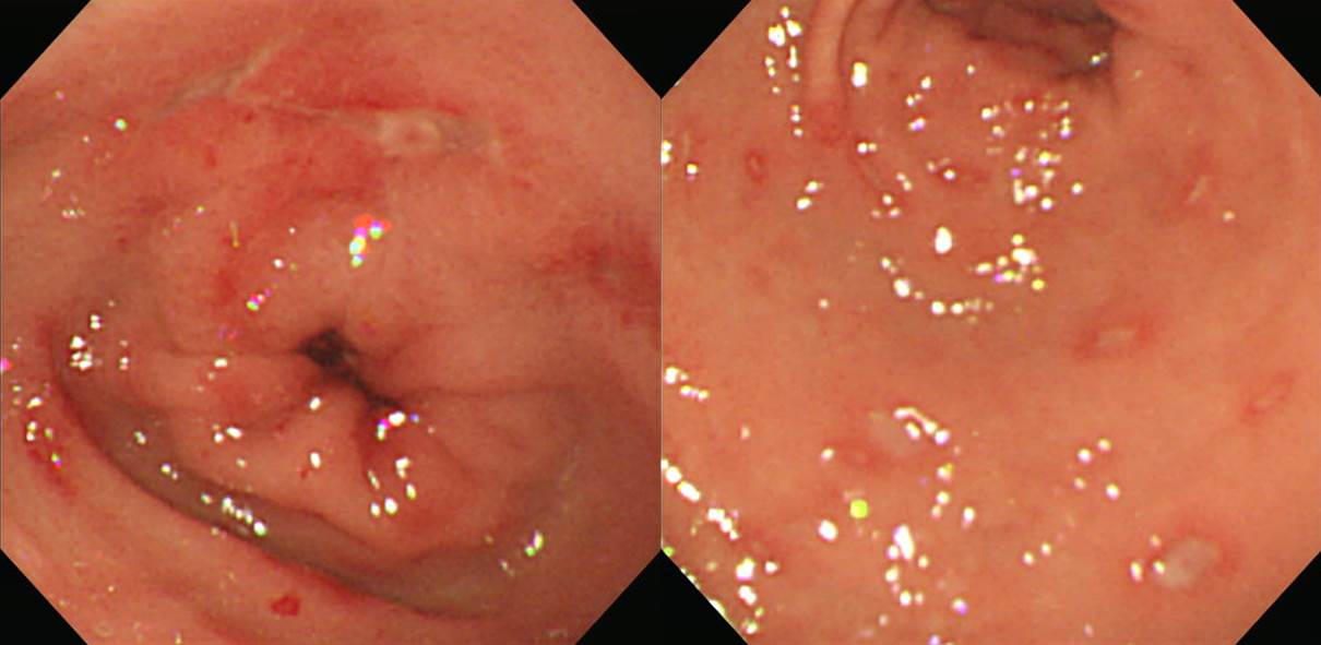
![]() 4. 피부 melanoma 치료 중 시행한 PET에서 우연히 발견된 상행결장 결핵
4. 피부 melanoma 치료 중 시행한 PET에서 우연히 발견된 상행결장 결핵
처음 진단할 무렵부터 cecum은 old tuberculosis로 인한 scar change가 있었으며 상행결장에 circular 한 erosive/ulcerative lesion이 있었음.
© 일원내시경교실 바른내시경연구소 이준행