EndoTODAY 내시경 교실
Beginner | ESA | Schecule | OPD
Seminars | Atlas | Recent | Links
![]() [Histologic classification of gastric cancer. 위암 조직형과 분화도] - 終
[Histologic classification of gastric cancer. 위암 조직형과 분화도] - 終
1. Introduction
2. 관찰자간 차이
4. Immunohistochemistry는 도움이 되는가?
6. Symposiums
7. References
![]() 1. 위암 조직형과 분화도의 큰 줄기 - 우리나라에서는 대강 WHO 기준을 따르고 있습니다.
1. 위암 조직형과 분화도의 큰 줄기 - 우리나라에서는 대강 WHO 기준을 따르고 있습니다.
1998년 일본위암분류(1998)에서 일본 외과의사들이 이해하는 위암의 조직학적 분류는 아래 그림과 같습니다. "The histological classification should be based on the prominant pattern of tumor"라고 언급하고 있습니다. 여기서 "prominant"는 우리나라 병리 가이드라인(2005)에서 언급한 "암세포의 면적이 가장 많은 유형"과 비슷한 의미로 생각됩니다.
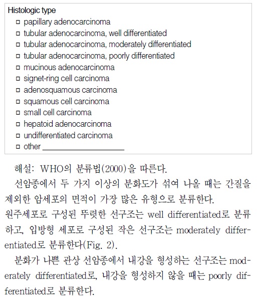
2005년 병리 가이드라인 (대한병리학회 소화기병리학연구회, 위암 병리보고서 기재사항 표준화, PDF 0.3M)은 (1) 위암의 histologic type은 2000년 WHO 분류를 따름, (2) 두 가지 이상의 분화도가 섞여 나올 때는 간질을 제외한 암세포의 면적이 가장 많은 유형으로 분류함, (3) 선구조나 편평상피 분화가 없는 경우에는 undifferentiated carcinoma로 분류한다고 명시했습니다.
2010년 일본위암취급규약 제14판에 따르면 위암의 분화도는 선관형성 상태에 따라 고분화형, 중분화형, 저분화형으로 분류된다. 여기에는 세포형질의 분화 경향과 핵이형도는 고려되지 않는다 (위와장 2010년 6월호 857쪽). → [이준행 생각] Structural atypia에 따라 분화도를 나누고 cellular atypia는 고려되지 않는다는 의미인 것 같다. 그런데 보통 structural atypia는 cellular atypia와 함께 발생한다. 간혹 Cellular atypia는 거의 없는데 structural atypia만 보이는 경우 '저이형도 분화형 위암'이라고 부른다. 예를 들어 foveolar type, fundic gland type, intestinal type, crawling type (WHYX lesion)등이 여기에 해당한다.
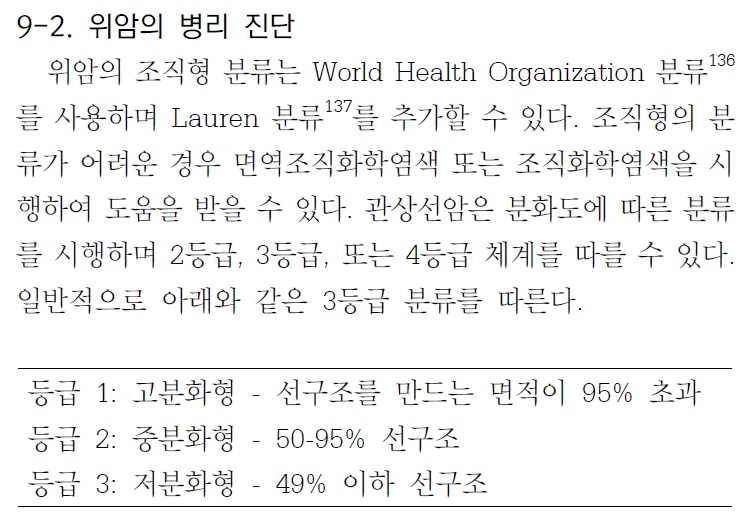
2014년 우리나라 다학제 위암진료권고안(제가 간사로 참여하였습니다)에서는 (1) 선구조를 만드는 면적이 95% 초과하면 고분화형, (2) 선구조를 만드는 면적이 50-95%면 중분화형, (3) 선구조를 만드는 면적이 49%이하면 저분화형으로 구체적인 수치를 언급하였습니다.
2023년 병리 가이드라인에서는 2019년 WHO classification을 거의 그대로 제시하였습니다.
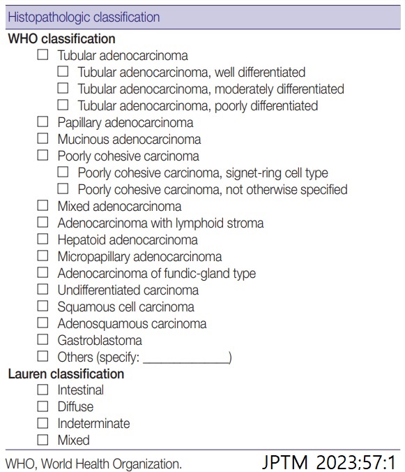
Fig. 7. Representative pictures of each histologic subtype of gastric carcinoma. Tubular adenocarcinoma (A), papillary adenocarcinoma (B), mucinous adenocarcinoma (C), poorly cohesive carcinoma, not otherwise specified (D), poorly cohesive carcinoma, signet-ring cell type (E), adenocarcinoma with lymphoid stroma 림프구버팀질동반샘암종 (F), hepatoid adenocarcinoma (G), micropapillary adenocarcinoma (H), adenocarcinoma of the fundic-gland type (I, J), undifferentiated carcinoma (K), and crawling-type adenocarcinoma (L).
2023년 병리 가이드라인의 tubular adenocarcinoma의 differentiation에 대해 아래와 같은 설명이 있습니다. 비전문가인 저는 다소 혼동스럽습니다. MD는 (1) cuboidal or flat cells 이거나 (2) distinct하지만 frequent luminal structures라는 것입니다. 면적뿐 아니라 세포형도 고려하는 것 같습니다.
"Tubular adenocarcinoma and papillary adenocarcinoma can be graded. When two or more differentiations are mixed in an adenocarcinoma, the differentiation grade reflects the largest tumor area. A distinct glandular structure composed of columnar cells is classified as WD, and a small glandular structure composed of cuboidal or flat cells is classified as MD. In a tumor with an in distinct glandular structure, carcinoma forming frequent luminal structures is classified as MD, and that with a rare luminal structure is classified as PD. Although the WHO recommends a two-tier grading system of low- (WD and MD) and high-grade (PD), most pathologists and clinicians use a three-tier grading system. We have agreed to used a three-tier grading system that can be easily switched to a two-tier grading system."
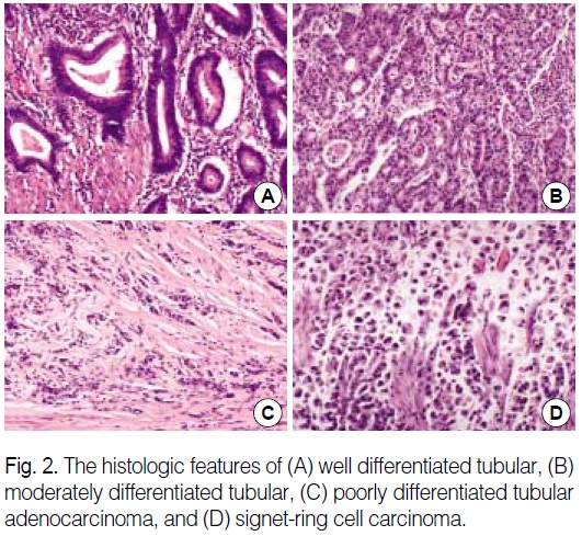
Grading of gastric tubular adenocarcinoma. Well-differentiated adenocarcinoma showing glandular structures composed of columnar tumor cells (A). Moderately differentiated adenocarcinoma exhibits more complex tubular structures with cuboidal and/or flat epithelial cells (B). Tubular structure is unclear in most tumor glands in poorly differentiated adenocarcinoma (C).
[2023년 종설] 샘암종의 분화도는 주로 관샘암종과 유두모양샘암종에 적용된다. 뚜렷한 샘 구조를 만드는 종양으로 주로 원주형 세포로 구성된 경우 고분화(well differentiated)로, 샘구조가 뚜렷하지만 샘들의 크기가 작고 입방형이나 납작한 세포가 주 구성세포인 경우 중등도 분화(moderately differentiated)로 분류되고, 내강 구조를 거의 형성하지 않을 때는 저분화(poorly differentiated)로 분류된다. 한 종양 내에서 두 가지 혹은 그 이상의 분화가 혼합되어 관찰될 경우, 일반적으로 가장 많은 부위에서 관찰되는 분화도에 따라 등급을 결정한다. (위암 병리 소견의 이해 김백희, 이성학. 헬리코박터학회지 종설, 2023)
![]() 2. 관찰자간 차이
2. 관찰자간 차이
Histological classification of gastric adenocarcinoma for epidemiological research: concordance between pathologists. Shibata et al. Cancer Epidemiol Biomarkers Prev 2001
Two pathologists, each blinded to the other's assessment, reviewed H&E-stained slides of gastric tumor... Concordance for tumor grade was 87%, with a kappa coefficient of 0.72 (95% confidence interval, 0.57-0.87).
Diagnosis of gastric epithelial neoplasia: Dilemma for Korean pathologists. Kim JM et al. World J Gastroenterol 2011 (PDF)
Korean pathologists experience much difficulty making a diagnosis because we are influenced by Japanese pathologists as well as Western medicine. Japan is geographically close to Korea, and academic exchanges are active. Additionally, Korean doctors are familiar with Western style medical terminology. As a result, the terminology, definitions, and diagnostic criteria for gastric intraepithelial neoplasia are very heterogeneous in Korea.
![]() 3. 조직검사와 수술 후 병리결과의 분화도 차이
3. 조직검사와 수술 후 병리결과의 분화도 차이
Differences between biopsy- or specimen-related Lauren and World Health Organization classification in gastric cancer. (World J Surg 2002)
Out of 48 tumors with preoperative diagnosis of an intestinal type, 10 tumors (20.8%) exhibited a diffuse growth pattern in the gastrectomy specimens; and 16% of the cases showed a disagreement of the pre- and postoperative histopathological type according to the WHO classification.
![]() 4. Immunohistochemistry는 도움이 되는가?
4. Immunohistochemistry는 도움이 되는가?
Expression of E-cadherin, beta-catenin, CD44s and CD44v6 in gastric adenocarcinoma: relationship with lymph node metastasis. (Anticancer Res 2003)
Expressions of CD44s and CD44v6 play an important role in tumor progression; especially, CD44v6 expression may be a useful predictor of lymph node metastasis, while the expressions of E-cadherin and beta-catenin complex are more probably related to tumor morphology than to tumor progression.
![]() 5. Histological heterogeneity
5. Histological heterogeneity
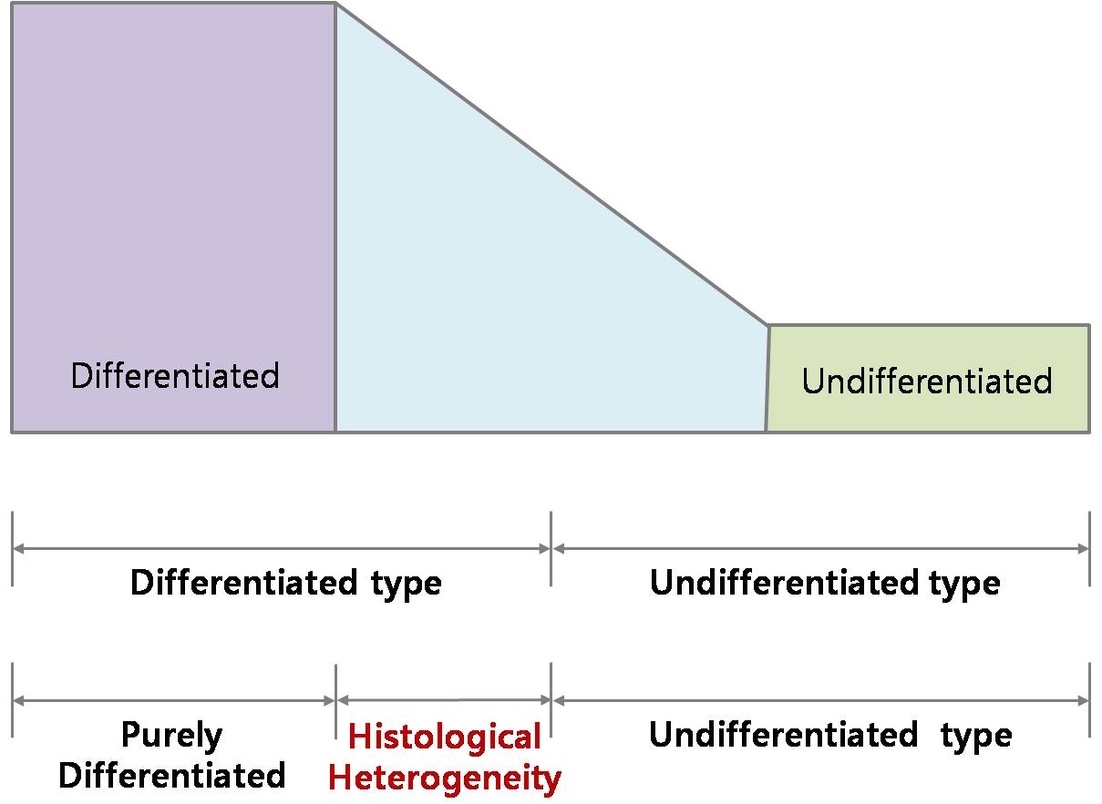
This is my conceptual model of histological differentiation of early gastric cancer. There are two dominant types of histology. One is differentiated type, and the other is undifferentiated type. However, there are a lot of cases in the middle. We don’t know exactly how many patients are included in this mixed area.
Usually, the histological grouping is made by the major area of histological differentiation. For example, if 70 percent of area shows undifferentiated histology, we call it undifferentiated type of early gastric cancer like the blue arrow. If 80 percent of area shows undifferentiated histology, we call it differentiated type of early gastric cancer like the red arrow. The concept of histological heterogeneity comes from this area. EGC cases mixed with undifferentiated component less than 50 percent of area can be called as histological heterogeneity. And we evaluated cases in this unique group of patients.
점막하암에서 histological heterogeneity가 있으면 림프절 전이의 위험이 높다는 것은 동경암센터(J Gastroenterol 2001)에서 발표한 바 있습니다. 당시의 결론입니다.
Conclusions: When the extension of endoscopic surgery to differentiated Sm-ca is considered, this therapeutic technique should be limited to the differentiated type of Sm-ca without histological heterogeneity. The Ki-67 labeling index provides useful information for identifying those patients with a high risk of lymph node metastasis.
[섞인 것은 순수한 것보다 나쁜가? Risk of lymph node metastasis in cases with histological heterogeneity]
ESD 후 병리결과에서 histological heterogeneity가 있다고 나온 경우 수술을 권할 것인가는 논란거리입니다. 일반적으로 점막에 국한되어 있고 complete resection이면 경과관찰을 권합니다. 그러나 submucosal invasion이 있다고 나온 경우는 고려해야 할 것이 많습니다. 병리과 선생님을 찾아가 함께 슬라이드를 보지 않을 수 없는 상황입니다.
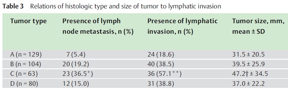
C: undifferentiated predominant mixed type, D: undifferentiated type. Undifferentiated-type-predominant mixed type (Type C) were independent risk factor for lymph node metastasis (Endoscopy 2009)
두 번째 패턴이 가장 흔합니다. 그러나 림프절 전이율은 큰 차이가 없습니다. Lymphatic이 있으면 더 나쁘지 않을까 생각됩니다만... 숫자가 적어서 결론을 내리기 어렵습니다. 데이타는 아래 표를 보십시요.
관심있게 봐야 할 부분은 histological type of the invasion front (undifferentiated type)입니다. 가장 깊게 침윤된 부위에 어떤 세포가 있는가에 따라 예후가 달라진다는 것입니다.
강남세브란스병원의 자료입니다. Signet ring cell carcinoma가 섞여 있으면 더 나쁘다는 것입니다. 섞인 것은 순순한 signet ring cell carcinoma보다 나쁩니다 (J Surg Oncol 2013).
[2014-6-18. 강남세브란스병원 김지현 교수님 의견] 저희 논문과 관련하여 메일을 드립니다^^ 조기위암을 대상으로 분석하였으며 partly-SRC(signet ring cell histology가 50% 이내로 mix된 경우)가 가장 공격적인 형태였습니다. 즉, partly-SRC는 SRC보다 behavior가 나쁜 것뿐만 아니라 저분화 위선암(poorly differentiated adenocarcinoma)보다도 나쁘게 나왔습니다. 다시 말씀드리면, pure SRC가 조기위암 중에서 가장 덜 공격적인 행태를 보였고, partly-SRC 즉, SRC가 일부 mix된 경우가 가장 공격적인 행태를 보이는 흥미로운 결과였습니다. (Partly-SRC는 mixed histology 혹은 histological heterogeneity와 유사한 개념으로 생각됩니다.) 이 때문에 ESD 적응증을 변경해야 하는지 의문이 있을 수 있습니다. 위암학회 때 발표한 바와 같이 partly-SRC 즉, SRC가 일부 mix된 경우더라도 main histology가 ESD indication을 만족한다면 lymph node metastasis는 없었습니다. 즉, partly-SRC로 인하여 indication을 따로 정할 필요는 없을 것 같습니다.
![]() 6. Symposiums
6. Symposiums
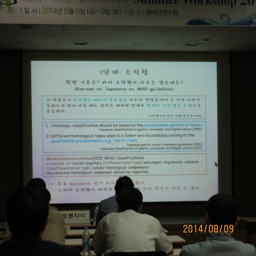
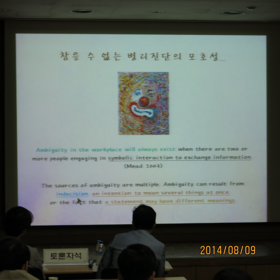
[2014-8-9. 대한헬리코박터상부위장관학회 서머워크샵. 인제대학교 주미. 위암 분화도. 관찰자간 차이 및 시술 전후 차이]
PDF 0.3 M
조직검사는 이와 같습니다. 일부만 보고 전체를 알기는 어렵습니다.
ESD 후 병리검사는 다릅니다. 정확히 구분할 수 있습니다.
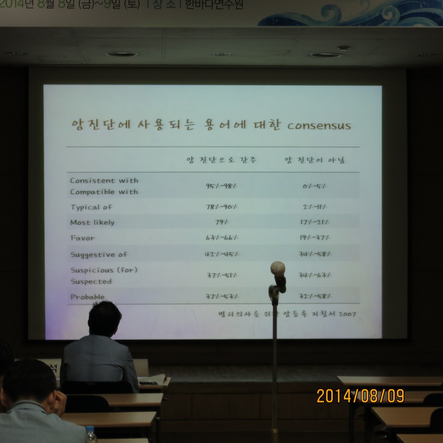
Word PDF 0.3 M. 2008년 5월 병리의사를 위한 암등록 지침서입니다. 이렇게 언급되고 있습니다. 'Comparable with’, ‘consistent with’, ‘compatible with’, ‘favors’, ‘most likely’, ‘typical of’와 함께 'malignant appearing'과 ‘suggests'도 암진단으로 간주되는 용어로 생각할 수 있다. 가능하면 암으로 의심되는 경우 상기한 용어를 사용하도록 권장하며, 그 이외의 용어에 대해서는 진단한 병리의사와 상의를 할 것을 권장한다.
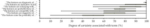
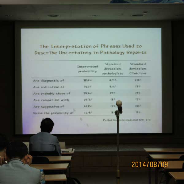
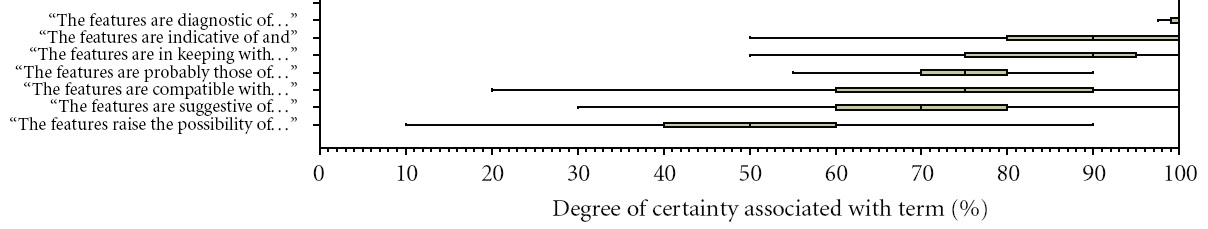
주미 교수님께서 소개한 두 자료를 고려할 때 대강 아래와 같이 정리할 수 있을 것 같습니다. Compatible with, probable, suggestive of 등에 대한 영국 병리의사와 우리나라 병리의사 사이의 온도차는 상당히 컸습니다.
용어 영국
병리의사우리나라
병리의사diagnostic 98.4% consistent with 95-98% indicative of 92.1% typical 78-90% most likely 79% probably 75.6% 37-53% compatible with 74.3% 95-98% suggestive of 69.8% 42-45% favor 63-66% possibility 52.9 suspicious
suspected37-51%
![]() [2015-4-3. 위암학회 KINGCA 2015. 주미 선생님의 강의]
[2015-4-3. 위암학회 KINGCA 2015. 주미 선생님의 강의]
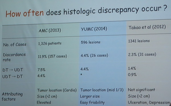
인제대 주미 주미 선생님은 histological discrepancy between endoscopic biopsy and surgical/ESD specimen을 강의하셨습니다. 수술/ESD 후 differentiate와 undifferentiated가 바뀌는 빈도는 서울아산병원 연구에서 11.9%, 연세대학교 연구에서 4.4%, 일본 Takao의 연구에서 2.3%였습니다.
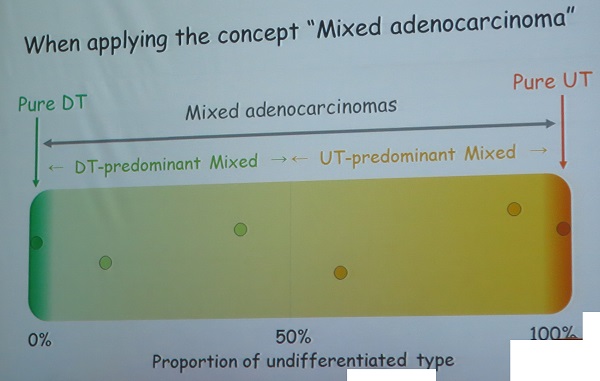
주미 선생님은 Histological heterogeneity와 mixed adenocarcinma는 다르다는 점을 지적하셨습니다. 아직 통일된 정의는 아닌 것 같지만... 그 중 mixed type의 림프절 전이가 많다는 점을 지적하셨습니다.
- Histological heterogeneity: morphologi diversity with >= 2 histologic subtypes regardless of their differentiation types
- Mixed adenocarcinoma: a mixture of differentiation type and undifferentiated type histology
(1) Mixed adenocarcinoma comprised od 10.7-44.4% of EGCs and correlated with tumor size, depth of invasion, and lymph node metastasis
(2) In particular, undifferentiated predominant mixed type is a significant risk factor for lymph node metastasis.
(3) Mixed adenocarcinoma is one of major tumor factors contributing histologic discrepancy between endoscopic biopsy and resection specimen.
Floor로부터의 질문에 대하여 삼성서울병원 병리과 김경미 선생님께서 (1) differentiated/undifferentiated 를 예측할 수 있는 면역염색이나 기타 방법은 없다, (2) histological heterogeneity는 submucosal invasion의 중요한 예측인자임을 지적하셨습니다.
![]() [images]
[images]
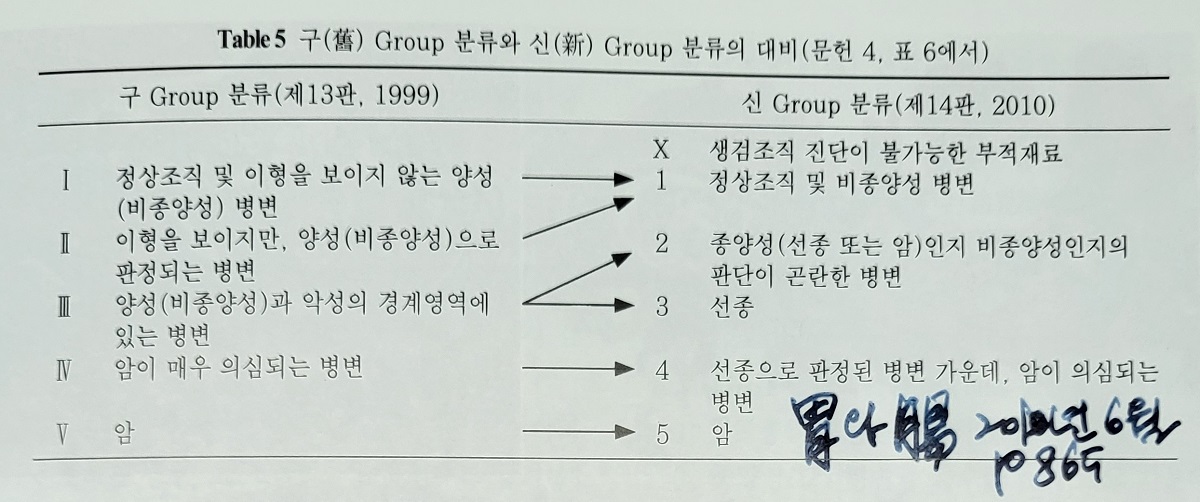
이 표의 2019 WHO 부분은 정확하지 않은 것 같습니다.

면적을 중요시하는 입장이고 퍼센트로 수치화되었습니다. 실제로 이렇게 하는 것 같지는 않습니다. 면적과 세포형을 모두 고려하는 것 아닌가 싶습니다.
![]() [References]
[References]
![]() © 일원내시경교실 바른내시경연구소 이준행. EndoTODAY Endoscopy Learning Center. Lee Jun Haeng
© 일원내시경교실 바른내시경연구소 이준행. EndoTODAY Endoscopy Learning Center. Lee Jun Haeng