EndoTODAY 내시경 교실
Beginner | ESA | Schedule | OPD
Seminars | Atlas | Recent | Links
 [조기위암 내시경 절제 후 국소 재발. Local recurrence after endoscopic resection of EGC] - 終
[2025-8-5. EndoTODAY 생각] ESD는 크게 해야 국소 재발이 적게 생깁니다. ESD를 크게 하면 시간도 오래 걸리고 합병증도 많습니다. ESD 시술 의사에 대한 사법 risk가 증가하면서 ESD 시술 의사들이 심적으로 위축되고 있습니다. 안타까운 일입니다.
 1. 조기위암 내시경 절제 후 국소재발 (국립암센터, 2012)
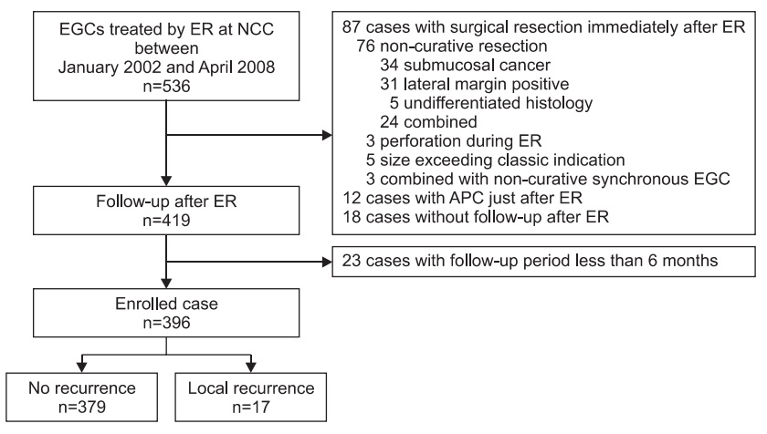
2012년 국립암센터에서는 조기위암 내시경치료 후 국소재발에 대하여 발표하였습니다 (Lee JY. J Gastric Cancer 2012). 2002년부터 2008년까지 비교적 초기에 시술된 환자 396명의 성적이었습니다. 17명이 국소재발을 보였는데 EMR 118예 중 14예(11.7%), ESD 278예 중 3예(1.1%)였습니다.
내시경 시술 후 추적관찰 방법은 아래와 같이 기술되어 있었습니다.
To evaluate local recurrence, two to four biopsy specimens were routinely obtained from the ER ulcer scar during each examination with standard fenestrated open-cup forceps (FB-25K-1; Olympus Co. Ltd) or ellipsoid fenestrated cup forceps with needle (FB-24K-1; Olympus Co. Ltd).
국소재발 17명 중 12명은 4개월 이내에 발견되었고, 나머지 5명은 1년 이후에 발견되었습니다. 대부분의 환자는 단순 궤양 반흔으로 보이거나 미세한 발적만 있었고 조직검사로 국소재발이 진단되었습니다. 2명은 mass로 발견되었는데 모두 incomplete resection으로 수술이 필요했는데 수술을 거부하여 경과관찰 하던 중 발견된 예였습니다.
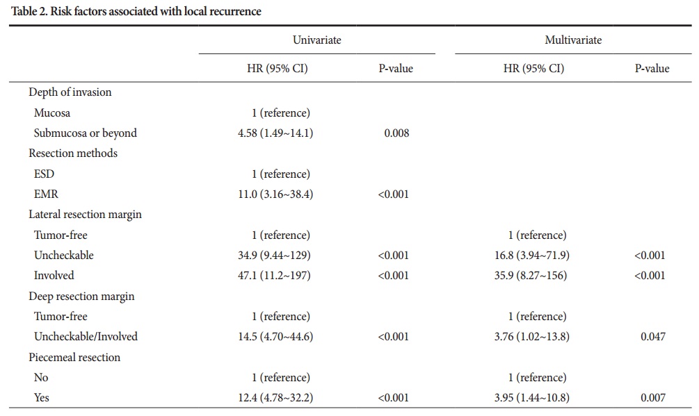
재발 위험인자 분석입니다. 위험인자가 하나라도 있는 경우 5년 국소재발률이 27%인 반면, 위험인자가 하나도 없는 경우는 국소재발이 전혀 없었습니다.
2. Endoscopic prediction of recurrence after margin-negative endoscopic resection (서울아산병원, 2016)
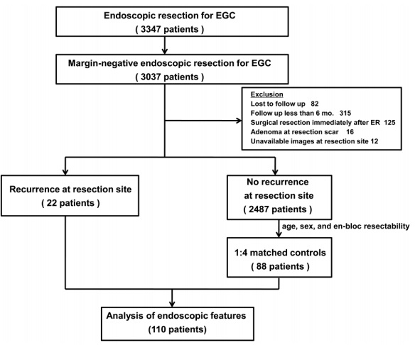
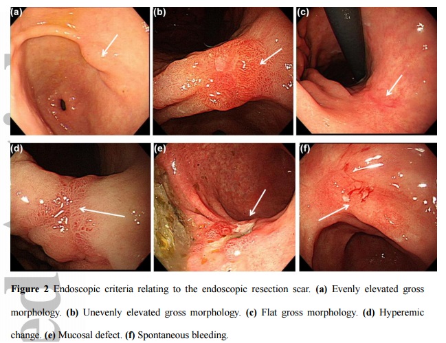
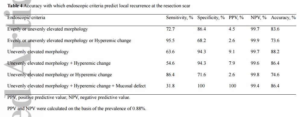
서울아산병원 최기돈 교수님 팀에서 위암 내시경치료 후 국소재발을 분석하였습니다 (Na HK. JGH 2016) . 1995년부터 2011년까지 3,347예의 조기위암 내시경치료 중 3,027예(90.7%)가 margin-negative EGC였습니다. 22예의 국소재발과 4배수의 control을 비교하였습니다. Differentiated type EGC의 en-block margin negative resection scar가 hyperemic change 없이 flat 하면 조직검사를 할 필요가 없다고 결론짓고 있습니다.
3. 국소재발을 발견하기 위한 전략 (2015년 12월 24일 잠정안)
1) 조기위암 EMR/ESD: 5년까지 추적 내시경마다 scar에서 조직검사 (2점) → 5년 후부터는 재발 의심 소견이 있을 때만 조직검사 (단, 조기위암 EMR/ESD 후 첫 추적 내시경에서는 H. pylori 조직검사도 함께 시행한다.)
2) 선종 EMR/ESD: 1년까지 추적 내시경마다 scar에서 조직검사 (2점) → 1년 후부터는 재발 의심 소견이 있을 때만 조직검사
참고. 'EMR/ESD 후 첫 추적 내시경에서는 scar에서 조직검사를 하지 말자'는 과거의 결정은 취소합니다. 첫 추적검사에서도 scar 조직검사를 해 주시기 바랍니다. 약간 편하자고 정한 결정인데 괜한 혼동만 있다고 판단되기 때문입니다. 아직 좀 더 경험과 자료 축적이 필요하다고 생각되므로 당분간 국립암센터 및 서울아산병원에서 제안한 것보다 조금 tight하게 검사하는 전략을 유지합시다.
4. Recurrence after ESD for pyloric ring cancer. 날문위암 내시경절제술 후 국소재발
들문 (cardia) 위암과 날문 (pylorus) 위암은 내시경 절제술 시 절제여분을 확보하기 어렵습니다. 그나마 들문 위암은 식도에서 접근하고 위에서 retroflection으로 접근하여 넉넉하지는 않지만 약간의 negative resection margin을 만들 수 있습니다. 그러나 날문 위암은 십이지장에서 retroflection을 하여 시술할 수 있는 경우가 많지 않아서 시술이 매우 어렵습니다. One piece resection을 하지 못하거나 원위부 positive resection margin이 되기 쉽습니다. 절제변연이 부족한 부분에 대하여 monopolar coagulation이나 APC를 이용한 소작술을 추가하기도 하지만 마음이 편치 않은 것은 사실입니다.
날문에 걸친 위암을 가진 환자에게는 다음과 같이 설명하고 있습니다.
"재발률을 낮게 유지하기 위하여 병소의 4 방향에 어느 정도의 여분을 두고 절제하고 있습니다. 그런데 병소의 위치가 음식이 나가문, 즉 위와 십이지장의 접합부인 날문에 근접한 경한는 한두 방향의 여분은 확보할 수 없습니다. 여분이 부족하다고 모두 재발하는 것은 아니며 무조건 수술하는 것도 아닙니다. 다만 재발률이 보통의 경우보다 약간 더 높다고 생각하시면 무난합니다. 통상의 재발률은 5% 정도로 보고 있는데, 이보다 약 2배 가량 위험하다고 추정하고 있습니다. 날문은 좁은 공간입니다. 여분을 확보한 상태에서 내시경점막하절제술(ESD)가 곤란하면 내시경점막절제술(EMR)이라는 조금 다른 방법으로 분할절제를 하기도 합니다. 날문 근처에서 시술하면 그 부분이 다소 좁아지게 됩니다. 간혹 음식물이 통과하지 못할 정도의 협착이 발생할 수 있습니다. 내시경 확장술 혹은 수술로 치료하고 있습니다." (2024년 12월 24일 현재)
2024년 현재 날문 위암의 재발률에 대한 대규모 자료는 발표된 바 없지만, 개인적으로도 드물게 국소재발을 경험하고 있습니다.
2010년 시술한 60대 여성입니다. 날문에 걸친 병변이었습니다. 시술 전 미리 설명드렸습니다만, ESD 시 절제변연을 충분히 확보할 수 없었습니다. 경과관찰을 하였는데 아쉽게 십이지장 쪽으로 재발하여 수술을 권하였습니다. 타병원에서 치료받으셨으므로 최종 병리 결과 자료는 없습니다.
날문 위암 재발 증례입니다. (EndoTODAY 위암 949)
5. 국소 재발에 대한 내시경 절제술이 가능했던 경우
6. 국소 재발에 대한 내시경 절제술이 불가능했던 경우
아래 증례는 국소 재발 후 ESD를 시도하다가 circumferential cutting 상태에서 중단하고 의뢰된 분입니다. 내시경 절제술이 도저히 불가능한 상황이었습니다.

[Cases]
위각부 조기위암 ESD 후 local recurrence로 의뢰된 분입니다. 내시경 검사에서 scar 점막 주변이 평탄하지 않아 여러 곳 조직검사를 했는데 모두 암이 나와 ESD를 시도하지 않고 수술을 하였습니다.
Stomach, subtotal gastrectomy:
Early gastric carcinoma
1. Location : middle third, Center at antrum, angle and lesser curvature
2. Gross type : EGC type IIb
3. Histologic type : tubular adenocarcinoma, moderately differentiated
4. Histologic type by Lauren : intestinal
5. Size : 2.7x2.3 cm
6. Depth of invasion : invades mucosa (lamina propria) (pT1a)
7. Resection margin: free from carcinoma, safety margin: proximal 3.3 cm, distal 3.4 cm
8. Lymph node metastasis : no metastasis in 17 regional lymph nodes (pN0)
9. Lymphatic invasion : not identified
10. Venous invasion : not identified
11. Perineural invasion : not identified
12. AJCC stage by 7th edition: pT1a N0
First ESD for pyloric ring EGC
Secon ESD for pyloric ring EGC (1 year after the first ESD)
Local recurrence 4 years after the first ESD
경계가 불분명한 위암에 대한 ESD 후 lateral margin 양성으로 ablation 치료를 추가하였으나 1년 반 후 local recurrence
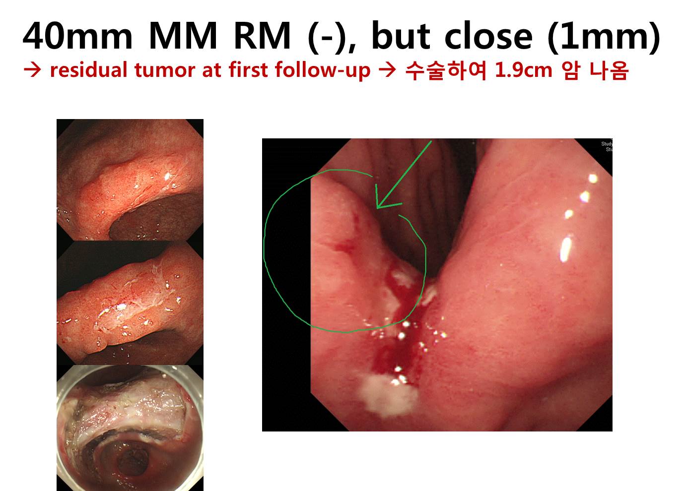
Stomach, ESD:
Early gastric carcinoma
1. Location : angle, lesser curvature
2. Gross type : EGC type IIc
3. Histologic type : tubular adenocarcinoma, well differentiated
4. Histologic type by Lauren : intestinal
5. Size of carcinoma : (1) longest diameter, 40 mm (2) vertical diameter, 25 mm
6. Depth of invasion : invades mucosa (muscularis mucosa) (pT1a)
7. Resection margin : free from carcinoma(N), safety margin : distal 2 mm, proximal 1 mm, anterior 4 mm, posterior 6 mm, deep 1000 ㎛
8. Lymphatic invasion : not identified(N)
9. Venous invasion : not identified(N)
10. Perineural invasion : not identified(N)
11. Microscopic ulcer : absent
12. Histologic heterogeneity: absent
Stomach, subtotal gastrectomy:
Early gastric carcinoma
1. Location : lower third, Center at antrum and lesser curvature
2. Gross type : EGC type IIc
3. Histologic type : tubular adenocarcinoma, well differentiated
4. Histologic type by Lauren : intestinal
5. Size : 1.9x1 cm
6. Depth of invasion : invades mucosa (muscularis mucosae) (pT1a)
7. Resection margin: free from carcinoma, safety margin: proximal 4 cm, distal 6.3 cm
8. Lymph node metastasis : no metastasis in 34 regional lymph nodes (pN0), (0/34: "1", 0/2; "3", 0/10; "4", 0/5; "4sb", 0/2; "5", 0/4; "6", 0/0; "7", 0/5; "8a", 0/3; "9", 0/1; "11p", 0/2; "12a", 0/0)
9. Lymphatic invasion : not identified
10. Venous invasion : not identified
11. Perineural invasion : not identified
12. AJCC stage by 7th edition: pT1a N0
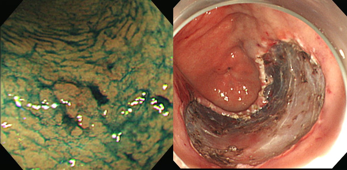
4년 전 ESD를 받고 경과관찰 중 내시경 육안소견에서 scar의 발적이 보여 조직검사를 하고 local recurrenc로 의뢰된 분입니다. 환자와 잘 상의하여 ESD를 하였고 13mm lamina propria 암으로 나왔습니다. ESD 후 local recur로 의뢰되면 늘 고민입니다. 아무래도 첫 소견을 정확히 알기 어렵기 때문입니다. Available한 정보를 잘 활용하고 환자와 오래 상의하는 수 밖에 없을 것 같습니다.
Resection margin을 확보하기 어려운 위치의 위암에서는 아무래도 국소 재발이 많습니다. 들문암 ESD 후 추적검사에서 국소 재발 발견.
[위암 805]
ESD for EGC was recommened by a surgeon. The margin of the lesion was not discrete, but the forceps biopsy showed "tubular adenocarcinoma, moderately differentiated (WHYX lesion)". ESD was done.
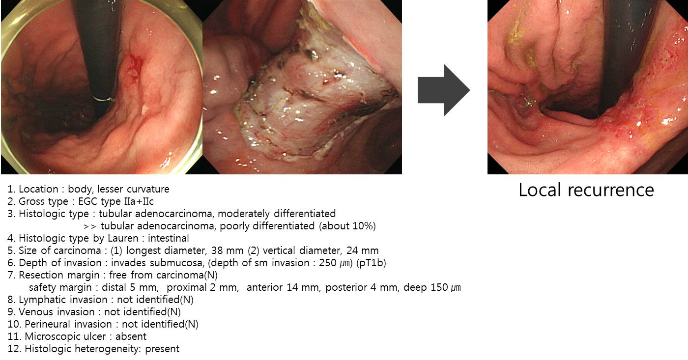
ESD: Early gastric carcinoma
1. Location : antrum, greater curvature
2. Gross type : EGC type IIc
3. Histologic type : tubular adenocarcinoma, moderately differentiated > poorly differentiated (30 %)
4. Histologic type by Lauren : mixed
5. Size of carcinoma : (1) longest diameter, 38 mm (2) vertical diameter, 30 mm
6. Depth of invasion : invades mucosa (muscularis mucosa) (pT1a)
7. Resection margin : free from carcinoma(N) but very close to proximal margin, safety margin : distal 4 mm, proximal less than 1 mm, anterior 2 mm, posterior 18 mm
8. Lymphatic invasion : not identified(N)
9. Venous invasion : not identified(N)
10. Perineural invasion : not identified(N)
11. Microscopic ulcer : absent
12. Histologic heterogeneity: present
The ESD pathology showed multiple risk factors of recurrence (size larger than 3 cm, mixed type histology with 30% of poorly differentiated component, negative resectiona margin but very close). I explained to the patient that there is no definite reason for surgey right now but the chance of recurrence is higher than usual, so careful follow-up is recommended.
Six years after the ESD, local recurrence was found. I recommended surgery.
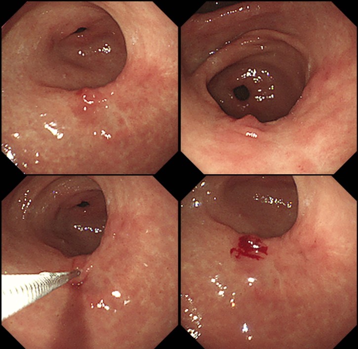
Stomach, radical subtotal gastrectomy: Early gastric carcinoma
1. Location : middle third, Center at angle and greater curvature
2. Gross type : EGC type IIb
3. Histologic type : tubular adenocarcinoma, moderately differentiated
4. Histologic type by Lauren : intestinal
5. Size : 1.5x0.6 cm
6. Depth of invasion : invades mucosa (lamina propria) (pT1a)
7. Resection margin: free from carcinoma, safety margin: proximal 5.0 cm, distal 3.7 cm
8. Lymph node metastasis : no metastasis in 41 regional lymph nodes (pN0)
9. Lymphatic invasion : not identified
10. Venous invasion : not identified
11. Perineural invasion : not identified
12. AJCC stage by 8th edition: pT1a N0
Careful follow-up is a very important part of endoscopic treatment of early gastric cancer.
2021-8-22. 내시경학회 세미나에서 연세대 윤영훈 교수님께서 멋진 동영상 강의를 하셨습니다.
[References]
1) Local recurrence associated with an adjacent neoplastic precursor lesion. Kikuchi S. Acta Med Okayama. 2016
Dysplasia-like atypia
© 일원내시경교실 바른내시경연구소 이준행. EndoTODAY Endoscopy Learning Center. Lee Jun Haeng.