EndoTODAY 내시경 교실
Beginner | ESA | Schedule | OPD
Seminars | Atlas | Recent | Links
![]() [확대내시경. Magnifying endoscopy] - 終
[확대내시경. Magnifying endoscopy] - 終
[Position statement, 2021-7-4] 내시경 3사의 최근 내시경은 확대내시경이 아니더라도 근접 관찰에 유리하게 렌즈의 초점거리가 설정되어 있습니다. 일반 내시경만으로 통상적인 진단과 치료를 시행하는데 거의 아무런 문제가 없습니다. Zoom 내시경을 이용한 NBI magnification으로 일반 내시경에서는 볼 수 없는 흥미로운 소견을 관찰할 수 있지만 아직 임상적 유용성은 명확하지 않습니다. 위암 내시경 전문가 입장에서 확대내시경은 전문가들의 고급스런 장난감입니다. 식도에서도 비슷하다고 생각합니다. 대장에서는 다소의 유용성이 있습니다.
2. Intestinal metaplasia의 확대내시경 소견
3. 위암의 확대내시경 관찰 - 3단계 접근법 (MESDA-G, magnifying endoscopy simple diagnostic algorithm for EGC)
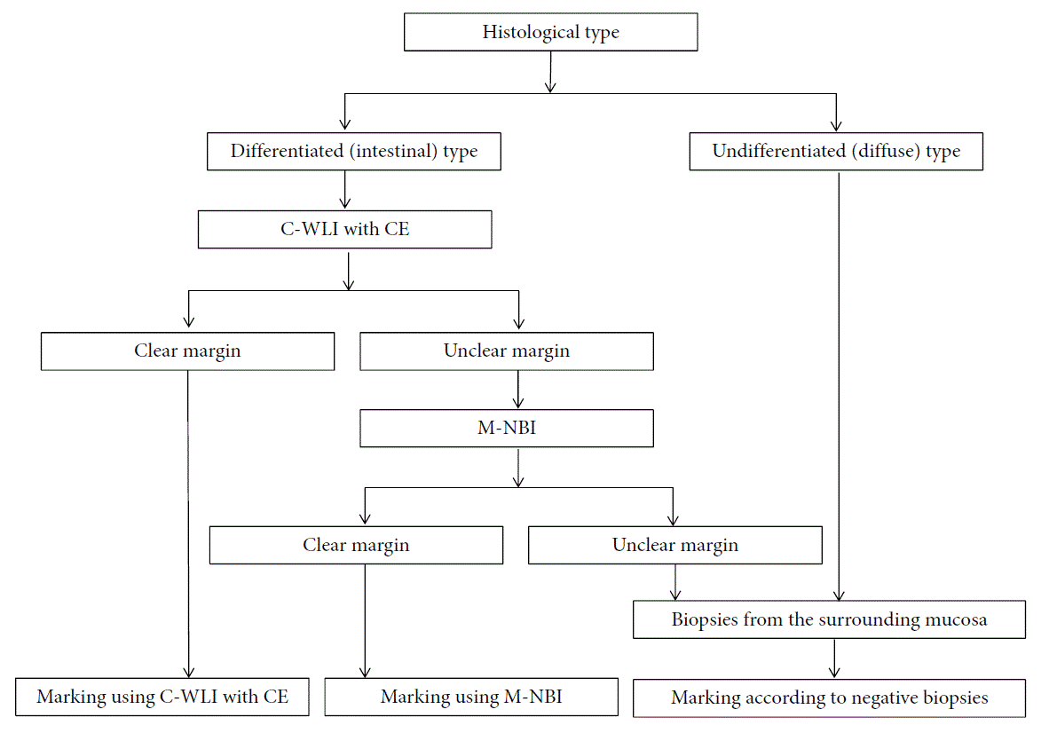
4. 조기위암 ESD 전 lateral margin의 확인
5. Undifferentiated-type 조기위암 ESD 전 NBI 확대내시경의 유용성
6. Near Focus
7. 식도 esophagus
8. 대장 image-enhanced endoscopy - PENTAX
9. References
PPT PDF 1.2M (GIE review, 이소정)

YouTube
![]() 1. 정상 위의 확대내시경 소견
1. 정상 위의 확대내시경 소견
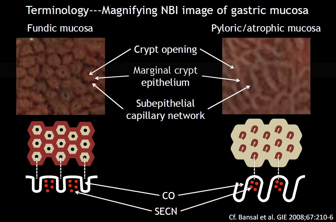
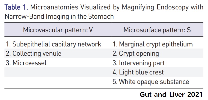
위의 정상 및 비정상 확대내시경 소견은 Fukuoka 대학의 Yao 교수가 2013년 Ann Gastroenterol와 2015년 Clin Endosc에 잘 정리한 것을 참고하십시오. VS (vessel plus surface) classification system이라고 부르는 것인데 microvascular pattern과 microsurface pattern을 한꺼번에 기술하고 있으므로 다소 혼란스럽습니다. 이 둘을 따로 또 같이 생각하면 이해가 쉽습니다.
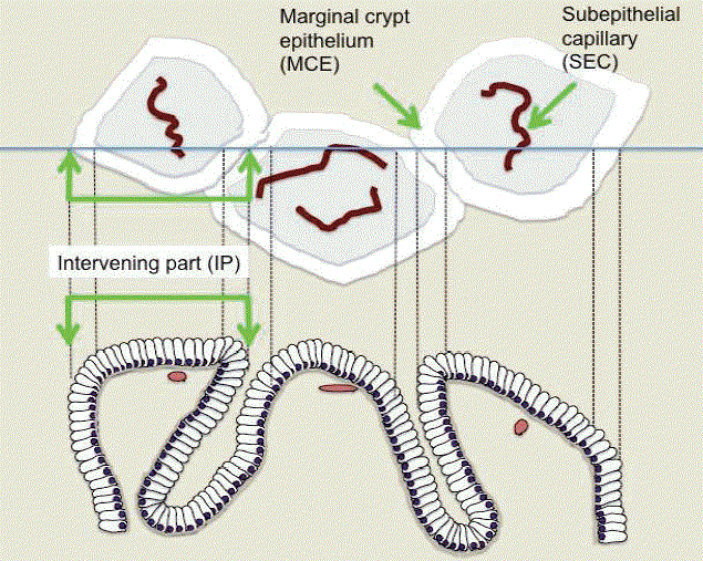
(1) microvascular pattern (V) : subepitherial capillary network (SECN), CV (collecting venule), MV (The microvessel is a term used for vessels that appear in pathological mucosa.)
(2) microsurface pattern (S): marginal crypt epithelium (MCE), crypt opening (CO)
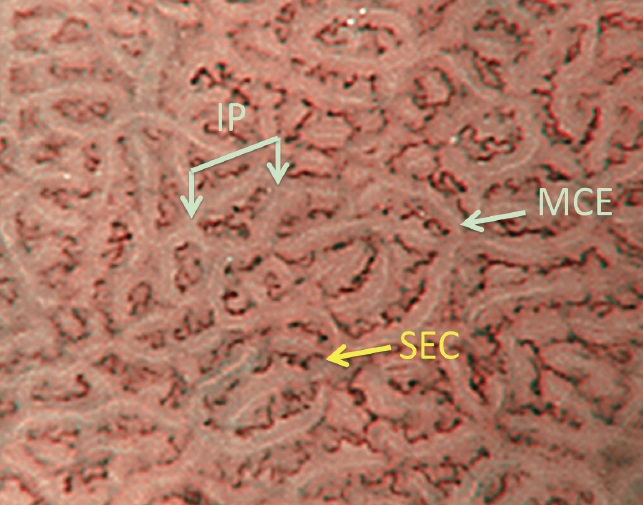
1) 정상 fundic gland: microvascular pattern은 SECN (subepitheliral capillary network)가 벌집모양을 이루며, microsurface pattern은 marginal crypt epithelium (MCE)에 둘러쌓인 crypt opening (CO)이 SECN 중앙에 위치합니다. Helicobacter 감염이 있거나 위축성 변화가 현저할 때는 이런 정상 소견이 보이지 않게 됩니다.
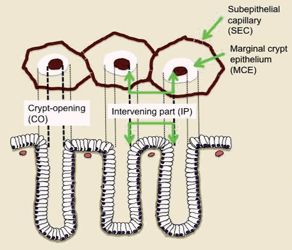
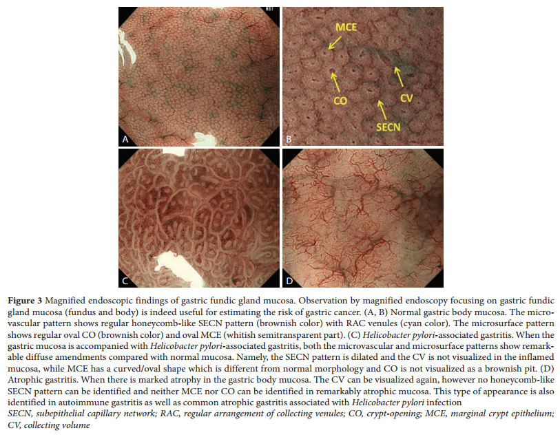
(a) Schematic diagram of the microvascular architecture and the microsurface structure of the normal gastric fundic gland mucosa corresponding to the surface morphology as visualized by magnifying endoscopy (ME) with narrow-band imaging (NBI). The microvascular architecture is formed by the capillaries and collecting venules. The morphology of each capillary is that of a polygonal closed loop. These loops anastomose repeatedly with each other, forming a regular honeycomb-like subepithelial capillary network pattern. The microsurface structure is made up of the marginal crypt epithelium/white zone (MCE/WZ), and the intervening part in between. The epithelial morphology is visualized as a semitransparent white belt-like structure (the MCE/WZ), showing a circular or oval shape at the center of which lies the crypt opening. (b) ME with NBI of normal fundic gland mucosa. (Muto M. Digest Endosc 2016)
2) 정상 pyloric gland: microvascular pattern은 dark brown colored coil-shaped open loop를 이루며, microsurface pattern은 regular polygonal 또는 curved marginal crypt epithelium pattern을 이룬다.
Dark brown coil-shaped open loop capillary & polygonal or curved MCE (a) Schematic diagram of the microvascular architecture and the microsurface structure of the normal gastric pyloric gland mucosa corresponding to the surface morphology as visualized by magnifying endoscopy (ME) with narrow-band imaging (NBI). The microvascular architecture is formed by capillaries and collecting venules, but the latter are rarely observed from the mucosal surface. The morphology of each capillary is that of coil-shaped open loops. The mucosal surface structure is made up of the marginal crypt epithelium/white zone (MCE/WZ) and the intervening parts surrounded by MCE/WZ. The MCE/WZ morphology usually shows polygonal structures but may be curved or linear. (b) ME with NBI of normal pyloric gland mucosa. (Muto M. Digest Endosc 2016) Some arteroles penetrate the mucularis mucosa and branch the capillary within the lamina propria. (Fundic gland mucosa에서는 arteriole이 점막하층에 국한된 반면 pyloric gland mucosa에서는 arteriole이 lamina propria에서도 관찰된다는 의미입니다.)
![]() 2. Intestinal metaplasia의 확대내시경 소견
2. Intestinal metaplasia의 확대내시경 소견
(A) Magnifying endoscopy with narrow-band imaging (M-NBI) findings of light blue crest (LBC) in gastric mucosa with intestinal metaplasia. Fine light blue (light cyan colored) linear reflections are located on the epithelial margins, visualized using M-NBI. LBC is a marker of brush border on the epithelial surface of intestinal metaplasia. (B) M-NBI findings of white opaque substance (WOS) in gastric mucosa with intestinal metaplasia. WOS visualized by reflections/strong scattering of whole projected lights located in the surface epithelium of the intervening part. WOS is lipid micro-droplet that is densely accumulated within the epitheliuam or beneath the mucosal epithelium.
![]() 3. 위암의 확대내시경 관찰 - 3단계 접근법 MESDA-G, magnifying endoscopy simple diagnostic algorithm for EGC)
3. 위암의 확대내시경 관찰 - 3단계 접근법 MESDA-G, magnifying endoscopy simple diagnostic algorithm for EGC)
1 단계. 정상 소견 익히기 - 위암 내시경 진단을 위해서는 fundic mucosa와 pyloric mucosa의 정상 NBI 확대내시경 소견을 잘 알아야 합니다. Microvascular structure와 microsurface structure가 위치에 따라 다르기 때문입니다.
2 단계. demarcation line 찾기 - 백색광 내시경으로 관찰하다가 의심스러운 함몰부위가 있으면 NBI 확대내시경을 적용합니다. 정상과 비정상의 경계가 보이면 이를 demarcation line이라고 부릅니다 (Gut Liver 2021). 한 임상연구에 의하면 검진 환자의 20% 정도에서 suspicious lesion이 보여 NBI 확대내시경을 했다고 합니다.
Digest Endosc 2015년 7월호 WEO Upper GI Cancer Committe
좌측 사진은 demarcation line이 명확한 반면 우측 사진에서는 demarcation line을 찾을 수 없습니다.
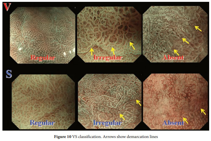
3 단계. IMVP와 IMSP 확인 - 일단 demarcation line이 있으면 microvascular pattern과 microsurface pattern을 관찰합니다 (VS classification). Irregular microvascular pattern (IMVP)이나 irregular microsurface pattern (IMSP)이 있으면 위암으로 진단할 수 있습니다.
4 단계 (optional). Other M-NBI findings useful for diagnosis of EGC
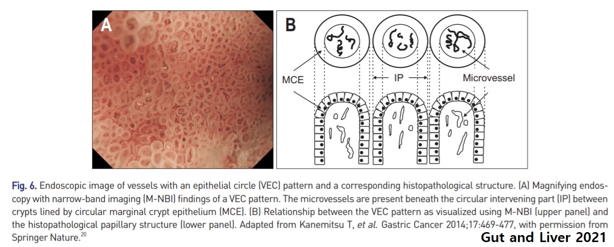
1) VEC (vessels within an epithelial circle) - papillary structure or coexisting undifferentiated-type carcinoma and submucosal invasion
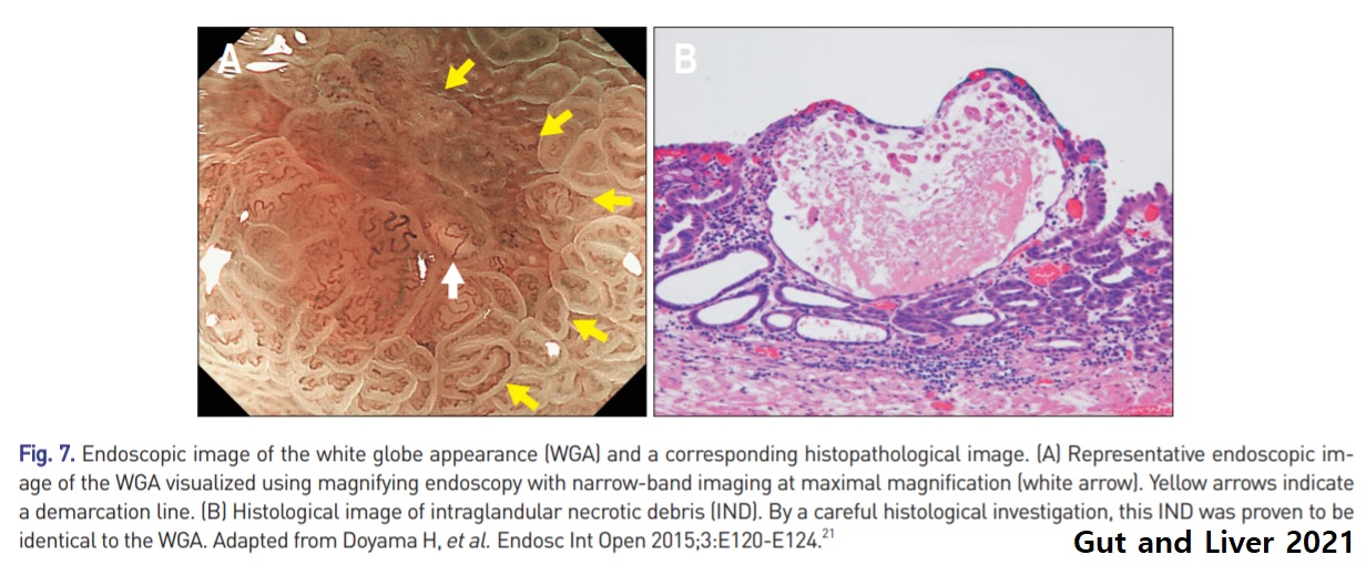
2) WGA (white globe appearance) - intraglandular necrotic debris
3) MCDL (multiple convex demarcation line) - non-cancerous depressed lesion
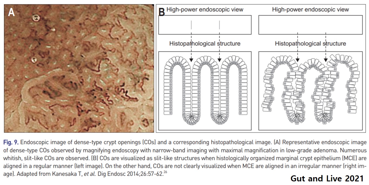
4) Dense-type crypt openings - low grade adenoma
5) Diagnosis of histological differentiation of EGC by M-NBI
![]() [Cases and articles]
[Cases and articles]
순천향대학교의 최근 논문의 증례입니다.
Korean J Helicobacter Up Gastrointest Res. 2015 Mar;15(1):39-43
![]() Takashi Kanesaka. Endosc Int Open 2015
Takashi Kanesaka. Endosc Int Open 2015
Takashi Kanesaka. Endosc Int Open 2015
Takashi Kanesaka. Endosc Int Open 2015
각 소견의 진단 정확도 (Takashi Kanesaka. Endosc Int Open 2015)
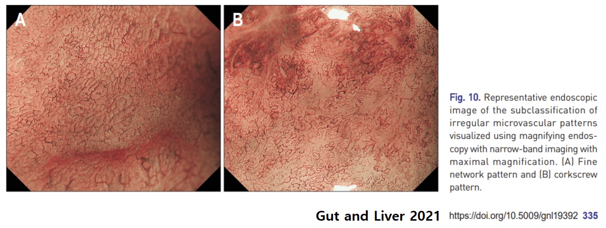
Representative cases for each endoscopic microvascular finding. Target lesions indicated with white arrows. a Case 1: dilation and tortuosity were present, but difference in caliber and variation in shape were absent. This lesion was histologically diagnosed as noncancerous. b Case 2: tortuosity was present but dilation, difference in caliber, and variation in shape were absent. This lesion was histologically diagnosed as noncancerous. c Case 3: dilation, difference in caliber and variation in shape were present, but tortuosity was absent. This lesion was histologically diagnosed as cancerous. d Case 4: tortuosity and variation in shape were present but dilation and difference in caliber were absent. This lesion was histologically diagnosed as cancerous. (Takashi Kanesaka. Endosc Int Open 2015)
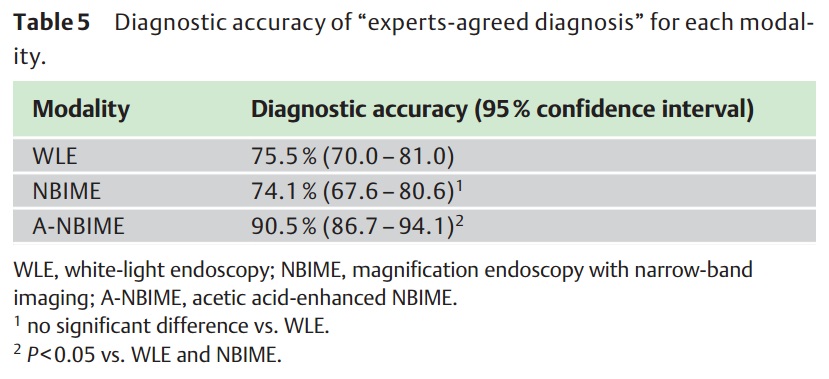
![]() 2016년 1월호 Endoscopy 지에 일본 연구자들이 위암 확대내시경에 대한 상세한 논문을 발표하였습니다 (Shibagaki K. Endoscopy 2016). WLE (white light endoscopy), NBIME (magnification endoscopy with narrow-band imaging), A-NMIME (NBIME with acetic acid enhancement)를 이용하여 macroscopic pattern에 따라 M1/M2/M3, capillary pattern에 따라 C1/C2/C3/C4, surface pattern에 따라 S1/S2/S3으로 나누었고 각각 adenoma/differentiated type EGC/undifferentiated type EGC로 간주하였습니다.
2016년 1월호 Endoscopy 지에 일본 연구자들이 위암 확대내시경에 대한 상세한 논문을 발표하였습니다 (Shibagaki K. Endoscopy 2016). WLE (white light endoscopy), NBIME (magnification endoscopy with narrow-band imaging), A-NMIME (NBIME with acetic acid enhancement)를 이용하여 macroscopic pattern에 따라 M1/M2/M3, capillary pattern에 따라 C1/C2/C3/C4, surface pattern에 따라 S1/S2/S3으로 나누었고 각각 adenoma/differentiated type EGC/undifferentiated type EGC로 간주하였습니다.
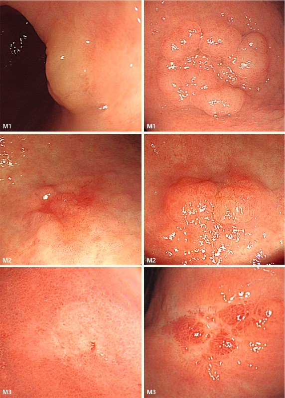
White-light endoscopy (WLE) images illustrating the macroscopic pattern classification of gastric mucosal neoplasms. Type M1, suggestive of adenoma, is a protruding or flat elevated whitish lesion with a roundish edge and a smooth or often nodular surface. Type M2, suggestive of differentiated adenocarcinoma, is an irregularly shaped and depressed, flat, or elevated lesion either with a red color or without discoloration. Type M3, suggestive of undifferentiated adenocarcinoma, is a depressed whitish lesion with or without variously sized reddish nodules.

Magnification endoscopy with narrow-band imaging (NBIME) images illustrating the capillary pattern classification of gastric mucosal neoplasms. Type C1, suggestive of adenoma, has capillaries with a homogenous diameter and distribution, which form round or oval networks (C1-a, network form) or grow within regular mucosal microstructures (C1-b, intra-microstructure form). Type C2, suggestive of differentiated adenocarcinoma, has capillaries with a heterogeneous diameter and distribution, which form a polygonal or incomplete network (C2-a, network form) or grow within irregular mucosal microstructures (C2-b, intra-microstructure form). Type C3, suggestive of undifferentiated adenocarcinoma, has capillaries with a heterogeneous diameter and distribution, which grow in a disordered fashion with an unclear mucosal microstructure. Type C4, which is not related to a specific histologic type, has capillaries that are invisible or obviously decreased in number.
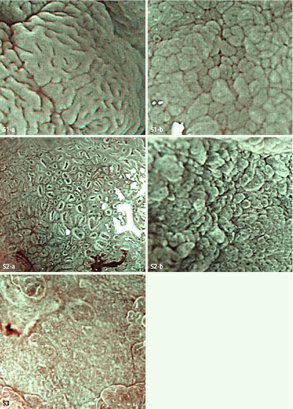
Magnification endoscopy with narrow-band imaging and acetic acid enhancement (A-NBIME) images illustrating the microstructure pattern classification of gastric mucosal neoplasms. Type S1, suggestive of adenoma, has glandular crypts present, with homogeneously sized, shaped and arranged foveolae (S1-a, foveola form) or grooves (S1-b, groove form). Type S2, suggestive of differentiated adenocarcinoma, has glandular crypts present, with heterogeneous foveolae or grooves (S2-a, foveola form; S2-b, groove form). Type S3, suggestive of undifferentiated adenocarcinoma, has absent or severely decreased numbers of glandular crypts.
저자들은 아래 Table 5 결과를 바탕으로 "A-NBIME showed statistically significantly higher diagnostic accuracy for gastric mucosal neoplasms, with good reproducibility, compared with WLE and NBIME, which provided similar lower accuracy."라고 결론짓고 있습니다. 어떻게 해석되어야 할지 고민입니다. Acetic acid를 사용한 확대내시경이 도움이 된다는 것은 인정할 수 밖에 없을 것 같습니다. 하지만 white light endoscopy와 NBI 확대내시경의 차이가 없다는 이번 결과는 기존에 많은 일본 연구자들이 NBI 확대내시경이 진단에 유용하다고 주장했던 것과는 상반되는 것입니다. 저는 이번 연구 결과를 'white light endoscopy도 잘 보면 상당히 좋다'는 방향으로 해석하고 싶습니다. 향후 어떤 방향으로 결론이 모일지 지켜볼 일입니다.
![]() 4. 조기위암 ESD 전 lateral margin의 확인
4. 조기위암 ESD 전 lateral margin의 확인
Differentiated-type에서는 white light endoscopy, chromoendoscopy, magnifying NBI 소견으로 범위를 정할 수 있습니다. 그러나 undifferentiated-type에서는 이 모든 것이 크게 도움되지 않아서 병소 주변 조직검사에 의존할 수 밖에 없습니다. 2016년 일본 암연구회병원 연구자들이 undifferentiated-type에서도 어느 정도 도움이 된다는 발표를 하고 있지만... (Horiuchi Y. Gastric Cancer 2016).
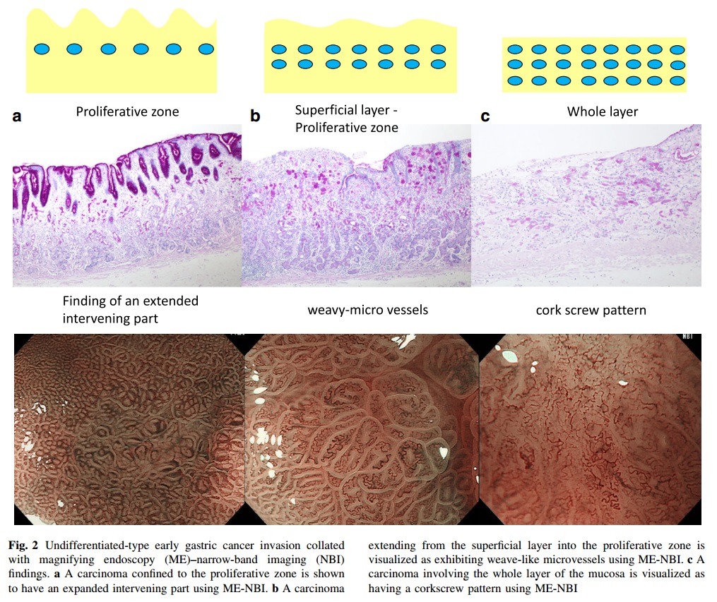
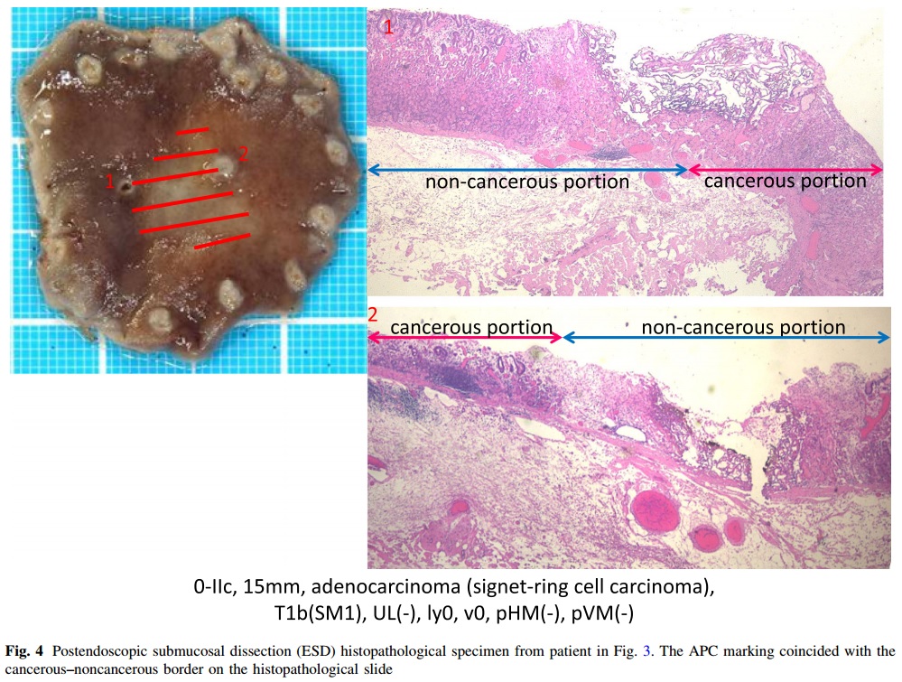
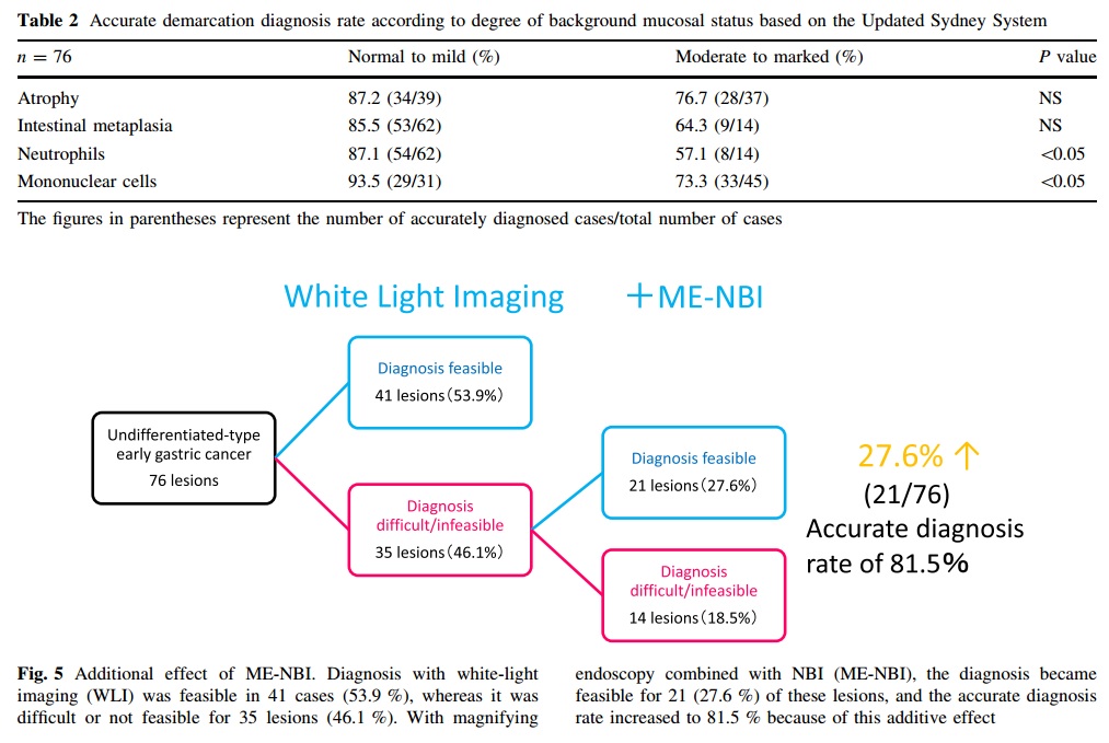
![]() 5. Undifferentiated-type 조기위암 ESD 전 NBI 확대내시경의 유용성
5. Undifferentiated-type 조기위암 ESD 전 NBI 확대내시경의 유용성
전통적으로 NBI 확대내시경은 differentiated-type EGC에서 도움이 되고 undifferentiated-type EGC에서는 별로 도움되지 않는 것으로 알려졌습니다. 2016년 일본 암연구회병원 연구자들이 undifferentiated-type EGC에서도 어느 정도 도움이 된다고 보고하였습니다 (Horiuchi Y. Gastric Cancer 2016).
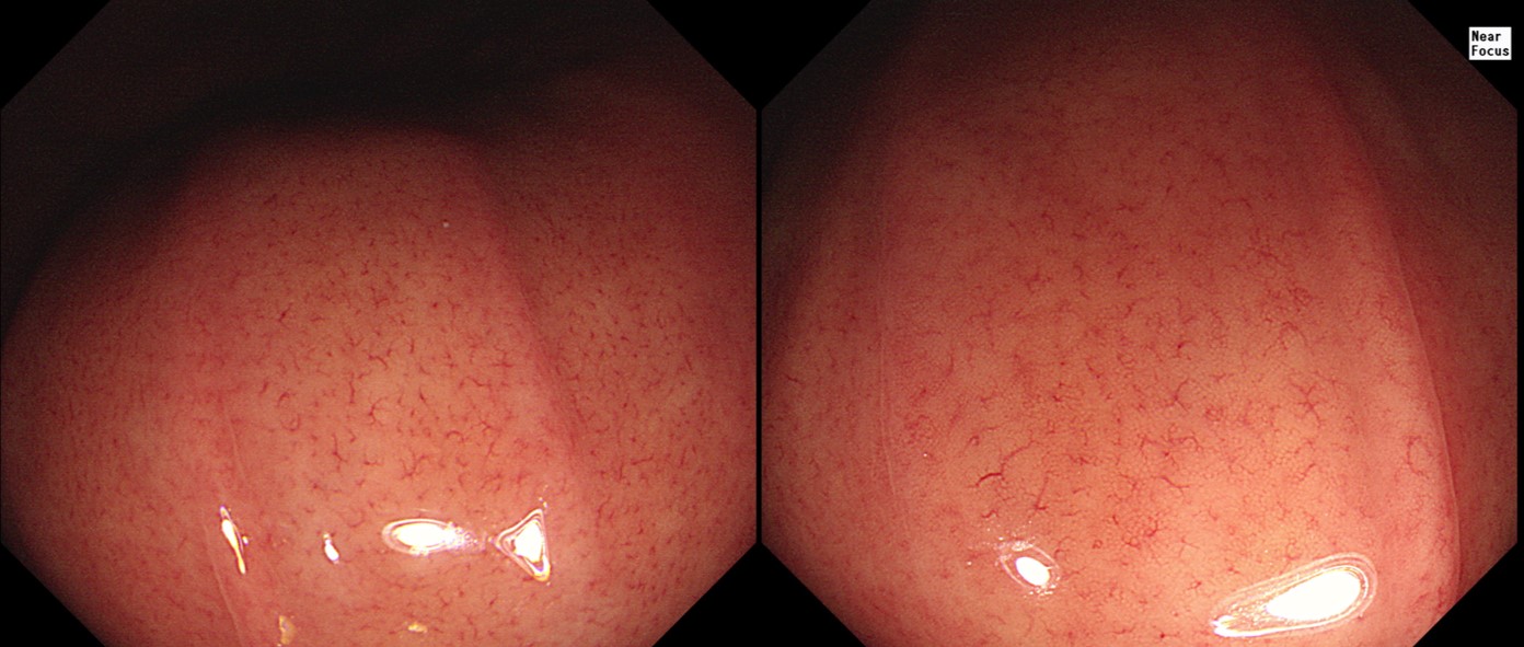
![]() 6. Near focus
6. Near focus
확대 내시경을 통하여 microvascular pattern과 microsurface pattern을 관찰하는 것이 유행입니다. Zoom lens의 이동을 이용한 정식 확대 내시경이 아닌 모델 중 Near Focus라는 기능을 가진 내시경이 있습니다 (초장기에는 dual focus라고 불렀습니다). Near Focus는 약간 digital 적으로 button을 누르면 렌즈 하나가 위치를 바꿔 focus가 가깝게 바뀌는 기능입니다. 좀 더 접근하려 fine surface pattern을 볼 수 있다는 것입니다.
Olympus Near Focus의 작동 원리 (https://medical.olympusamerica.com/technology/dual-focus)
아래 사진은 정상 위점막입니다. 우측 Near Focus 영상에서 RAC이 더 잘 보니다. 약간의 surface pattern과 함께.
Near Focus를 켜면 zoom 정도는 아니지만 보통 내시경보다는 훨씬 확대된 영상을 보게 됩니다. 진단적 유용성은 아직 잘 모르겠지만, 흥미로운 장난감인 것은 틀림 없습니다. 아래 증례의 우측 아래 사진이 Near focus입니다. Solid 부분이 30% 정도 있는 Lauren diffuse type 위암이었습니다.

Stomach, subtotal gastrectomy: Early gastric carcinoma
1. Location : lower third, Center at antrum and postero-greater curvature
2. Gross type : EGC type IIc
3. Histologic type : tubular adenocarcinoma, poorly (solid) differentiated with signet ring cell component (20%)
4. Histologic type by Lauren : diffuse
5. Size : 2x1.5 cm
6. Depth of invasion : invades submucosa (sm3) (pT1b)
7. Resection margin: free from carcinoma, safety margin: proximal 4.1 cm, distal 6.6 cm
8. Lymph node metastasis : no metastasis in 37 regional lymph nodes (pN0)
9. Lymphatic invasion : not identified
10. Venous invasion : not identified
11. Perineural invasion : not identified
12. AJCC stage by 8th edition: pT1b N0
![]() 7. 식도 esophagus
7. 식도 esophagus
식도 확대내시경에서는 IPCL을 관찰해야 합니다.
IPCL (자료 제공: 이선영 교수님)
몇 개의 분류법이 있었는데 이를 통합하여
ANBIIG Tailand 2020에서 일본 Toranomon Hospital의 Daisuke Kikuchi 선생의 강의를 꼭 들어보시기 바랍니다.
![]() 8. 대장 image-enhanced endoscopy (IEE)
8. 대장 image-enhanced endoscopy (IEE)
| Image-enhanced endoscopy (IEE) | |
| Conventional chromoendoscopy | Virtual chromoendoscopy |
| Dye-based | Equipment-based |
| Contrast: indigocarmine Absorptive: crystal violet | Optical: NBI (Olympus), OE (Pentax) Electronic: FICE, i-scan SE & TE (Pentax) Others: BLI, LCI (Fujifilm) |
| Pit pattern | Capillary pattern |
색소내시경을 이용한 Kudo 분류는 pit pattern을 보는 것이고, NBI 등을 이용한 Sano 분류는 capillary pattern을 보는 것입니다. Hiroshima, NICE, JNET은 capillary pattern을 주로 보면서 surface pattern을 조금 참조하는 방법입니다.
Pit는 intestinal gland의 opening입니다. Indigocarmine을 분무하면 dye가 pit에 고이므로 pit가 검게 보입니다. Crystal violet 염색을 하면 pit 주변 상피세포의 핵이 염색되므로 pit 자체는 하얗게 보입니다.
[Conventional IEE (pit pattern)]
Kudo 분류.

Hyperplastic polyp이나 serrated polyp에서는 crypt 상부 절반이 serration 되면서 하부가 확대되기 때문에 pit의 변화가 옵니다.
[Equipment-base IEE]
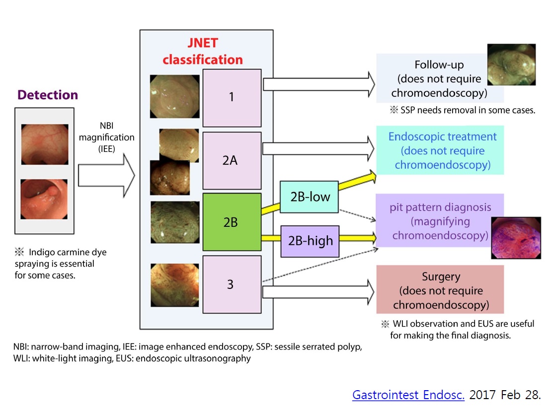
색소 분무 후 확대내시경 소견인 pit pattern은 일찍부터 Kudo 분류로 통일되었습니다. 반면, NBI를 이용한 대장내시경 확대내시경 소견에 대해서는 매우 많은 분류법이 난립하였습니다 (Utsumi T. Clin Endosc 2015). 마침내 2016년 여름 JNET 분류가 나오면서 총정리되는 분위기입니다 (Sano Y. Digest Endosc 2016).
Sano 분류. Capillary pattern만 관찰하므로 비교적 간단한 분류입니다. 그러나 Sano 분류는 확대내시경 관찰을 전제로 적용하는 방법입니다.
NICE 분류. Capillary pattern을 주로 보면서 색조나 surface pattern을 관찰하는 방법입니다. NICE II는 Sano II-IIIA와 비슷하고 NICE III는 Sano IIIB와 비슷합니다. Sano capillary pattern classification은 확대내시경을 전제로 적용한 것인 반면, NICE 분류는 확대내시경이 아닌 일반 내시경을 전제로 만들어진 방법입니다.
JNET 분류. NICE II를 JNET 2A, 2B로 나눈 것으로 이해하면 되겠습니다. NICE 분류의 한계를 극복하기 위한 JNET 분류는 확대내시경을 전제로 만들어진 방법입니다.
JNET 분류 reference panel
Depth of invasion 관찰에는 equipment-base IEE (Sano, NICE, JNET 분류)보다 색소내시경 후 확대내시경으로 관찰한 pit pattern 분류가 우월한 것으로 인정되고 있습니다. 따라서 strategy라는 것이 제안되었습니다. 간단히 말하자면 일반 내시경(white light endoscopy)에서 이상 소견이 보이면 equipment-based IEE를 적용(가장 간단한 것은NBI를 켜는 것입니다)하고 안 좋은 소견이 보이면 pit pattern을 보기 위하여 crystal violet 염색 및 확대내시경을 하는 것입니다.
Three step strategy.
실제 환자에서는 애매한 경우가 아주 많습니다. 2023년 8월 20일 KSGE seminar의 토론을 살펴보시기 바랍니다.
KSGE seminar. Log-in 要
[i-scan과 optical enhancement (Pentax)]
Pentax는 비록 광학적 확대내시경을 제공하지 않고 있으나 i-Scan이라는 image enhancement technology를 제공하던 회사입니다 (World J Gastroenterol. 2010 - full text free). 최근에는 i-Scan의 mode를 바꿔서 surface enhancement (SE)와 tone enhancement (TE) 기능으로 이해하기 쉽게 만들었다고 합니다. 여기에 optic filter를 적용하여 좁은 band를 이용한 optical enhancement (OE)를 더하여 세 가지 mode가 가능하게 되었습니다. SE, TE를 비교할 수 있는 mini atlas와 OE의 기술적 특징을 설명한 white paper를 소개합니다.
- SE, TE를 비교할 수 있는 mini atlas. PDF 4.7M
- OE의 기술적 특징을 설명한 white paper. PDF 0.6M
보다 자세한 정보는 아래 링크에 있습니다.
* 참고: i-scan technology
![]() [References]
[References]
1) EndoTODAY image enhanced endoscopy IEE
2) Magnifying endoscopy in upper GI tract 김광하 (IDEN 2019)
3) NBI magnifying endoscopic classification of colorectal tumors proposed by the Japan NBI Expert Team Sano Y. Digest Endosc 2016 (PDF)
4) Image-enhanced endoscopy and its corresponding histopathology in the stomach Doyama H. Gut Liver 2021
5) 2017년 5월 20일 내시경기기/스텐트연구회 심포지엄 (그랜드앰배서더호텔)
6) Olympus 사의 magnifying endoscopy training phantom
2019-11-16 KSGE 경주
2019-11-16 KSGE 경주
7) JDDW 2024에서 Yao 확대내시경 모델의 upgrade version을 보았습니다.

8) 長野확대내시경연구회
© 일원내시경교실 바른내시경연구소 이준행. EndoTODAY Endoscopy Learning Center. Lee Jun Haeng.