EndoTODAY ГЛНУАц БГНЧ
Beginner | ESA | Schedule | OPD
Seminars | Atlas | Recent | Links
![]() [Esophageal melanocytosis - malignant melanoma ЙпЛ§РЬ АЁДЩЧбАЁ?] - №ћ
[Esophageal melanocytosis - malignant melanoma ЙпЛ§РЬ АЁДЩЧбАЁ?] - №ћ
[РЬСиЧрРЧ РсСЄРћРЮ ПфОр (2017-11-28)]
Melanosis (melanin РЬГЊ lipofuscinРЧ ФЇТјПЁМ ЛчПыЕЧДТ ПыОю)ДТ benignРЬ ЦВИВОјДТ АЭ ААСіИИ, melanocytosis (melanin Л§МК ММЦїРЧ СѕАЁ)ПЁМДТ malignant melenoma ЙпЛ§РЬ АЁДЩЧб АЭРИЗЮ УпСЄЕЫДЯДй. ЙЎЧх КИАэЕЕ РжАэ АГРЮРћ АцЧшЕЕ РжНРДЯДй. ЙЎСІДТ НФЕЕ СЖСїАЫЛчПЁМ melanocytosisПЭ melanosisАЁ ИэШЎШї БИКаЕЧСі ОЪАэ КИАэЕЧДТ АцПьАЁ ИЙДйДТ АЭРдДЯДй. ОЯЙпЛ§ РЇЧшРЧ ТїРЬАЁ РжДТ ЕЮ ПыОюДТ БИКаЧиМ ЛчПыЧв АЭРЛ СІОШЧеДЯДй.
![]() 1. Melanocytosis СЄРЧ (melanocytosisАЁ ИТДТАЁ melanosisАЁ ИТДТАЁ?)
1. Melanocytosis СЄРЧ (melanocytosisАЁ ИТДТАЁ melanosisАЁ ИТДТАЁ?)
MelanocytosisДТ ЧЧКЮПЁ ИЙРК ЧіЛѓРЬЙЧЗЮ ЧЧКЮАњ РЧЛчПЁАд ЙЎРЧЧЯПДНРДЯДй.
ЧЧКЮАњ РЧЛч ДфКЏ: "MelanocyteАЁ СѕАЁЧЯИщ melanocytosisРЬАэ melanin pigmentАЁ СѕАЁЧЯИщ melanosisРдДЯДй. ДыКЮКаРЧ melanosisДТ melanocytosisПЁ РЧЧб АЭРЬЙЧЗЮ ЛчНЧ ЕбРК ИХПь КёНСЧеДЯДй. БзЗЏГЊ АЃШЄ melanocyteДТ СѕАЁЕЧОю РжСі ОЪАэ melaninИИ СѕАЁЕШ melanosisЕЕ АЁДЩЧеДЯДй. Melanoma(atypical melanocytesИІ КИРг)АЁ ОЦДЯАэ ДйИЅ РЬЛѓРЬ ЕПЙнЕЧОю РжСі ОЪРИИщ УпРћАќТћРК ЧЪПфЧЯАкСіИИ КА ЙЎСІДТ ОјРЛ АЭ ААНРДЯДй."
НФЕЕ СЖСїАЫЛчПЁМ melanosisЗЮ ГЊПРИщ ДыКЮКа melanocytosisРдДЯДй. H & E ПАЛіРИЗЮ melanocyte БИКаРЬ НБСі ОЪБт ЖЇЙЎПЁ КДИЎАњ МБЛ§ДдЕщРЬ melanocytosisЗЮ СјДмРЛ КйРЬБтКИДйДТ melanosisЗЮ СјДмРЛ СжДТ АцПьАЁ ИЙРК АЭ ААНРДЯДй. ДйИИ "melanocytisis = melanosis"ДТ ОЦДеДЯДй. Чб ИЎКф(Chang. Arch Pathol Lab Med 2006)ПЁМ ЧиДч ГЛПыРЛ ПХБщДЯДй.
Although the term melanosis was used in most of the previous reports, this term does not accurately describe the increased number of melanocytes found in this condition. In addition, this term is a generic term describing conditions in which there is an abnormal grayish black or brownish black pigmentation and does not imply that the underlying pigment is specifically melanin.For example, the melanosis described in the duodenum, ileum, and colon is characterized by the presence of pigmented macrophages containing ЁЎЁЎpseudomelaninЁЏЁЏ in the lamina propria without the participation of melanocytes. Pseudomelanin has been shown to be lipomelanin, a mixture of lipofuscin and melanin, which presents as aggregates of electron-dense granules within macrophages. Therefore, the unhelpful term esophageal melanosis should be avoided in pathology reports.
![]() 2. Melanocytosis КДИЎ
2. Melanocytosis КДИЎ
![]() 3. Melanocytosis АЁ melanomaРЧ precursorРЮАЁ?
3. Melanocytosis АЁ melanomaРЧ precursorРЮАЁ?
СЄЛѓРћРИЗЮ НФЕЕПЁДТ ОрАЃРЧ melanocyteАЁ СИРчЧв Мі РжНРДЯДй. РЬАїПЁМ melanocytosisЕЕ Л§БтАэ melanomaЕЕ АЁДЩЧеДЯДй. БзЗЏГЊ ГЛНУАцРИЗЮ ШЎРЮЧв Мі РжДТ melanocytosisАЁ melanomaЗЮ СјЧрЧЯДТАЁДТ КаИэЧЯСі ОЪНРДЯДй. МвМіРЧ СѕЗЪКИАэИИ РжРЛ ЛгРдДЯДй. ОеМ МвАГЧб ИЎКф(Chang. Arch Pathol Lab Med 2006)ПЁМДТ ОрАЃ КЮСЄРћРЮ РЧАпРЬ МвАГЕЧАэ РжНРДЯДй.
Endoscopic melanocytosis of the esophagus has been reported in patients with anal melanoma and esophageal squamous cell carcinoma in situ. Notably, melanocytosis has been described in 25% to 30% of surgical specimens of esophagus containing primary malignant melanomas, and this lesion has been suggested to be a precursor of melanoma by some authors. However, no follow-up information is currently available, and there is no documented case in the literature in which esophageal melanocytosis progressed to esophageal melanoma.
Upper aerodigestive tractРЧ squamous cell carcinomaПЭ АќЗУЕЧОю РжДйДТ ПЌБИАЁ РжНРДЯДйИИ ОЦСї СЄМГРК ОЦДеДЯДй.
Anal melanomaПЭРЧ АќЗУМКРК КвИэШЎЧеДЯДй. РиОюЙіЗСЕЕ ССРЛ АЭ ААНРДЯДй.
СІАЁ АцЧшЧб melanocytosis Сп АЁРх ЧіРњЧб АцПьИІ МвАГЧеДЯДй. УЙ СјДм 7Гт ШФ melanomaЗЮ progressionРЛ ЧЯПДНРДЯДй.
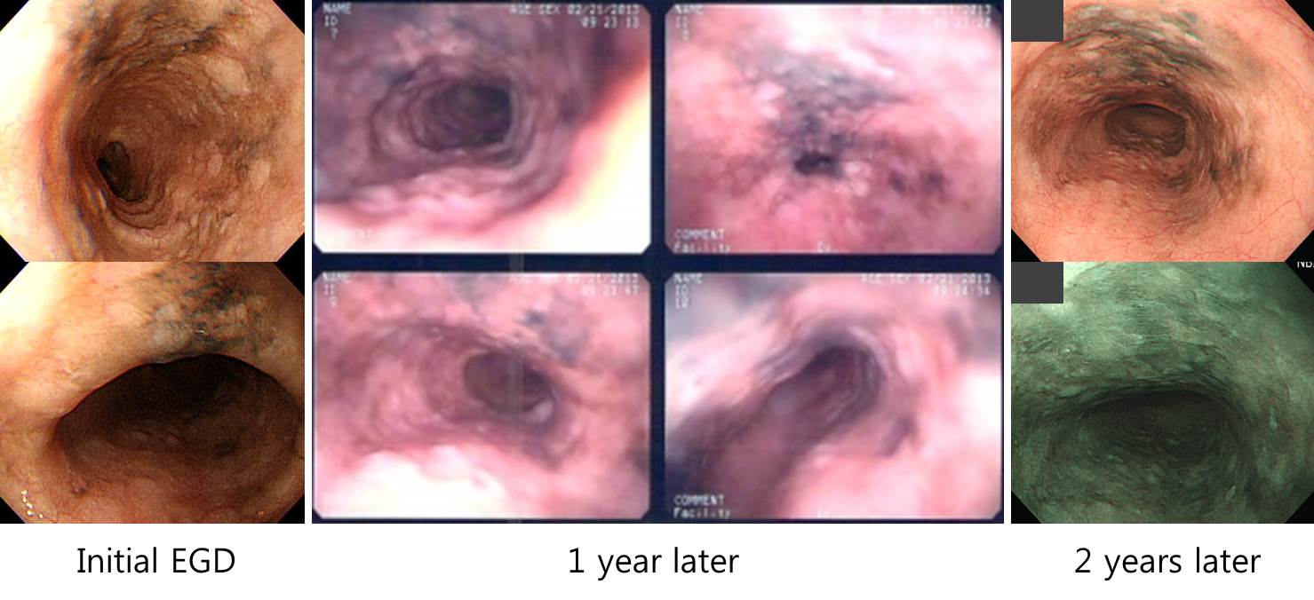
6Гт ШФ. MassАЁ ИИЕщОюСГАэ СЖСїАЫЛчПЁМ malignant melanomaЗЮ ГЊПШ.
ДйРНРК СЖСїАЫЛчПЁМ melanosisЗЮ КИАэЕЧОњРИГЊ melanocytosisЗЮ Л§АЂЕЧДТ ПЙРдДЯДй. РЬЗБ АцПьАЁ ДыКЮКаРдДЯДй.
ОЦЗЁ ГэЙЎЕЕ melanosisЗЮ ЕЧОюРжСіИИ melanocytosisАЁ ИТДТ ЧЅЧіРЮ АЭ ААНРДЯДй.
![]() 5. АЈКАСјДм
5. АЈКАСјДм
Primary malignant melanoma of the esophagus is also rare, with an incidence of 0.0036 in 100,000. It represents approximately 0.1% to 0.2% of all esophageal neoplasms. So far, less than 200 cases of esophageal melanoma have been published worldwide. Endoscopically, it often presents as pigmented or nonpigmented polypoid mass in the middle or lower esophagus. Histologically, melanoma is composed of epithelioid cells arranged in nests or spindle cells arranged in fascicles, with or without deposition of melanin pigment. The nuclei of the melanocytes were typically large and round or oval with a vesicular chromatin pattern and distinct or prominent nucleoli. Nuclear pseudo-inclusions were often readily identified. Most of the tumors are highly cellular and contain numerous mitotic figures. If a tumor is amelanotic, it may be difficult to recognize as malignant melanoma without ancillary immunohistochemical staining. Lentiginous or pagetoid intraepithelial spread is often present in esophageal mucosa adjacent to the invasive melanoma. These intraepithelial melanocytes are apparently atypical and frankly malignant in nature and are readily distinguishable from the simple melanocytosis. ence of heavily pigmented dendritic melanocytes in stromal tissue differentiate the lesion from melanoma and melanocytosis. (Chang. Arch Pathol Lab Med 2006)
2) Pseudomelanosis - Anthracosis, exogenous dye ingestion, hemosiderosis, lipofuscin deposition. easily excluded after histologic and histochemical examination.
3) Black esophagus - Dark-pigmented esophagus with ulcerations which corresponds to severe acute inflammation with mucosal necrosis seen on histologic examination. The etiological factor involved seems to be ischemic injury caused by arteriolosclerosis, arterial thrombosis, or aortic dissection.
4) Melanocytic nevus
Melanocytic nevi are uncommonly seen in the esophageal mucosa. To our knowledge, only a single case of blue nevus is found in the literature, and this was reported by Lam et al from a 52-year-old Chinese woman who presented with linear patches of bluish pigmentation in her lower esophagus. Like its cutaneous and mucosal counterparts, this is characterized by the presence of dendritic melanocytes in the subepithelial connective tissue without junctional melanocytic activity. The absence of cytologic atypia and the presence of heavily pigmented dendritic melanocytes in stromal tissue differentiate the lesion from melanoma and melanocytosis. (Chang. Arch Pathol Lab Med 2006)
![]() 6. КДИЎАњ МБЛ§ДдРЧ РЧАп (PКДПј JБГМіДд)
6. КДИЎАњ МБЛ§ДдРЧ РЧАп (PКДПј JБГМіДд)
СІАЁ АЁВћ РкЙЎРЛ БИЧЯДТ Чб КДИЎАњ МБЛ§ДдВВ melanocytosisПЁ ДыЧб РЧАпРЛ ПЉТоОњНРДЯДй. БйЛчЧб ДфКЏРЛ ЙоОвНРДЯДй. АЈЛчЧеДЯДй.
"ЧЧКЮАњ МБЛ§ДдРЧ РЧАпРЬ ИТДТ АЭ ААНРДЯДй. БзЗЏГЊ ЧЧКЮИІ СІПмЧб РхБт, ДЋРЬГЊ НФЕЕ ЕюПЁМДТ melanocytosisПЭ melanosisИІ ААРЬ ЛчПыЧЯДТ АЭ ААНРДЯДй. БзЗЏГЊ РЮПыЧЯНХ Arch Pathol Lab MedРЧ ГэЙЎ ГЛПыАњ ААРЬ, ДыРхПЁМ ШчШї ЛчПыЕЧДТ melanosis coliЖѓДТ КДКЏРК melanin pigment ЖЧДТ melanocyteРЧ СѕАЁПЭДТ ЛѓАќОјДТ lipofuscinРЛ СжМККаРИЗЮ Чб pseudomelanosisРЬОюМ, РЇРхАќПЁМ melanosisЖѓДТ ПыОюИІ ЛчПыЧв ЖЇ ШЅЕПРЬ ЕЩ Мі РжРЛ АЭРЬЖѓДТ Л§АЂРЬ ЕьДЯДй. ЧіРч НФЕЕПЁМДТ melanosisПЭ melanocytosisЖѓДТ ПыОюИІ ЕПРЧОюУГЗГ ЛчПыЧЯДТ АЭ ААНРДЯДйИИ, ШЅЕПРЛ ЧЧЧЯБтРЇЧиМДТ melanocytosisЖѓАэ ЧЯДТ АЭРЬ Дѕ РћР§Чи КИРдДЯДй."
![]() [FAQ]
[FAQ]
[2015-3-12. ОжЕЖРк СњЙЎ]
НФЕЕПЁМ ЛчСјАњ ААРК КДКЏРЛ ЙпАпЧЯПЉ СЖСїАЫЛчИІ ЧЯПДДТЕЅ, "mildly thickened esophageal mucosal tissue with increased melanin pigments in basal layer, suggestive of melanocytosis"ЗЮ ГЊПдНРДЯДй.
Esophageal melanocytosisИІ АЫЛіЧиМ Чб ИЎКф(Chang. Arch Pathol Lab Med 2006)ИІ КИДЯ, АсЗаРК benignРЬСіИИ anal melanomaГЊ esophageal squamous cell carcinoma in situПЁМ ЕПЙнЕЧБтЕЕ ЧбДйАэ ЧеДЯДй. НФЕЕПЁМ dark brown colorРЧ pigmentationРЬ РжРИИщ СЖСїАЫЛчИІ Чб РћРЬ ИюЙј РжНРДЯДйИИ, СЄРл СЖСїАсАњАЁ РЬЗИАд ГЊПТ АЭРК БзГЩ f/uЧиЕЕ ССРКСі, ОЦДЯИщ ЕПЙнЕЧОю РжДТ carcinoma in situ ААРК ЛѓШВРЛ ГѕУЦРЛ АЁДЩМКРЬ РжДТСі, 3Тї КДПјРИЗЮ РЧЗкЧиОп ССРЛСі АсСЄРЛ ГЛИЎБт ОюЗЦНРДЯДй. БГМіДдРЧ АэАп КЮХЙЕхИГДЯДй.
[2015-3-12. РЬСиЧр ДфКЏ]
ЧзЙЎРЬГЊ ДйИЅ АїРЛ АЦСЄЧв ЛѓШВРК ОЦДб АЭ ААНРДЯДй. 6АГПљ ШФ УпРћГЛНУАц СЄЕЕИщ ОюЖГБюПф?
[2017-11-28. УпАЁ] Esophageal melanosisАЁ melanomaЗЮ progressionЧб ПЕГВДы СѕЗЪКИАэАЁ РжНРДЯДй (НФЕЕРЧ melanosisПЁМ ОЧМККЏШЧб ПјЙпМК ОЧМК ШцЛіСО 1ПЙ. ДыЧбГЛАњЧаШИСі 2001). РњЕЕ АцЧшЧб РћРЬ РжНРДЯДй. Follow up РЬПмПЁ ДйИЅ ЙцЙ§РК ОјРИГЊ ШЏРкПЁАд Бз АЁДЩМКРЛ АцАэЧи Еб ЧЪПфДТ РжРЛ АЭ ААНРДЯДй.
![]() [References]
[References]
1) Esophageal melanoma НФЕЕ ШцЛіСО
© РЯПјГЛНУАцБГНЧ ЙйИЅГЛНУАцПЌБИМв РЬСиЧр. EndoTODAY Endoscopy Learning Center. Lee Jun Haeng.