EndoTODAY 내시경 교실
Beginner | ESA | Schedule | OPD
Seminars | Atlas | Recent | Links
 [위장관 병리. Pathology of GI tract] - 終
3. Esophagus
4. Benign diseases of the stomach
5. 위암의 병리 Pathology of gastric cancer
7. Lymphomas
8. Others
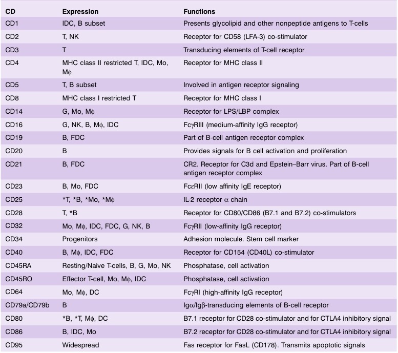
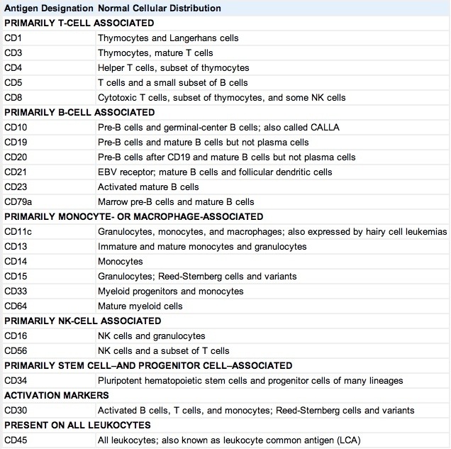
10. Clusters of differentiation (CD)
11. Symposiums
12. FAQs
13. References
PDF 12M (우리말 version)
 1. Histology of the GI tract
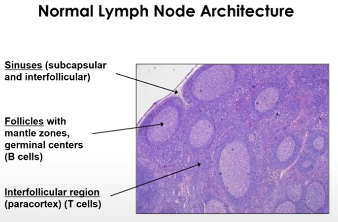
2. Pathology basics
3. Esophagus
PDF. 2026-9-23.
2026-9-23. EndoTODAY 수요내시경세미나
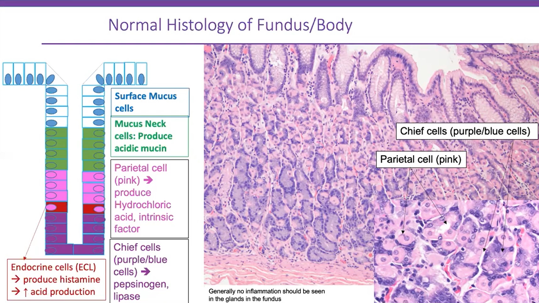
4. Benign diseases of the stomach
| Pathology of acute erosive gastritis |
| Acute phase Vascular engorgement Lamina propria hemorrhage Superficial necrosis Polymorpholeukocutes in pits and glands Ulcers and erosions Superficial fibrin deposits |
| Healing phase Epithelial regeneration with nuclear engorgement Pit elongation Mucus depletion Nuclear mitosis |
| Definition of atrophy | Loss of appropriated glands |
| Phenotype 1 of atrophic gastritis | Shrinkage or complete disappearance of glandular units, replace by expanded (fibrotic) lamina propria |
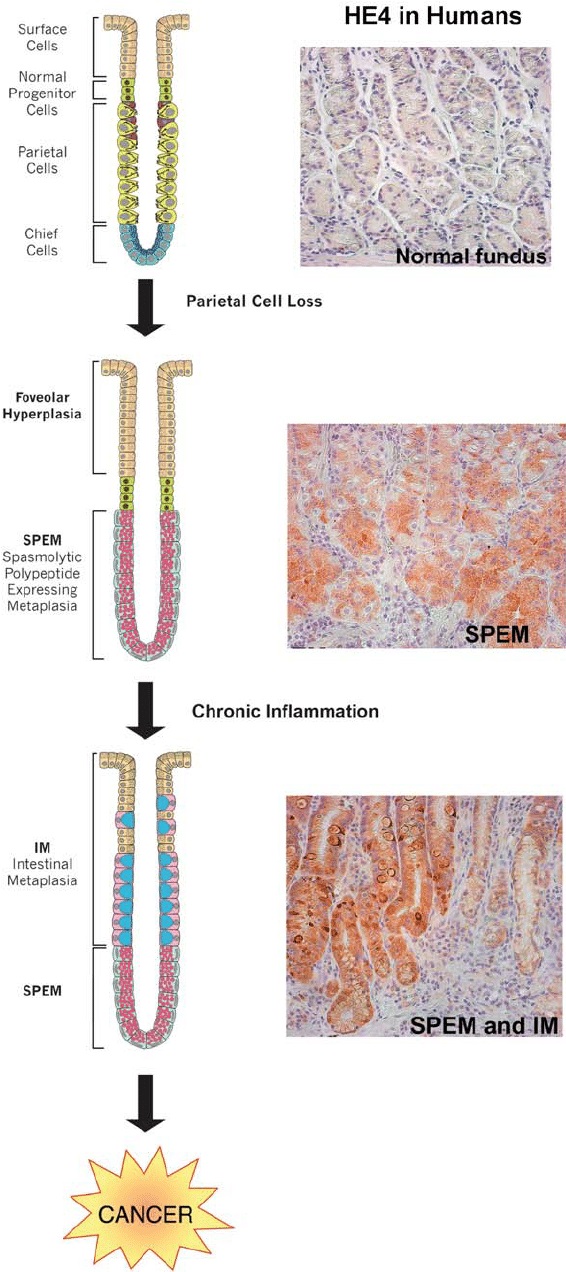
| Phenotype 2 of atrophic gastritis | Replacement of the native glands by other featuring a new commitment (i.e., intestinal metaplasia and/or pseudopyloric metaplasia = SPEM) |
| Intestinal metaplasia | IM may arise in either native mucus-secreting (antral) epithelia or previously antralized oxyntic glands (pseudopyloric metaplasia = SPEM) |
 그냥 gland가 없어지는 것과 intestinal metaplasia 또는 pseudopyloric metaplasia (= SPEM, spasmolytic polypeptide-expressing metaplasia)를 동반하는 종류가 있다는 설명. Antrum에서 intestinal metaplasia가 gland proper에서 발생하기보다는 mucus-secreting foveolar epithelium에서 발생하는 현상과 그림 B가 달라서 의문임
 Type 1은 mucus droplet이 없고 brush border가 있는 absorptive cell이 만들어지는 초기 현상이고 type 2,3는 mucus droplet이 만들어지고 brush border가 없는 non-absorptive cell (= Goblet cell)이 만들어지는 후기 현상이다. 처음에는 antral-corpus junction에서 atrophic, intestinalized mucosa가 나타나고 점차 이보다 proximal과 distal에서도 관찰된다.
5. 위암의 병리 Pathology of gastric cancer
1) 종설과 리뷰
위암 병리 소견의 이해 (김백희, 이성학. 2023) PDF 1.5M
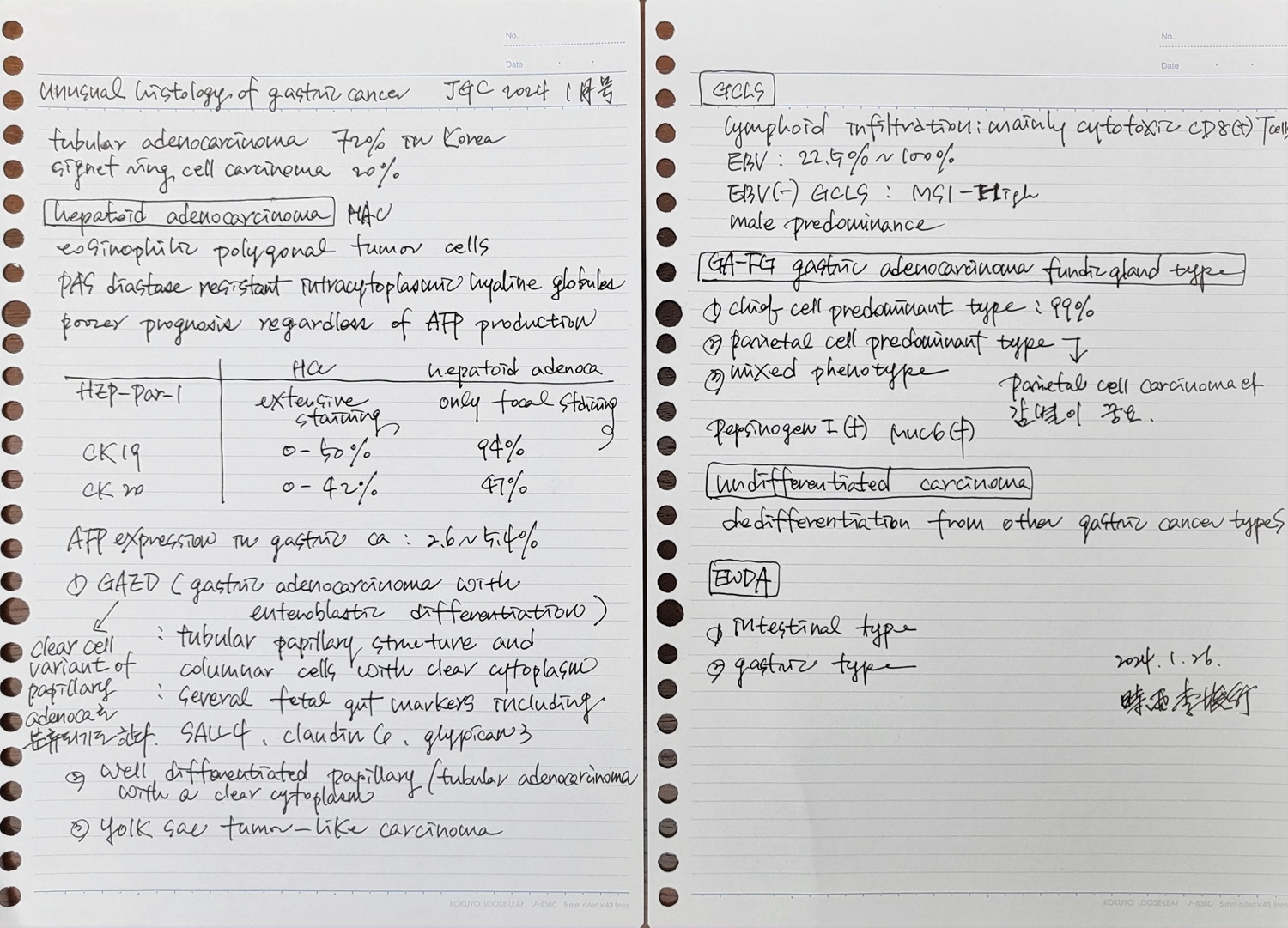
Unusual or uncommon histology of gastric cancer. JGC 2024. 메모
위 조직 생검과 병리학적 진단 김용일 (제3회 내시경세미나) PDF - 매우 classic한 강의. 병리 가이드라인 前
Polypectomy/ESD Specimen 김경미 (2007년 내시경세미나) PDF 0.5M - 2005년 위암 병리 가이드라인 후 삼성서울병원에서 주로 참고하였던 자료
2) 동영상 강의
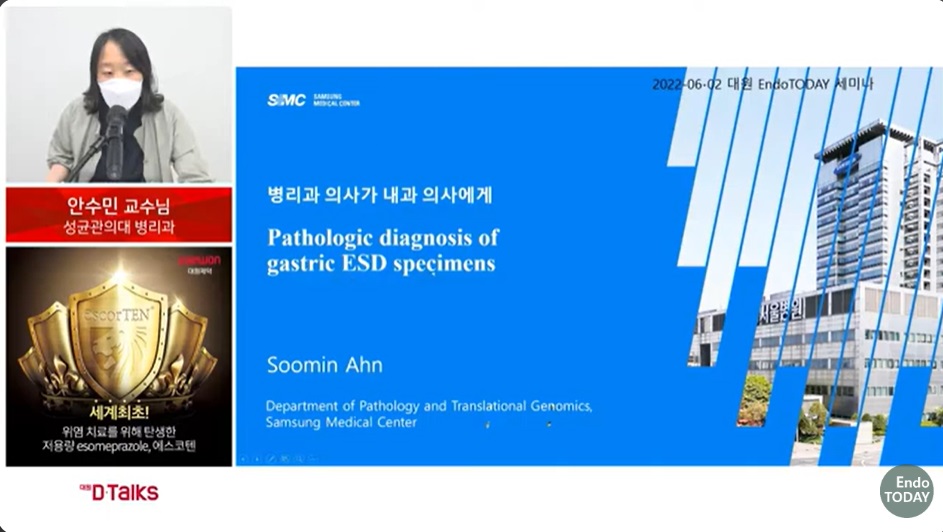
조기위암 내시경 치료 후 병리학적 평가 (주미). KSGE 57회 내시경세미나. log-in 要
아산병원 박영수 교수님 강의 2026-3-15 KSGE 내시경세미나. log-in 要
3) 개념을 잘 설명하는 그림
2년차 전임의들께 설명하는 그림
위암 조직형과 분화도
Intestinal type gastric cancer의 발생 Clin Microbiol Infect 2015
Diffuse type gastric cancer의 발생 Biochim Biophys Acta Rev Cancer 2022
Mucin phenotype
4) On-line 병리 증례
5) EndoTODAY 참고자료
6. Lower GI GI tract
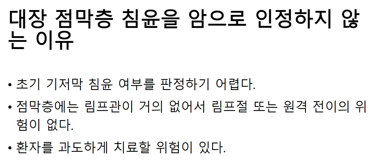 점막까지 침윤한 암을 high grade로 coding하도록 권하고 있다.
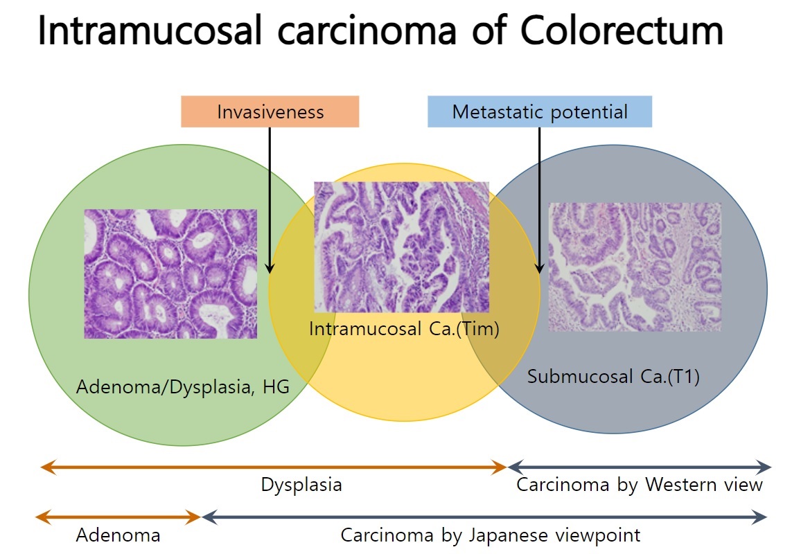 WHO와 일본은 다르다.
 위에서는 T1a, T1b가 있지만 대장에서는 그냥 T1만 있다. 위의 T1a (점막암)이라는 것을 대장에서는 인정하지 않는다.
7. Lymphomas
8. Others
9. Mucin staining - MUC2 (Goblet cell marker), MUC5AC (foveolar marker), MUC6 (antral/pyloric gland marker)
Mucin staining
MUC2 Epithelial mucin expressed in intestinal goblet cells and airway epithelium; gene is at 11p15.5
Relatively specific for predicting colorectal origin for Paget disease (Am J Surg Pathol 2001;25:1469)
Gel forming MUC2 mucin may act as barrier to prevent infiltration of malignant cells in breast mucinous / colloid carcinoma
Positive staining: intestinal and airway epithelium; mucinous carcinoma of colon, breast, pancreas, ovary and stomach
Negative staining: stomach, breast ductal and lobular carcinomasMUC3 Upregulated by steroid hormones in vitro
Positive staining: invasive breast carcinoma, gastric carcinoma (associated with poor prognosis)MUC4 Transmembrane or membrane bound mucin that provides a protective layer of mucus
Normally acts as barrier to apical surface of epithelial cells, playing a protective and regulatory role
Positive staining - normal: tracheobronchial mucosa, colon, stomach, cervix and lung; normal salivary glands
Positive staining - disease: pancreatic, colonic, pulmonary and gastric carcinoma; sclerosing epithelioid fibrosarcoma (Am J Surg Pathol 2014;38:1538, Am J Surg Pathol 2012;36:1444)MUC5AC. Also known as MUC5 (GeneCards) Protects epithelium Positive staining - normal: stomach (foveolar epithelium of body and antrum), tracheobronchial mucosa, endometrium (mucinous metaplasia, eosinophilic change / metaplasia (Mod Pathol 2005;18:1243), surface syncytial change, ciliated change in 52%)
Positive staining - disease: extramammary Paget disease but not mammary Paget disease or normal breast tissue (Am J Surg Pathol 2001;25:1469); mucinous carcinoma of ovary, diffuse type gastric carcinoma (83%)
Negative staining: normal breast tissueMUC6 Produces protective glycoprotein coat for gut epithelia
Positive staining - normal: stomach (pyloric glands), gallbladder, colon and endocervix
Positive staining - disease: invasive ductal carcinoma of breast, gastric carcinomas
Negative staining: normal breast tissue
In a cohort of 97 gastric cancers 30% expressed MUC5AC and MUC6 (gastric phenotype), 40% expressed MUC5AC, MUC6 and MUC2 (gastrointestinal phenotype), 10% expressed MUC2 alone (intestinal phenotype) and 20% lacked all markers (null phenotype). (J Surg Oncol. 2008;98:124-129)
10. Clusters of differentiation
11. Symposiums
1) 2022-7-2. 헬리코박터 위암연구회 워크숍. 위암 중개 연구를 위한 첫걸음
[남기택 교수님. 연세의대 의생명과학부]

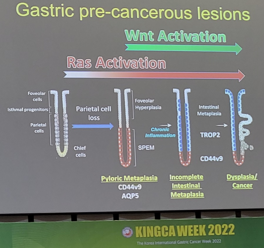 2022-9-1. KINGCA. James Goldenring 선생님 강의
[서인석 교수님. 서울대 생리학교실]
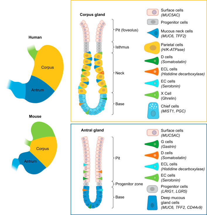

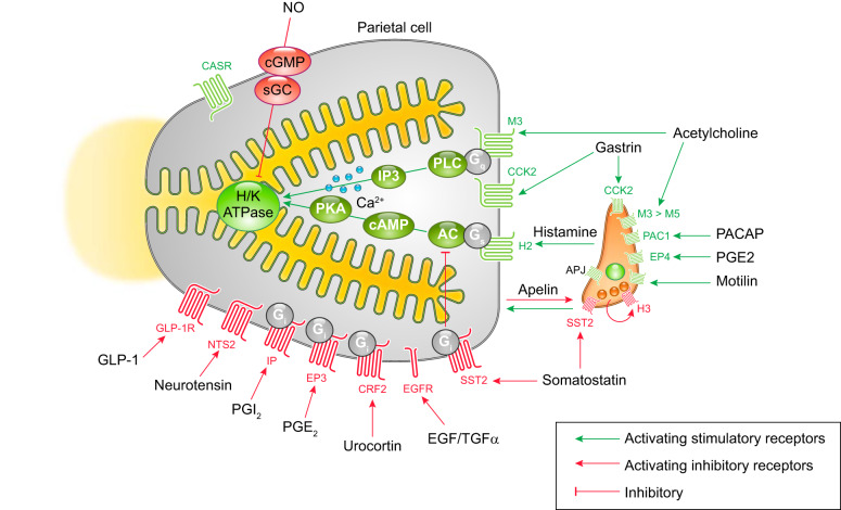
[김현기 교수님. 연세의대 병리학교실]
[FAQs]
[2021-10-16] 병리 선생님. 제발 다른 sample은 따로따로 결과를 내 주세요. 하바터면 실수할 뻔 했습니다.
첫 세 sample이 모두 암이었습니다. 세 개의 암이 있던 환자입니다. 그런데 병리 보고서는 실수하기 쉽게 씌여 있었습니다.
[2022-3-23. 애독자 질문]
교수님. 안녕하십니까. 지방 건진센터에서 근무하고 있습니다. 매우 사소한 질문이라 실례를 무릅쓰고 이메일을 보내 봅니다.
검사에서 이상 병변이 있으면 영상 CD, 내시경 결과지, 조직 검사 결과지, 조직 슬라이드를 진료 의뢰서에 동봉하여 상급 병원에 의뢰를 하는데요... 조직은 염색 슬라이드를 발급 받아 환자분께 드리는데... 최근에 adenocarcinoma M/D로 확인된 1cm 가량의 EGC IIc가 의심되는 환자분께서 진료보신 상급병원에서 파라핀 블록을 따로 요청하신 경우가 있었습니다.
예전에는 MALToma 의심되는 소견으로 염색 슬라이드를 동봉하여 보냈더니 염색 슬라이드를 20장 추가로 보내달라고 하셨던 경우도 있었는데 장당 2만원, 총 40만원이라서 환자분께서 10장만 받아가셨던 일도 있었습니다. 두 분 다 제대로 챙겨주지 않았다고 병원에 찾아와 심하게 컴플레인을 하셨었구요.
그 외에는 염색 슬라이드만 보냈어도 진료와 시술에 문제가 된 경우가 없어서 앞으로도 염색 슬라이드만 신청할 것 같은데 혹시 의뢰 받으시는 경우에 위와 같이 파라핀 블록이나 비염색 슬라이드가 필요한 경우가 있으신지요? 제가 모르고 부족하게 일을 하고 있지 않았는지 걱정이 됩니다. 늘 건강하시고 항상 감사드리고 있습니다.
[2022-3-23. 이준행 답변]
좋은 질문입니다. 현장의 어려움을 잘 알겠습니다.
이상과 현실은 다릅니다. 병리과 의사의 입장에서 좀 더 많은 slide가 있으면 좋으니 paraffin block이 있으면 도움이 되는 경우가 있을 것입니다. Unstained slide가 있어서 적절한 면역형광염색을 하면 또 얼마나 좋겠습니까? 그러나 의뢰되는 환자가 H&E slide 이외에 unstained slide, 혹은 paraffin block을 가지고 오는 경우는 거의 없습니다. 물론 큰 병원 의사가 unstained slide, paraffin block을 가지고 오라고 하면, 진단을 하는데 꼭 필요한 것이라고 말하면 환자는 다시 원래 병원에 가서 그런 것을 달라고 요청할 것입니다. 어마어마한 컴플레이는 눈에 선합니다.
그래서 저는 외부 슬라이드를 봐 주시는 병리 선생님께서 (가끔, 아주 가끔) unstained slide, 혹은 paraffin block을 요청하더라도 못 들은 척 하고 있습니다. 내시경 검사를 다시하고 있습니다. 외부 병원 자료는 H&E slide 정도에서 만족하고 있습니다. 보다 자세히 평가하는 것은 상급병원에서 재검하여 얻은 조직으로 시행하는 것이 절차도 매끈하고 신뢰감도 높이는 방법이라고 생각합니다. 절대 다시 얻을 수 있는 검체도 아닌 내시경 검체를 위하여 환자를 원래 병원으로 다시 가서 며칠 기다렸다가 unstained slide나 paraffin block을 받아오게 하는 것은 too much라고 생각합니다.
저와 다른 생각을 가진 clinician도 계실 줄로 압니다. 물론 unstained slide나 paraffin block까지 가져오라고 요청하는 그 분들의 의견을 존중합니다. 그러나, 만약에 제가 환자를 의뢰하는 입장이라면 그 분께는 다시는 환자를 의뢰하지 않을 것 같습니다. 의료는 이상과 현실의 타협이기 마련인데, 지나친 이상주의자는 저와 잘 맞지 않기 때문입니다.
요컨데, 의뢰하는 병원을 바꾸시기 바랍니다. 그것밖에 답이 없습니다. 생각은 거의 안 바뀌니까...
[2024-12-27] 제29회 병리세미나를 금요내시경집담회와 통합하여 진행하였습니다. 그간 안수민 교수님과 저의 개인 교습 스타일로 진행해왔었고 가끔 다른 교수님들이나 fellow들이 함께 한 적은 있습니다. 그러나 모든 분들에게 도움될 것으로 판단되어 처음으로 병리과 선생님을 모신 regular 병리집담회를 시작하였습니다. 20년 전부터 하고 싶던 것을 드디어 성사시켜 매우 기쁩니다.
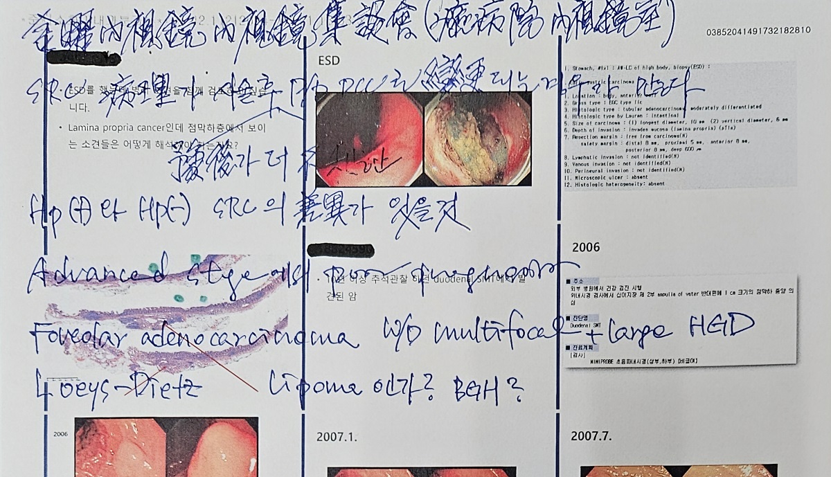
Signet ring cell carcinoma도 두 종류가 있다는 것이 저의 주장입니다. Hp negative이고 flat 하고 white 하고 예후가 매우 좋은 것과 Hp positive이고 AGC가 되기 쉽고 재발이 많은 종류입니다. 이 개념을 좀 더 연구해 볼 생각입니다.
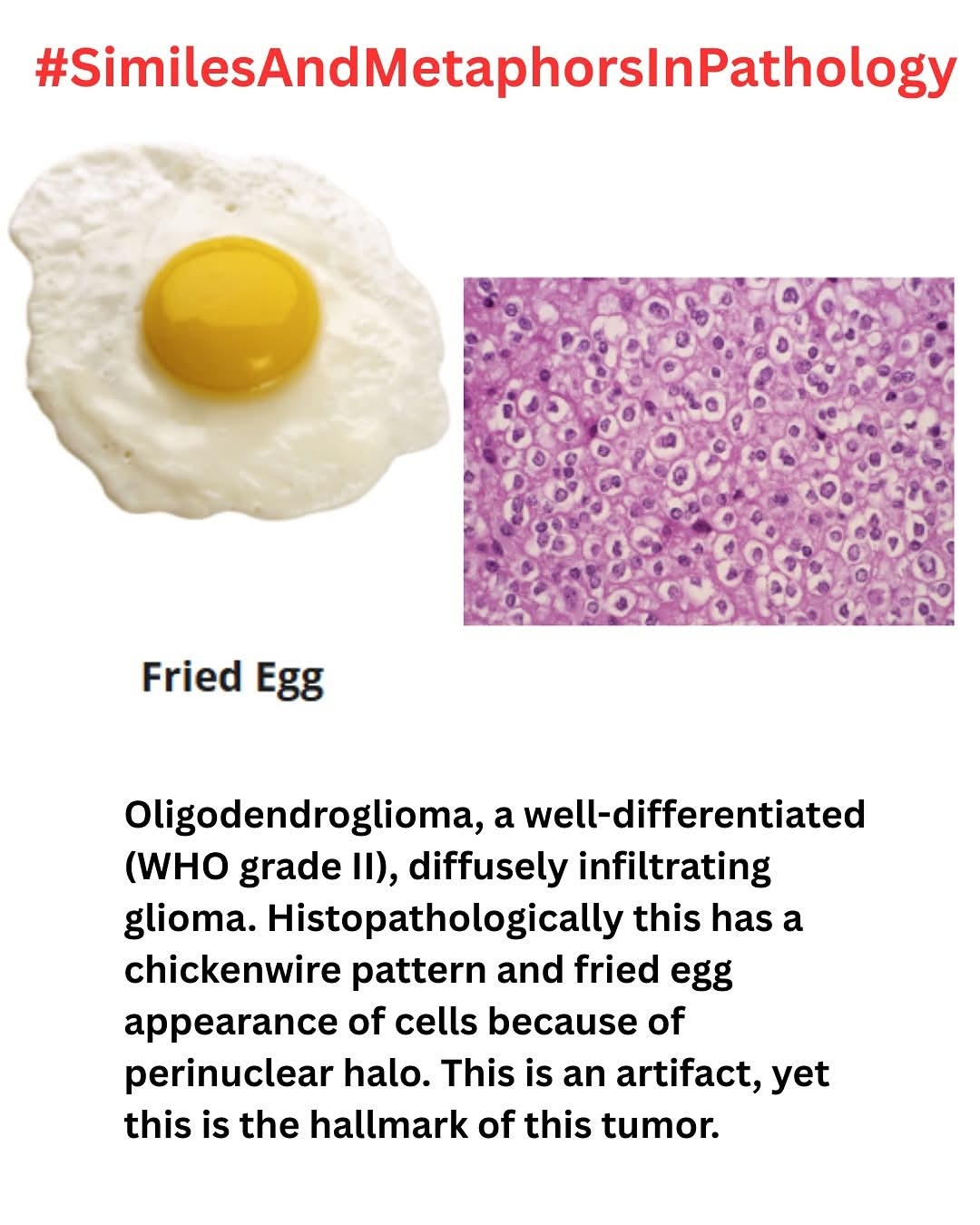
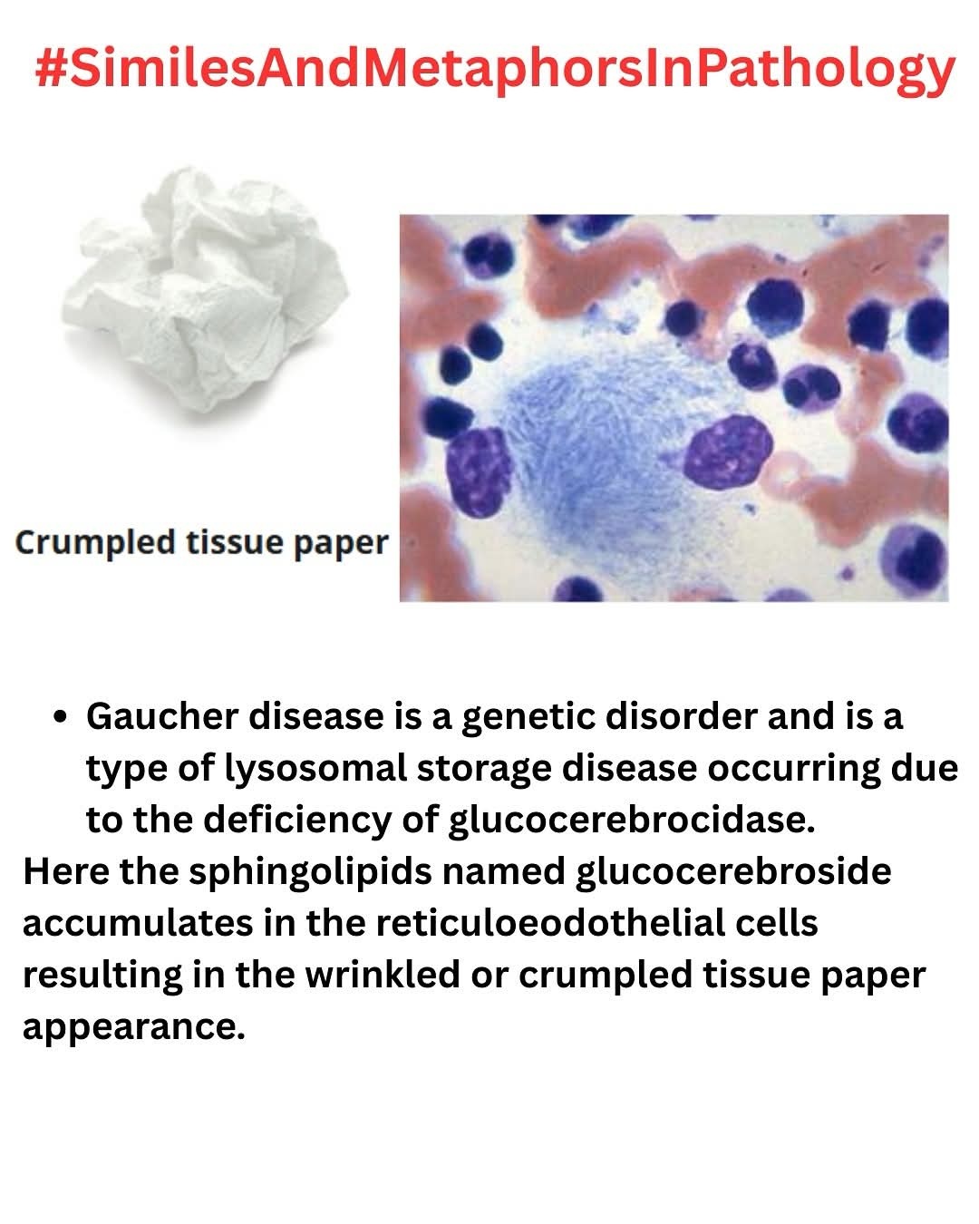
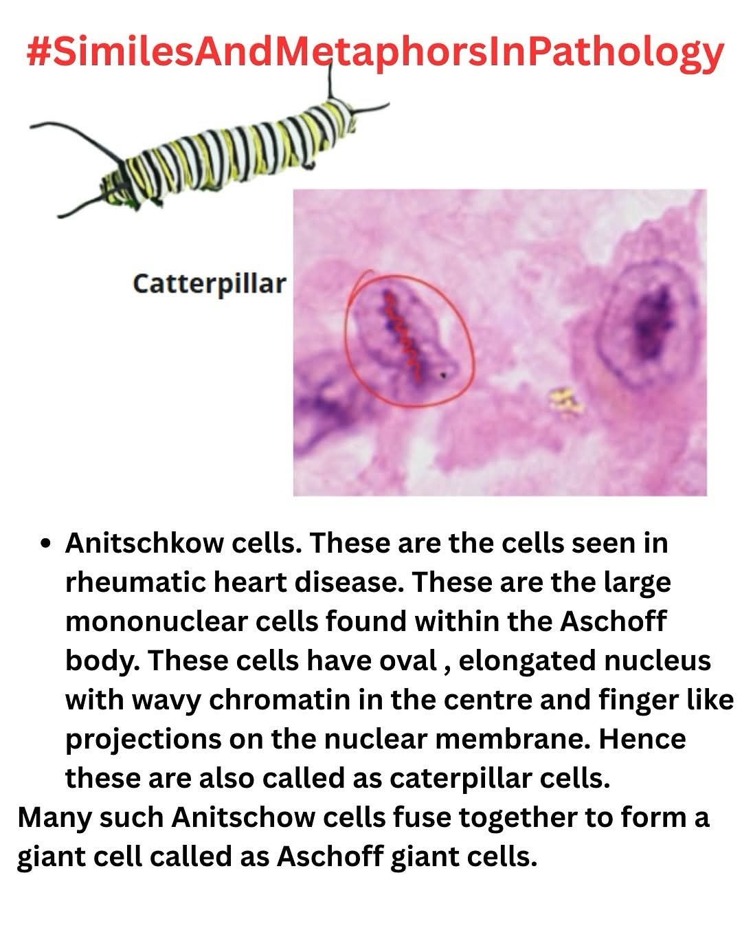
[2025-6-15] 병리에서 사용하는 여러 metaphor 중 일부입니다.
[2026-3-15] KSGE 내시경 세미나
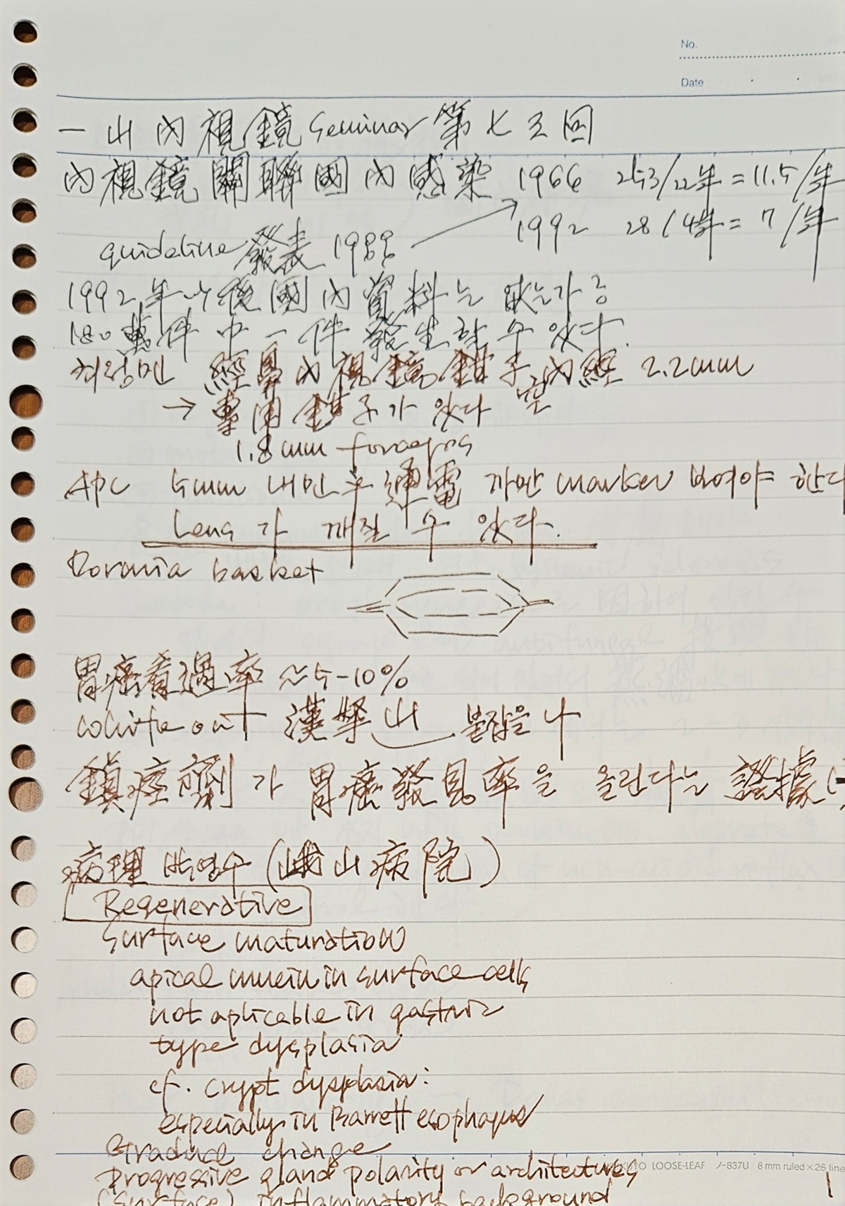
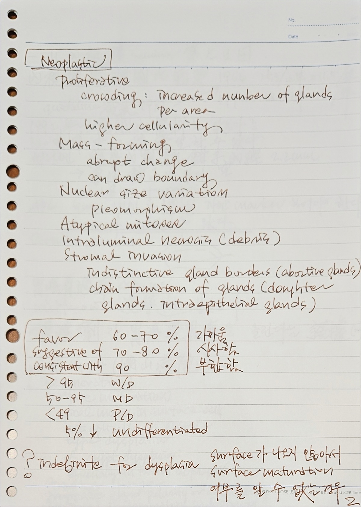


박영수 교수님
박영수 교수님
[References]
4) AJCC 8th PPT 인터넷 자료
5) 위내시경 조직검사 코드 - internal use only
6) 알기 쉬운 병리학 PDF 13.5M (PPT slide 135장)
7) 소화기계 종양의 병리 YouTube. 장기택
© 일원내시경교실 바른내시경연구소 이준행. EndoTODAY Endoscopy Learning Center. Jun Haeng Lee