EndoTODAY 내시경 교실
Beginner | ESA | Schedule | OPD
Seminars | Atlas | Recent | Links
 [StomachTODAY 054. Pyloric gland adenoma] - 終

 1. Pyloric gland adenoma 개요 (Pathology Outlines)
Pyloric gland adenoma는 misnomer 라는 주장이 있습니다 (Hashimoto. Histopathology 2015). 왜냐하면 이름과 달리 fundic gland에서 발생하기 때문입니다.
2. Pyloric gland adenoma 병리 소견
한 review article에서 옮깁니다 (Arch Pathol Lab Med 2015).
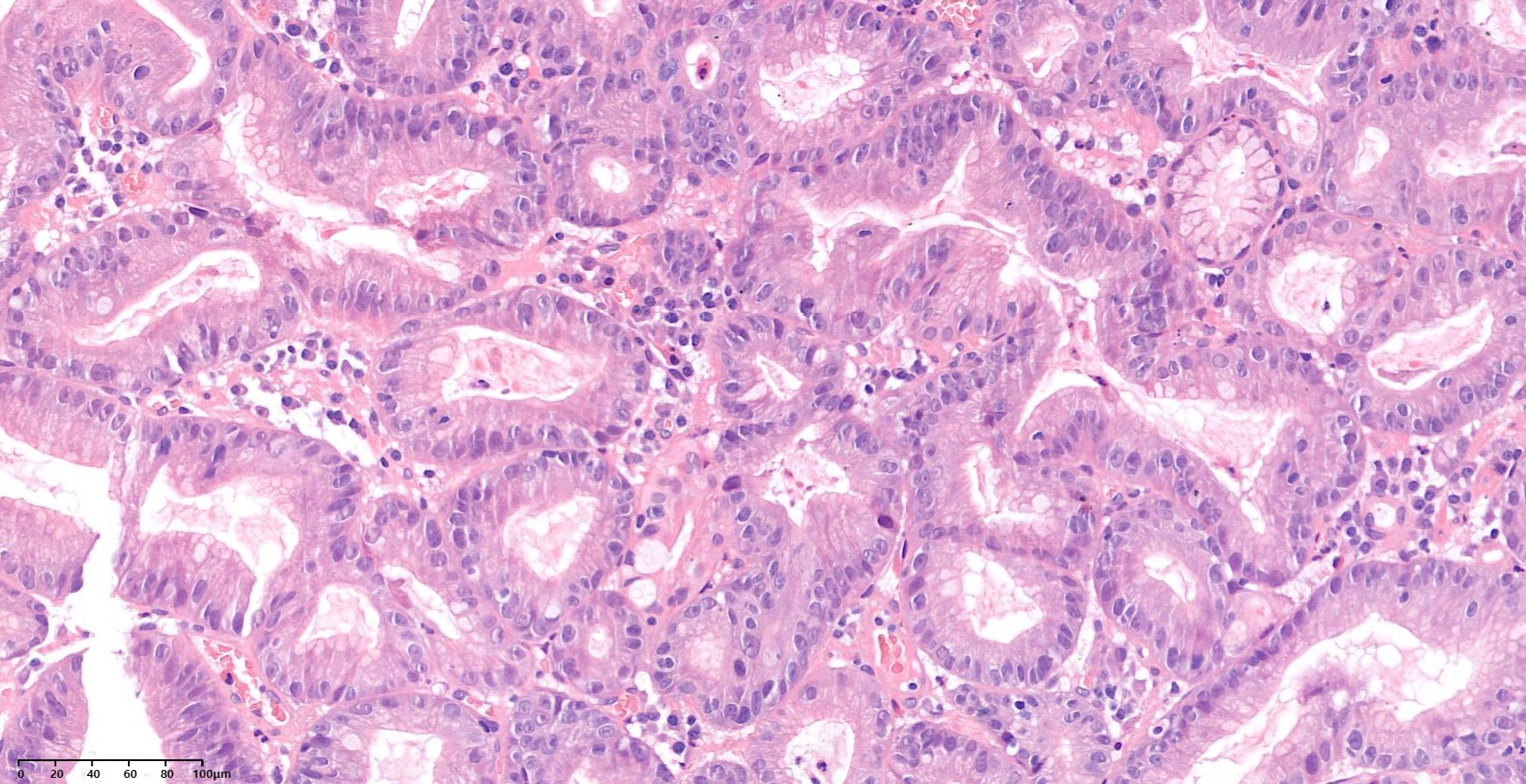
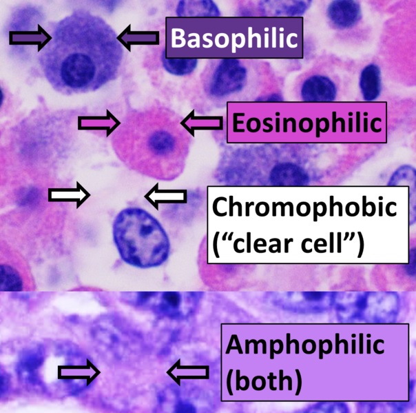
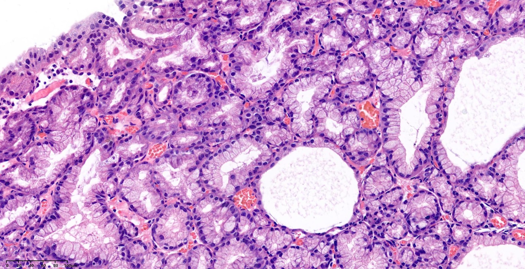
Pyloric gland adenomas are rare neoplasms of the gastrointestinal tract. Gastric pyloric gland adenomas have been shown to arise in chronically damaged mucosa. The neoplastic glands have gastric pyloric gland differentiation and have a tightly packed organization with occasional cystic dilatation. The individual cells are cuboidal to columnar, with eosinophilic to amphophilic cytoplasm and either no apical mucin cap or a poorly formed apical mucin cap. The nuclei are round to oval, with occasional prominent nucleoli. Immunohistochemically, the neoplastic cells label with markers of gastric pyloric gland differentiation, including MUC6 and MUC5AC. (foveolar epithelium marker인 MUC5AC와 pyloric gland marker인 MUC6가 둘 다 양성인 점이 특징)
3. 내시경 소견
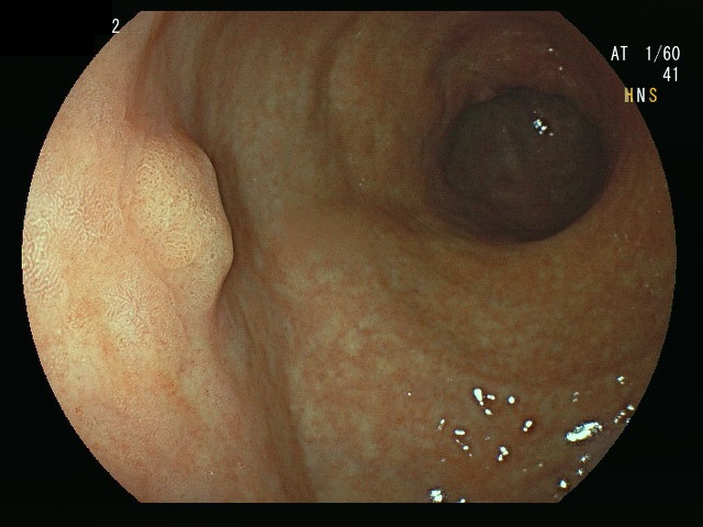
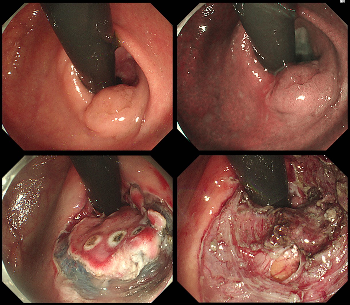
Pyloric gland adenoma는 대부분 proximal stomach 또는 cardia에 위치한 융기형 병소입니다. Dysplasia가 거의 없거나 경한 경우가 많습니다.
EMR을 시행 - 병리사진
Fundus에서 발견된 PGA → EMR + ablation
Antrum에서 발견된 PGA → EMR
간혹 위체부 소만에서도 발견됩니다.
위체부소만에서 발견된 PGA → EMR (background mucosa: autoimmune gastritis)
EMR이나 ESD 시술 후 high grade dysplasia를 동반한 것으로 나오는 경우가 있습니다.
A 55 years old male was referred due to a cardia polyp. The initial pathology was "indefinite for dysplasia", and there was a note that "there are slightly crowded glands with equivocal nuclear features of regenerating atypia versus low-grade dysplasia. Close follow up is recommended." Pathology review of the outside slide was "gastric type mucosa showing pyloric gland type adenoma." Endoscopic resection was done.
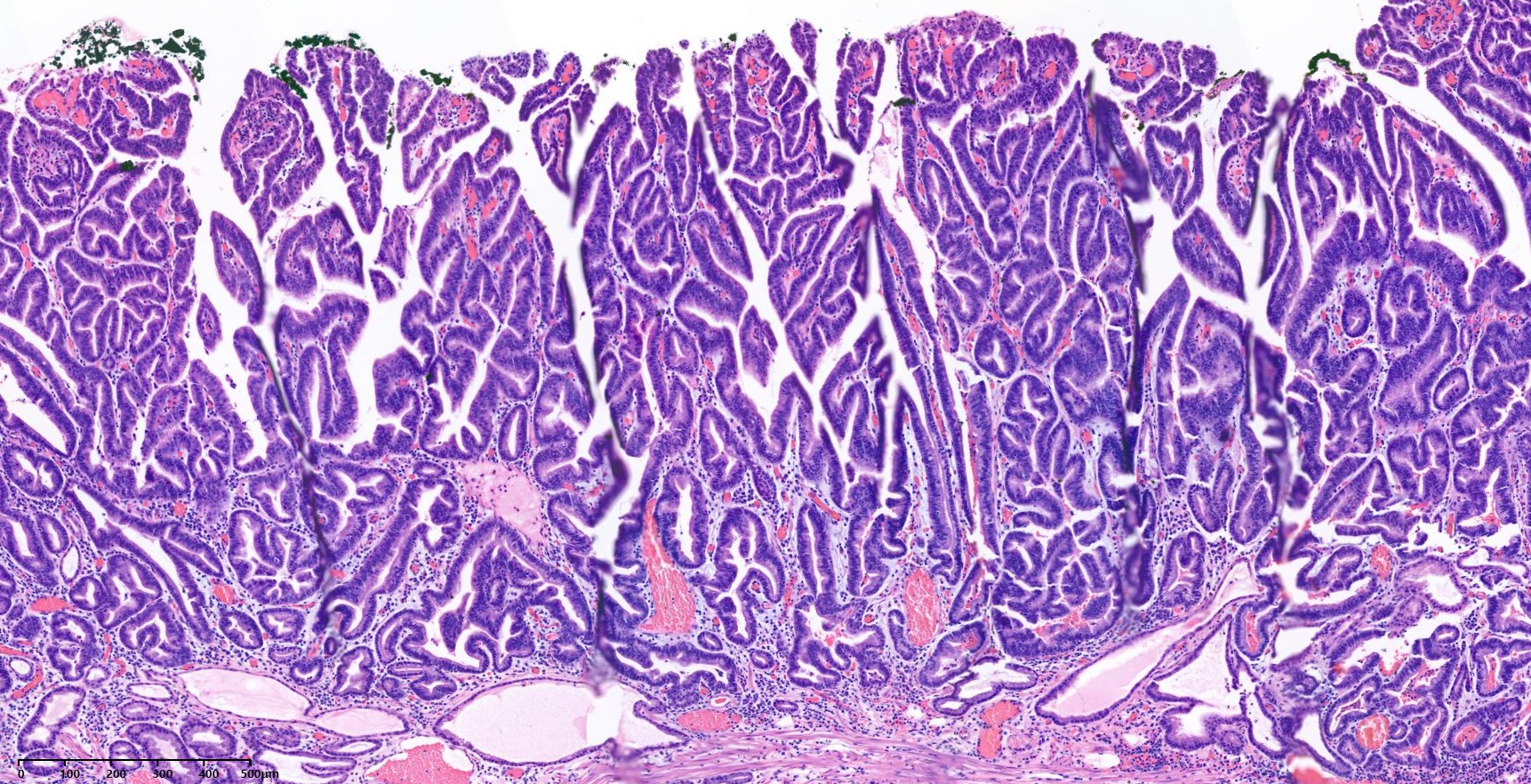
Pyloric gland adenoma with high grade dysplasia
1. Location : cardia
2. Gross type : elevated
3. Size of adenoma : (1) longest diameter, 10 mm (2) vertical diameter, 6 mm
4. Resection margin : negative resection margins

Stomach, endoscopic submucosal dissection:
Pyloric gland adenoma with high grade dysplasia
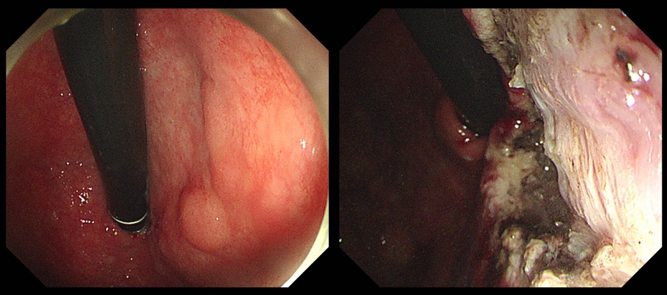
1. Location : mid body, greater curvature
2. Gross type : elevated
3. Size of adenoma : (1) longest diameter, 20 mm (2) vertical diameter, 16 mm
4. Resection margin : negative resection margins
ESD 시행한 PGA with HGD - 병리 사진
위암 수술 후 잔위에서 발견될 수도 있습니다.
잔위 들문에서 발견된 PGA → ESD → PGA with focal HGD (M/65, 2019)
PGA에서 암이 발생한 경우는 개인적으로는 한 증례밖에 경험하지 못했습니다.
조직검사에서 high grade dysplasia 소견이 없고 고령이면 치료 없이 경과관찰을 하기도 합니다.
Cardia 주변의 넓은 PGA. 고령으로 치료 없이 경과관찰
PGA와 역류성 식도염. PGA는 초고령으로 치료 없이 경과 관찰 - 병리사진
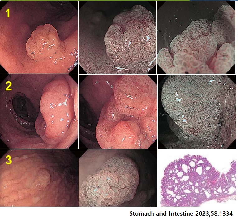
아래는 문헌에 나왔던 증례입니다.
대한내과학회지 2011;80:78
[References]
1) EsoTODAY - 십이지장질환 증례토의
2) SmallTODAY - 소장질환 증례토의
3) ColonTODAY - 대장질환 증례토의
4) Dr. Sinn's LiverTODAY - 간질환 증례토의
© 일원내시경교실 바른내시경연구소 이준행. EndoTODAY Endoscopy Learning Center. Lee Jun Haeng (Updated: 2025-2-17)