EndoTODAY 내시경 교실
Beginner | ESA | Schedule | OPD
Seminars | Atlas | Recent | Links
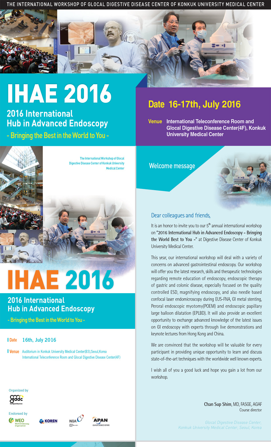
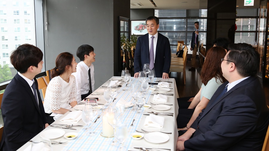
![]() [The International Workshop of Glocal Digestive Disease Center of Konkuk University Medical Center]
[The International Workshop of Glocal Digestive Disease Center of Konkuk University Medical Center]
일시: 2016년 7월 16일
한국, 일본, 중국, 홍콩, 싱가폴의 여러 유명 강사들이 총출동하였습니다.
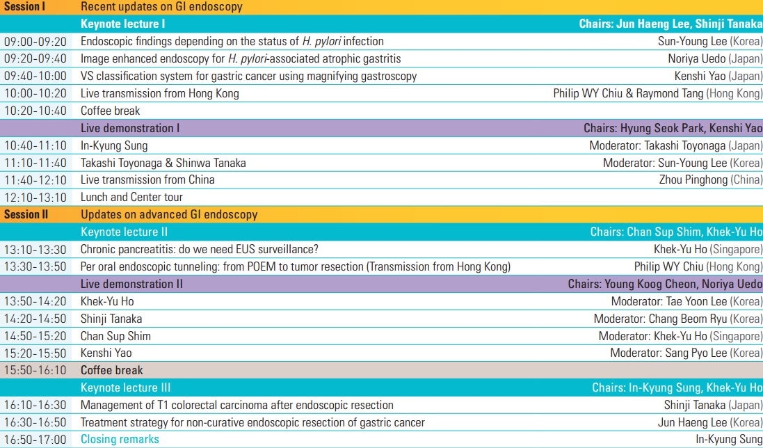
특히 7월 16일 토요일 오후 4층 International Teleconference Room에서 parallel session으로 열리는 Joint Symposium은 한일양국 정부의 지원을 받아 개최하는 첫 내시경 심포지엄이라 의미가 있었습니다.
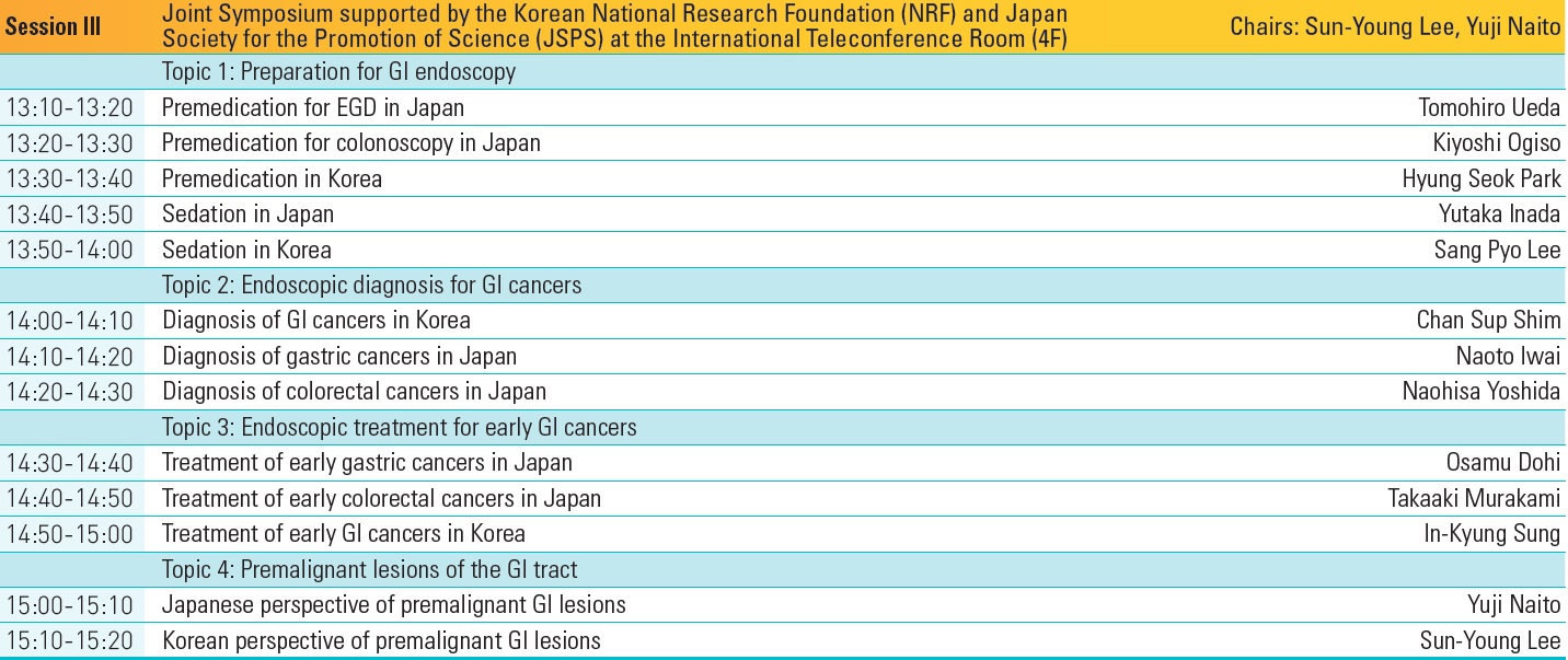
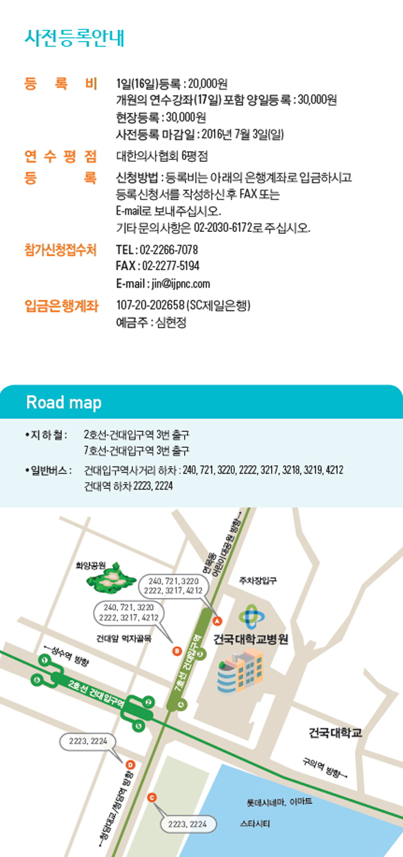
등록방법은 아래와 같았습니다. 팜플렛을 보시려면 여기를 누르십시오.

 건국대학교 fellow 선생님들과 함께
 건국대학교 천영국 교수님과 소화기내과 fellow 선생님들
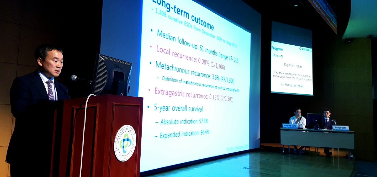
![]() 1. Treatment Strategy for Non-curative Resection of Early Gastric Cancer (Jun Haeng Lee)
1. Treatment Strategy for Non-curative Resection of Early Gastric Cancer (Jun Haeng Lee)
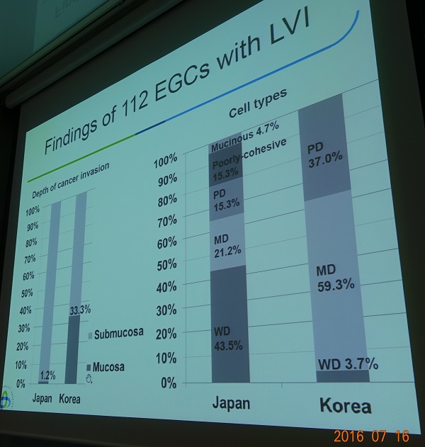
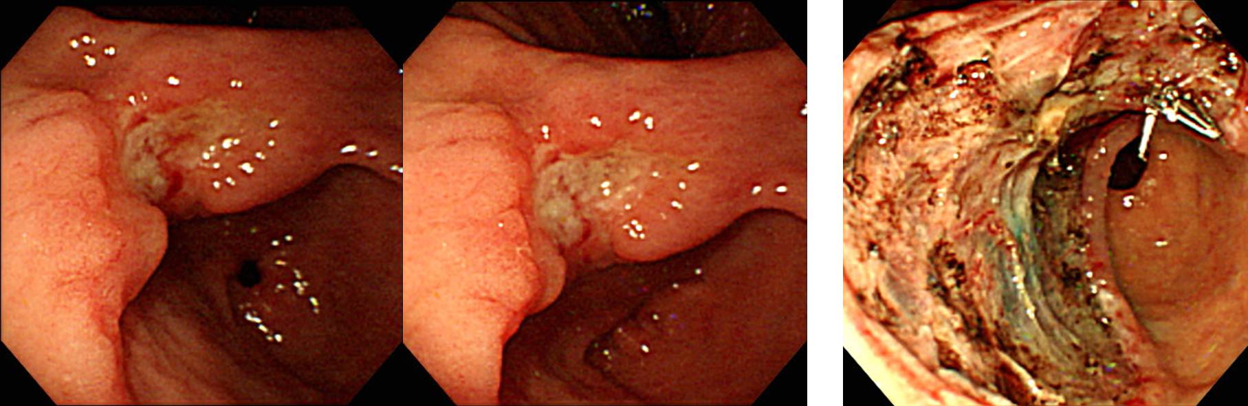
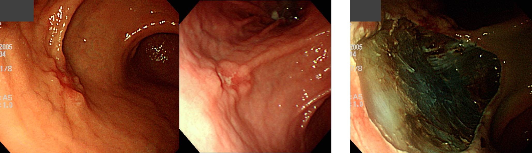
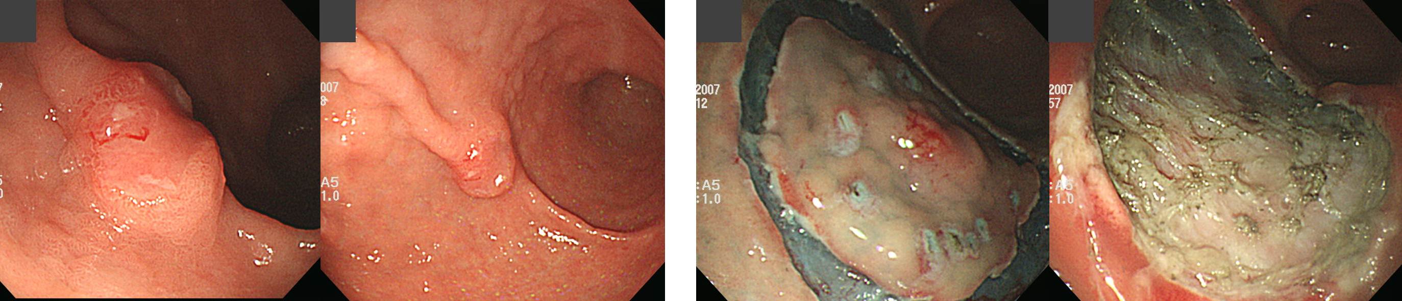
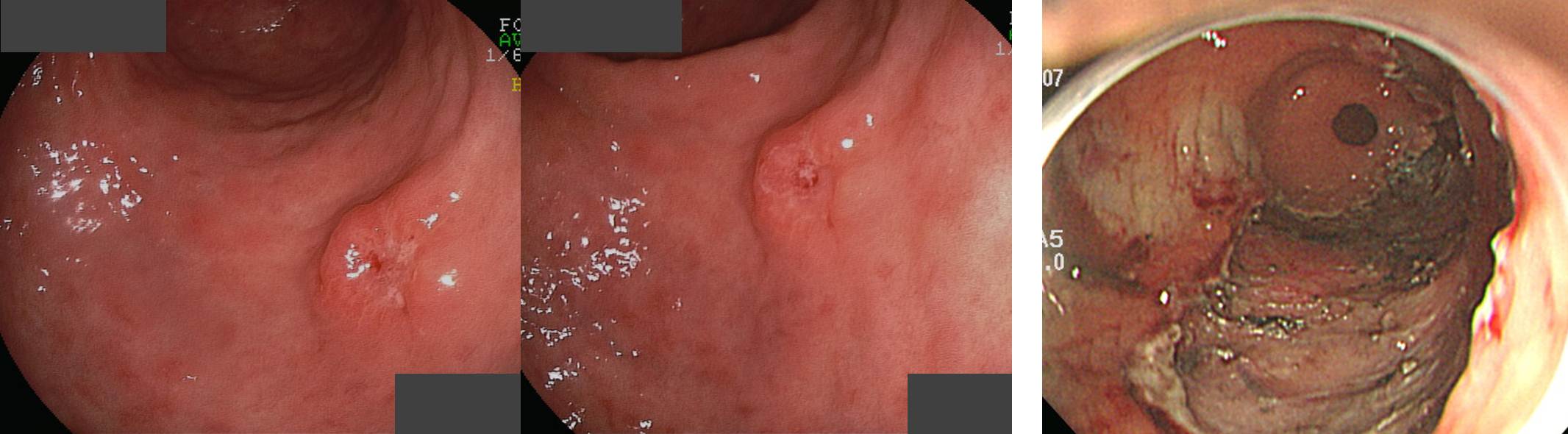
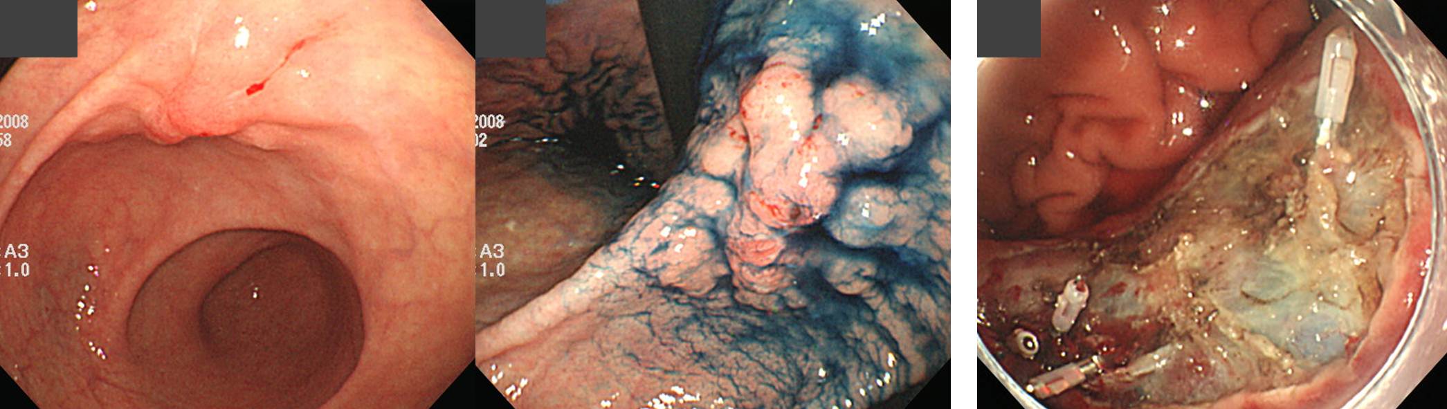
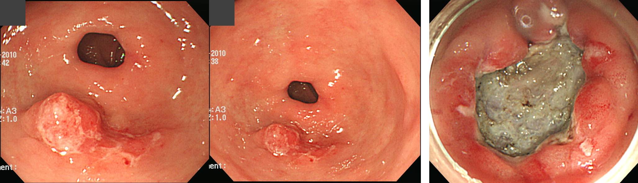
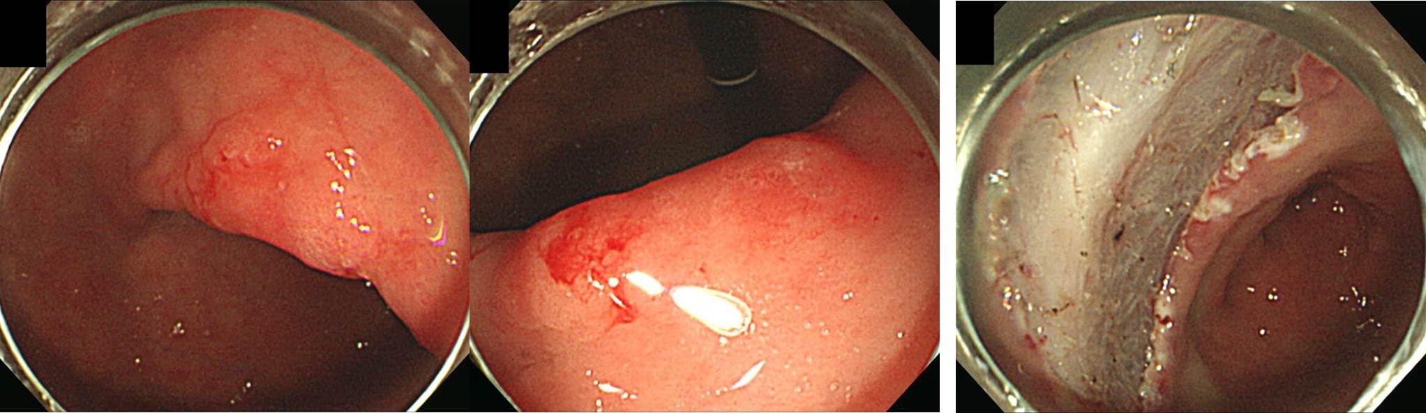
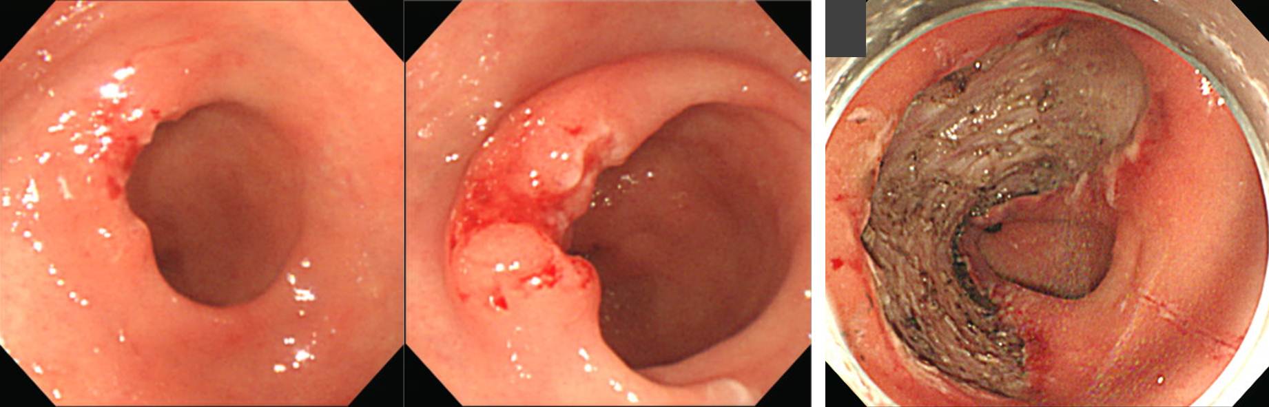
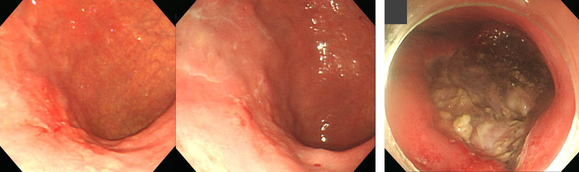
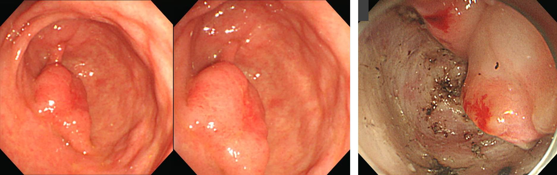
조기위암 내시경 치료 후 수술이 필요한 환자에서 수술 후 림프절 전이가 확인되는 경우는 10% 내외입니다 (Kim ER. Br J Surg 2015). 저희 기관에서는 수술이 필요한 환자 274명 중 194명(70.8%)에서 수술을 시행하여 local residual cancer가 있었던 환자가 10명 (5.2%), 림프절 전이가 있었던 환자가 11명(5.7%)이었습니다. 아래는 림프절 전이가 있던 11예의 사진입니다. 상당수는 beyond expanded indication 증례였음을 알 수 있습니다. ESD 도입 초기에 다소 무리하여 시술한 환자는 결국 수술을 하게 되었던 것입니다. 이 증례들에 대하여 많은 토론이 있었습니다. 내시경에 대한 연구나 강의에서는 내시경 사진이 많아야 토론이 활발해집니다.

![]() 2. VS classification system for gastric cancer using magnifying gastroscopy
2. VS classification system for gastric cancer using magnifying gastroscopy
Yao 교수님은 정상 위점막대한 확대내시경 소견을 상세히 설명한 후 위암에 대한 VS system을 소개하였습니다.
1) Magnifying endoscopy findings of the normal stomach
(1) microvascular architecture (V) : subepitherial capillary network (SECN), CV (collecting venule)
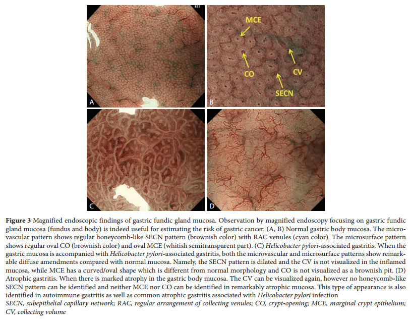
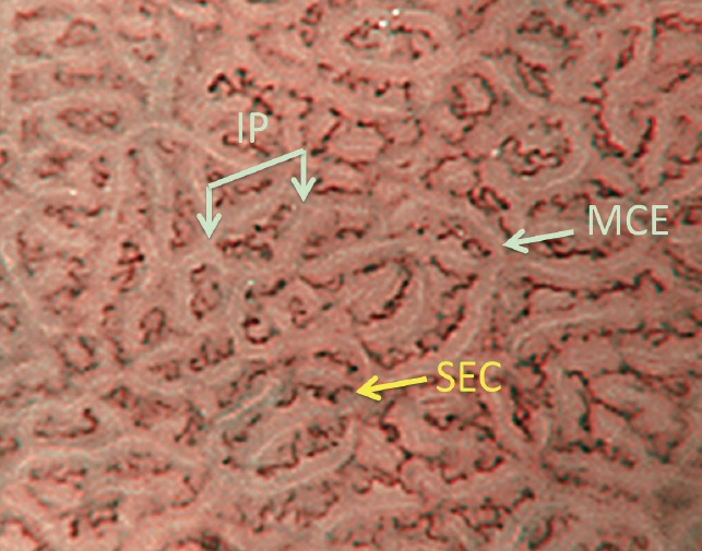
(2) microsurface structure (S): marginal crypt epithelium (MCE), crypt opening (CO)i. 정상 fundic gland: microvascular pattern은 SECN (subepitheliral capillary network)가 벌집모양을 이루며, microsurface pattern은 marginal crypt epithelium (MCE)에 둘러쌓인 crypt opening (CO)이 SECN 중앙에 위치합니다. Helicobacter 감염이 있거나 위축성 변화가 현저할 때는 이런 정상 소견이 보이지 않게 됩니다.
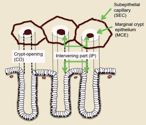
(a) Schematic diagram of the microvascular architecture and the microsurface structure of the normal gastric fundic gland mucosa corresponding to the surface morphology as visualized by magnifying endoscopy (ME) with narrow-band imaging (NBI). The microvascular architecture is formed by the capillaries and collecting venules. The morphology of each capillary is that of a polygonal closed loop. These loops anastomose repeatedly with each other, forming a regular honeycomb-like subepithelial capillary network pattern. The microsurface structure is made up of the marginal crypt epithelium/white zone (MCE/WZ), and the intervening part in between. The epithelial morphology is visualized as a semitransparent white belt-like structure (the MCE/WZ), showing a circular or oval shape at the center of which lies the crypt opening. (b) ME with NBI of normal fundic gland mucosa. (Muto M. Digest Endosc 2016)
ii. 정상 pyloric gland: microvascular pattern은 dark brown colored coil-shaped open loop를 이루며, microsurface pattern은 regular polygonal 또는 curved marginal crypt epithelium pattern을 이룬다.
(a) Schematic diagram of the microvascular architecture and the microsurface structure of the normal gastric pyloric gland mucosa corresponding to the surface morphology as visualized by magnifying endoscopy (ME) with narrow-band imaging (NBI). The microvascular architecture is formed by capillaries and collecting venules, but the latter are rarely observed from the mucosal surface. The morphology of each capillary is that of coil-shaped open loops. The mucosal surface structure is made up of the marginal crypt epithelium/white zone (MCE/WZ) and the intervening parts surrounded by MCE/WZ. The MCE/WZ morphology usually shows polygonal structures but may be curved or linear. (b) ME with NBI of normal pyloric gland mucosa. (Muto M. Digest Endosc 2016)
2) VS system for gastric cancer
위암의 확대내시경 진단에서는 VS classification이 활용됩니다. Microvascular pattern과 microsurface pattern을 별도로 평가하여 함께 고려하는 것입니다.
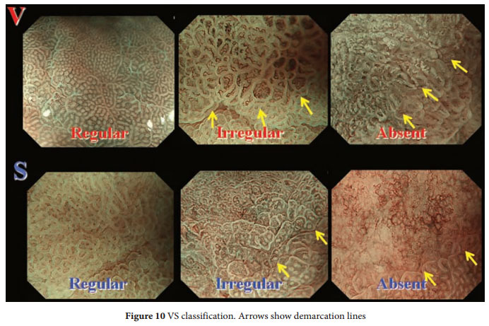
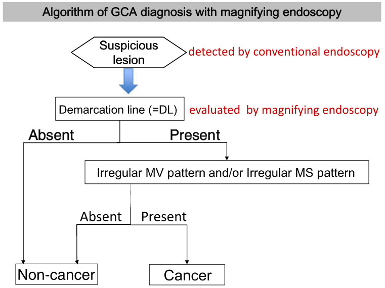
여기에 demarcation line을 더하면 분류가 완성됩니다. Digest Endosc 2015년 7월호에 WEO Upper GI Cancer Committee에서 확대내시경을 이용한 위암 diagnostic algorithm schema를 발표하였습니다.


* 참고: EndoTODAY 확대내시경
* 참고: Ann Gastroenterol 2013, Clin Endosc 2015, Digest Endosc 2016
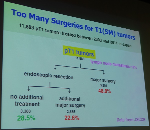
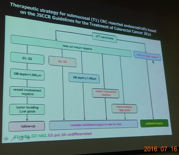
![]() 3. Management of T1 colorectal carcinoma after endoscopic resection (Shinji Tanaka, Hiroshima University)
3. Management of T1 colorectal carcinoma after endoscopic resection (Shinji Tanaka, Hiroshima University)
SM 대장암의 림프절 전이는 10% 정도입니다. 그런데 실제 임상 진료 현장을 살펴보면 70%가 수술을 받습니다. 어떻게하면 unnessary surgery를 피할 수 있을까요?

![]() 4. Joint syposium
4. Joint syposium
1) Premedication for EGD in Japan (Tomohiro Ueda)
Water please (r): 조직검사 channel을 통하여 물을 pumping 하는 장치
Minclea (r): 페퍼민드 오일이 주 성분인 새로운 antispasmodic agent (2011년 소개됨). 내시경 검사 도중 위가 움직이면 Minclea를 위점막에 뿌려주면 곧 stomach motility가 낮아진다고 합니다. 주사하는 것이 아니므로 편할 것 같습니다.
일본 대형 병원에서는 위내시경을 위하여 sedation을 하는 경우가 드물다고 합니다. 15% 미만이라고 합니다. 우리가 60% 이상에서 sedation을 한다고 말하니 매우 놀라는 눈치였습니다.
* 참고: EndoTODAY antispasmodics
2) Premedication for colonoscopy in Japan (Kiyoshi Ogiso)
PEG-ASC caused dehydration of high concentration.
150 US dollars for colonoscopy, 450 dollars for ESD
일본과 우리의 약제 종류는 비슷한데 일본에서의 용량이 조금 작았습니다. 농도가 더 높은지는 파악하기 어려웠습니다.
3) Premedication for endoscopy in Korea (박형석)
건국대의 최근 분석에 따르면 PEG + ascorbic acid로 대장 전처치를 하였을 때 3.2 %에서 renal impairment가 발생하였다고 합니다. 아마도 temporary 했을 것 같다고 합니다.
4) Sedation in Japan (Yutaka Inada)
일본에서는 propofol을 사용하는 경우가 적고 pentazosine 사용이 많습니다.
Deep sedationis not performed in colorectal ESD.
5) Sedation in Korea (이상표)
건국대 외래 내시경실에서 procedural sedation의 빈도는 70%를 넘지만 국가암검진프로그램에서는 procedural sedation이 15% 정도라고 합니다.
6) GI stent (심찬섭)
High cervical cancer에 대하여 SEMS를 적용한 예를 보여주셨습니다. Stent의 한쪽은 길고 한쪽은 짧아서 foreign body sensation이 적다고 합니다.
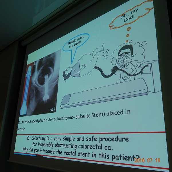
1988년 rectal stent가 없던 시절에 esophageal stent를 rectum에 삽입한 예를 보여주셨습니다. 환자는 "Thank you, my God!"이라고 말했고 의사는 "Oh, my God!"을 외쳤습니다. 심교수님은 이 증례를 UEGW에 하였는데 "간단히 colostomy를 하지 왜 stent를 하는가?"라는 질문을 받았다고 합니다. 이 경험을 바탕으로 심찬섭 교수님은 나중에 colon stent를 개발하였다고 합니다.
심교수님께서는 "Creativity is thinking up new things. Innovation is doing new things" (Theodor Levitt) 이라는 격언을 보여주시면서 강의를 마치셨습니다.
7) Diagnosis of gastric cancers in Japan (Naota Iwai)
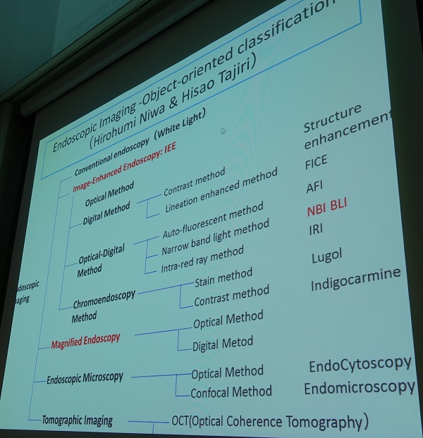
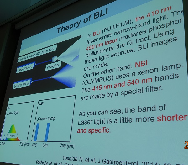
LASERSO(BLI)는 Fujifilm 사에서 개발한 450 nm laser와 410 nm laser를 사용한 새로운 내시경 system이라고 합니다.
8) Treatment of colorectal cancers in Japan (Naohisa Yoshida, Kyoto Prefectural University of Medicine, Kyoto, Japan)
여러 분류가 있지만 regular, irregular, destroyed로 나누는 것이 현실적입니다.
멀리서 보면 NBI와 BLI가 비슷해 보이지만 확대해보면 BLI에서 surface pattern과 vascular pattern이 좀 더 명확해 보입니다.
Surface pattern과 vascular pattern 중 어느 것이 더 중요한지 논의가 있었습니다. 이 둘을 비교하는 연구는 없었던 모양입니다. Yoshida 선생님은 surface pattern이 기존의 pit pattern과 비슷하여 좀 더 이해하기 쉽다고 하였습니다. Vascular pattern은 pit pattern과는 전혀 다르므로 좀 더 이해하기 어려울 것이라도 답했습니다. 아물러 "fist surface pattern, and then vascular pattern"이라고 강조했습니다.
참고: EndoTODAY 확대내시경
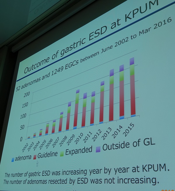
9) Treatment of early gastric cancers in Japan (Osamu Dohi)

Kyoto Prefectural Hospital에서는 지금까지 1300예 정도의 gastric ESD를 했는데 adenoma는 5% 정도 밖에 되지 않았습니다 (암 1249, 선종 52). 우리나라와는 상당히 다른 수치입니다. Pathology 철학 차이 아닐까 합니다. 일본에서는 웬만하면 암이니까요.
Abdominal compression method를 사용하면 병소로의 접근이 쉽다고 합니다.
10) Treatment of early colorectal cancers in Japan (Takaaki Murakami)
항혈응고제 사용환자에서 cold snare polypectomy(CSP)가 더 안전할 수 있습니다 (Horiuchi A. GIE 2014). Yoshida 선생님은 cold snare polypectomy에서는 전기 손상이 없고 cutting depth가 낮기 때문에 출혈이 적다고 답하였습니다. CSP에서 늘 피가 나지만 대부분 저절로 멎습니다.
연자는 Flush knife BT를 소개하였습니다. Flush knife는 Fujinon사의 endoknife이고 Flush knife-BT는 끝이 약간 뭉퉁한 형태입니다. Olympus사의 Kneedle knife나 Flex knife와 비슷한 방식으로 이용하는 절개도이지만, injection도 할 수 있습니다. ESD 도중 endoknife를 넣고 뺄 필요가 없다는 장점이 있습니다.

Broucher from Fujinon company (PDF, 0.8 M)
11) Japanese perspective of premalignant GI lesions (Yuji Naito)
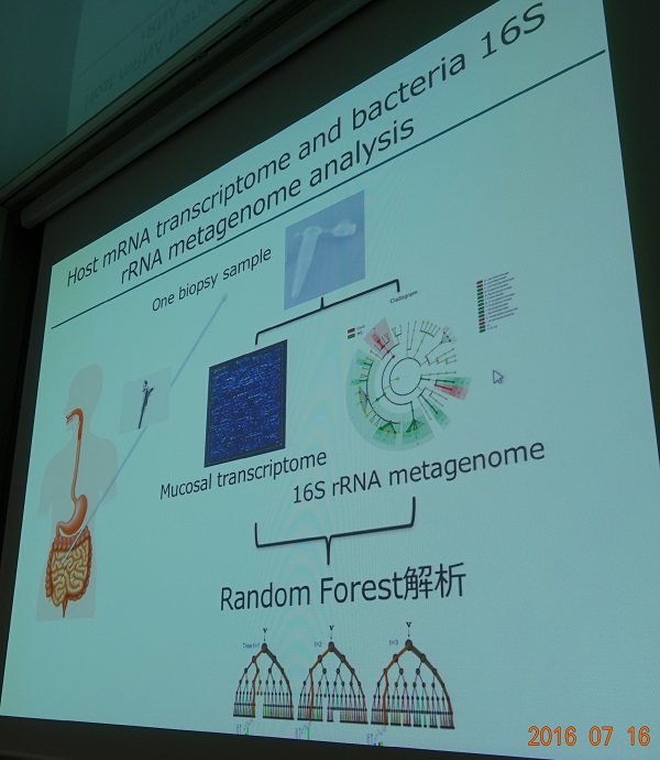
조직검사에서 선종이었던 176예 중 ESD 후 127명(72.2%)은 암으로 49명은 선종으로 최종 결론이 나왔다고 합니다. Naito 선생님은 조직검사에서 선종이 나왔더라도 확대내시경을 통하여 최종 결론이 암으로 나올 사람을 골라낼 수 있다고 강조하였습니다.
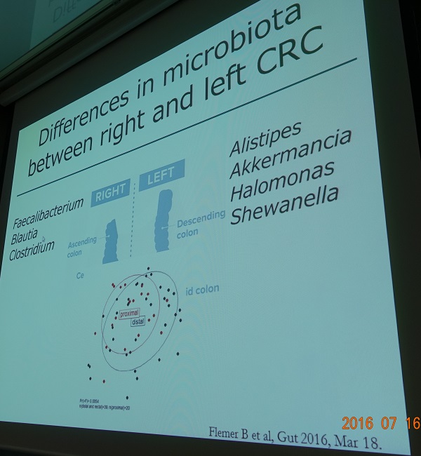
Fecal bacteria의 중요성을 강조하셨습니다. 좌측 대장과 우측 대장을 균이 다르다고 합니다.
Naito 선생님은 과거에는 자신이 선종이라고 진단한 병소를 병리과 의사도 선종이라고 진단했는데, 요즘은 자신의 선종을 자꾸 very well differentiated adenocarcinoma라고 진단한다면서 병리과 의사가 암으로 진단하는 기준이 낮아졌다고 설명하였습니다.
12) Korean perspective of premalignant GI lesions (이선영)
![]() 5. [2016-7-17] Evaluation of devices/accessories to improve ESD practice. Noriya Uedo (Osak Medical Center)
5. [2016-7-17] Evaluation of devices/accessories to improve ESD practice. Noriya Uedo (Osak Medical Center)
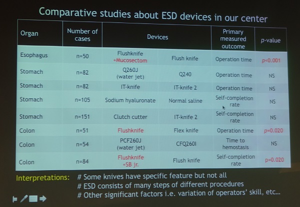
Water jet endoscope는 operation time을 유의하게 줄이지 못했지만 시술자는 항후 시술에서 water jet endoscope를 쓰고 싶다고 말합니다.
Water jet knife는 operation time을 유의하게 줄였습니다. 주로 device change 시간을 현저히 줄였습니다.
Mucosectom (partially insulated type knife)
Scissor-type knife는 기대와 달리 self completion rate가 높지 않았고 operation time은 유의하게 길었습니다.
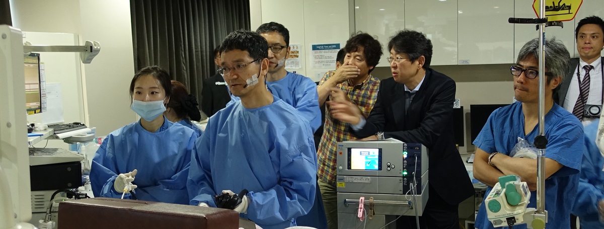
![]() 6. ESD for GI neoplasm - current status and new development. Shinawa Tanaka (Kobe University)
6. ESD for GI neoplasm - current status and new development. Shinawa Tanaka (Kobe University)
Advantage of submucosal dissection at lower level: (1) Smooth dissection with less hemorrhage is enabled. (2) Precise pathological diagnosis including lymphovascular invasion is possible.
Vessel sealing method: Medium size vessel에서 soft coagulation mode로 주변을 지혈한 다음 forced coagulation mode로 절제합니다 (Tanaka, Toyonaga. Dig Endosc 2013). 최근 연구에 의하면 one ten mode (Effect 1, 10 W)가 vessel sealing method에 가장 적합하지 않은가 생각됩니다.
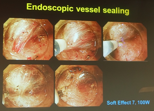
Advantage of endoscopic vessel sealing: (1) This technique makes it possible to continue the procedure without interruption for replacement of the device. (2) It also enables additional precoagulation for reperfused blood vessels after precoagulation.
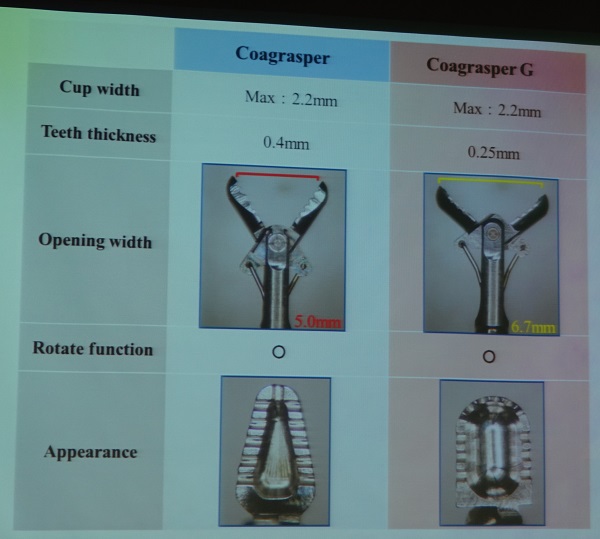
Coagrasper는 rotation function이 있어 큰 혈관 출혈에 유용하지만 artery나 deeper submucosal layer로부터의 출혈을 지혈하기 어려운 경우가 있습니다. 새로 개발된 Coagrasper G는 좀 더 넓게 벌어지고 끝이 뭉퉁하여 gastric ESD에서 좀 더 유용합니다 (G: gastric).
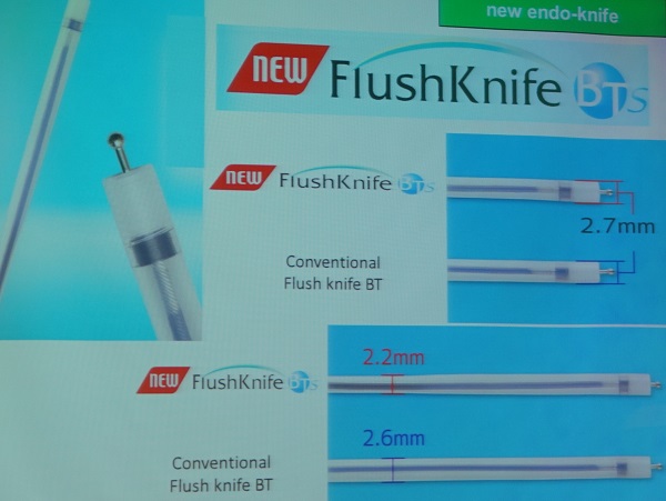
FlushKnife BTs (S: slim)는 좀 더 가늘어서 ESD 도중 공기나 물을 suction하기에 유리합니다. 좀 더 큰 각도로 retroflection할 수 있을 것 같습니다.
순천항대학교 류창범 교수님 comment: 어제 Toyonaga 선생님 시술을 유심히 보신 분은 느끼셨겠지만 coagulation을 찔끔찔끔 조금씩 합니다. 강한 파워로 꾹 눌러서 세게 coagulation하면 delayed bleeding 위험이 있습니다.
![]() 7. 확대내시경 live demonstration (Kenshi Yao)
7. 확대내시경 live demonstration (Kenshi Yao)
65배 확대내시경으로 microvascular structure 뿐만 아니라 RBC 움직임까지 볼 수 있습니다. Constant distance (2 mm)를 유지하기 위하여 short black cap을 사용합니다.
선종의 경우 margin은 non-neoplastic mucosa로 덮인 경우가 많으므로 demarcation line을 잡기 어려울 수 있습니다. 좀 더 중앙으로 들어가 보면 abrupt하게 microsuface pattern이 변한 부위가 있습니다.
White opaque substance가 regular하므로 선종으로 판단됩니다. White opaque substance 때문에 microvascular pattern을 관찰할 수 없는 경우가 있습니다. 대장에서 용종 주변의 lipid droplet과 비슷하다고 생각하면 됩니다. Intestinal metaplasia에서도 white opaque substance가 보이는 경우가 많은데 이는 incomplete type intestinal metaplasia에서 흔하고 adenoma나 cancer로 발전할 가능성이 높은 경우라고 생각하면 됩니다.
하얀 점액이 부착된 위암이 보였는데, 이는 헬리코박터와 관련된 위암과 관련된 소견이기도 한데 점액이 잘 떨어지지 않으면 확대내시경으로 정확한 관찰이 어려울 수 있습니다.
과형성 용종은 혈관이 균일하게 늘어난 모양입니다. 조직검사는 필요하지 않습니다 .
RAC가 없어지면 헬리코박터 감염의 확대내시경 소견이라고 생각할 수 있습니다.
확대내시경에서 water immersion을 사용하는 이유: (1) 빛 반사가 줄어서 보다 선명하게 관찰할 수 있습니다. (2) 물과 공기의 굴절률이 다릅니다.
![]() 8. Gastric ESD live demonstration
8. Gastric ESD live demonstration
| EUS setting for gastric ESD of Dr. Uedo | |
| Marking (Dual knife) | Forced Coag E2 25W |
| Mucosal incision (Dual knife) | EndoCut E2 D3 I3 |
| Submucosal dissection (IT-2 knife) | Swift Coag E3 80W |
* 참고: EndoTODAY 고주파전류발생장치 (원리부터 ERBE VIO 300D까지)
© EndoTODAY Endoscopy Learning Center. Jun Haeng Lee. 일원내시경교실 바른내시경연구소 이준행