EndoTODAY ГЛНУАц БГНЧ
Beginner | ESA | Schedule | OPD
Seminars | Atlas | Recent | Links
![]() [SGI 2016 - The annual meeting of Society of GI Intervention]
[SGI 2016 - The annual meeting of Society of GI Intervention]
РхМв: K Hotel
РЯНУ: 2016Гт 10Пљ 7-8РЯ
SCI meetingРЬ ЙњНс 10ЙјТАПДНРДЯДй. ЧбБЙ ЛчЖїКИДй ПмБЙРЮРЬ ИЙРЬ ТќМЎЧЯДТ international ЧаШИПДНРДЯДй.
![]() 1. Diagnosis and management strategy of refractory GERD: Endotherapy or Laparoscopic fundoplication? (МПяОЦЛъКДПј УжБтЕЗ)
1. Diagnosis and management strategy of refractory GERD: Endotherapy or Laparoscopic fundoplication? (МПяОЦЛъКДПј УжБтЕЗ)
When to consider surgery or endoscopic anti-reflux treatment?
1) pH impedance shows excessive reflux and positive symptom association
2) Symptom control is inadequate: severe regurgitation not controlled with optimal acid suppression
- Large hiatal hernia (+) --> Surgery
- Normal GE junction anatomy, reluctant to undergo surgery --> Endoscopy
ЧіРч ГЛНУАц ФЁЗсЙ§РК 4 АЁСіАЁ РжНРДЯДй.
![]() 2. Efficacy and long-term result of laparoscopic fundoplication (АЁХчИЏДыЧаБГ БшСјСЖ)
2. Efficacy and long-term result of laparoscopic fundoplication (АЁХчИЏДыЧаБГ БшСјСЖ)
ЧіРч laparoscopic surgeryПЭ ОрЙАФЁЗсИІ КёБГЧб ПЌБИ Сп LOTUS triak (Galmiche JP. JAMA 2011)ПЁМДТ ХЋ ТїРЬАЁ ОјОњСіИИ, REFLUX trial (Grant AM. BMJ 2013)ПЁМДТ laparoscopic surgeryАЁ Дѕ ССДйДТ АсАњПДНРДЯДй.
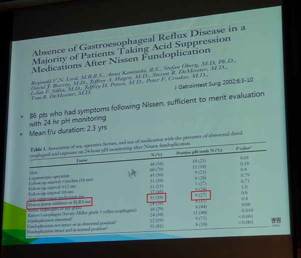
Nissen fundoplication ШФ GERDАЁ ОјДТЕЅЕЕ PPIИІ КЙПыЧЯДТ ШЏРкАЁ ИЙДйДТ ШяЙЬЗЮПю ПЌБИИІ МвАГЧи СжМЬНРДЯДй. (J Gastrointest Surg 2002;6:3-10)
![]() 3. The Stretta Procedure - Personalized medicine of GERD (Mark D. Noar. USA)
3. The Stretta Procedure - Personalized medicine of GERD (Mark D. Noar. USA)
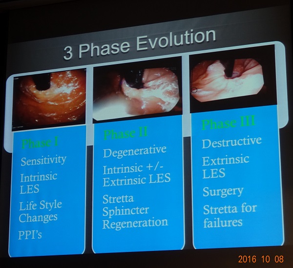
Disease progression mechanisms
Sphincter-target therapyРЮ StrettaДТ destructive procedureАЁ ОЦДЯАэ stimulating procedureРдДЯДй.
10 year durability study: Noar M. Surg Endosc 2014
RESULTS: The primary outcome was achieved in 72% of patients (95% confidence interval 65-79). For secondary outcomes, a 50% or greater reduction in PPI use occurred in 64% of patients, (41% eliminating PPIs entirely), and a 60% or greater increase in satisfaction occurred in 54% of patients. Both secondary endpoints were achieved. The most common side effect was short-term chest pain (50%). Pre-existing Barrett's metaplasia regressed in 85% of biopsied patients. No cases of esophageal cancer occurred.
Extrinsic sphincter therapyПЭ intrinsic sphincter therapyАЁ РжАэ РЬИІ ЧдВВ ОВИщ Дѕ ССНРДЯДй. Extrinsic sphincter therapyРЮ Nissen fundoplicationРЬ НЧЦаЧб АцПь intrinsic sphincter therapyРЮ Stretta therapyАЁ ССРК optionРдДЯДй.
* ТќАэ: EndoTODAY Stretta
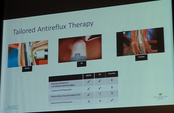
![]() 4. Trans-oral anterior fundoplication (Ali Lankarani. Borland Groover Clinic, Jacksonville, Florida, USA)
4. Trans-oral anterior fundoplication (Ali Lankarani. Borland Groover Clinic, Jacksonville, Florida, USA)
Anti-reflux barrierДТ ХЉАд LESПЭ flap valveРдДЯДй. Flap valve (= GE junction geometry) ДТ intra-abdominal esophageal locationАњ angle of HisЗЮ БИМКЕЫДЯДй. GE junction geometryАЁ ИСАЁСіИщ LESИИРЬ РЏРЯЧб barrierРдДЯДй.
Opposing sling and clasp muscle fibers. The longitudinal muscle layer of the stomach has been cut away to show the opposing sling and clasp muscle fibers. These fibers sit in tonic opposition until a swallow triggers receptive relaxation. It is thought that progressive stretching of these fibers leads to valve incompetence and subsequent GERD. (Jobe BA. Am J Gastroenterol 2004)
Intraabdominal esophagusРЧ angle of HisАЁ ПЙАЂРЛ РЏСіЧЯИщМ ШПАњРћРЮ flap valveИІ ЧќМКЧЯАэ РжДТ И№НР (medigus.com)
Angle of HisАЁ ЕаАЂРЬ ЕЧИщМ flap valve БтДЩРЛ РвРН (medigus.com)
Angle of HisАЁ ЕаАЂРЬ ЕЧАэ, hiatusАЁ ГаОюСіАэ, intraabdominal esophagusАЁ ЛѓНТЧЯИщ sliding hiatal herniaАЁ ЕЪ (medigus.com)
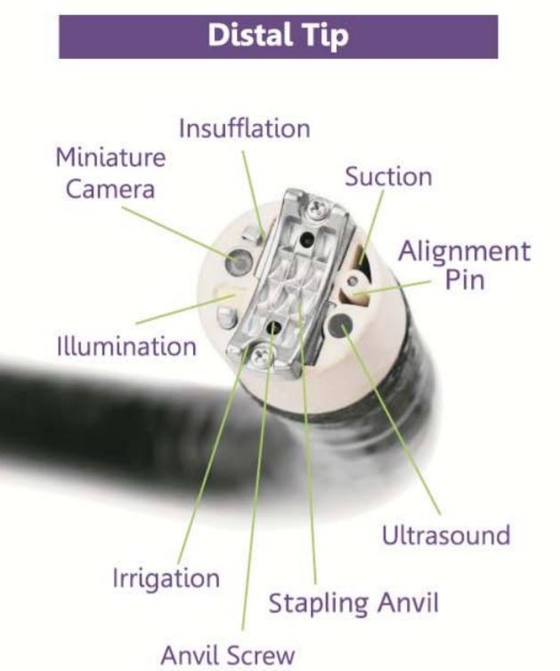
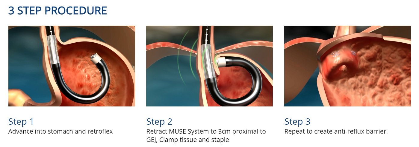
Medigus Ultrasonic Surgical Endostapler (MUSE)РЧ ММАЁСі РлПы БтРќ
АЛчДТ Winston ChurchillРЧ ИЛРЛ РЮПыЧЯИч АРЧИІ ИЖУЦНРДЯДй.
Now it is not the end.
It is not even the beginning of the end.
But, it is, perhaps, the end of the beginning.
* ТќАэ: Medigus Лч ШЈЦфРЬСі
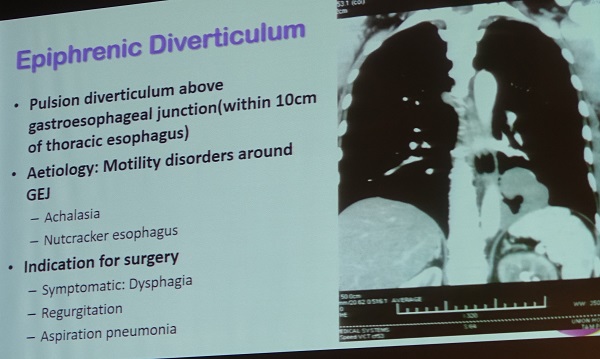
![]() 5. Surgical and endoscopic managemen of esophageal diverticulum: Zenker's and epiphrenic (Philip Chiu, The Chinese university of Hong Kong)
5. Surgical and endoscopic managemen of esophageal diverticulum: Zenker's and epiphrenic (Philip Chiu, The Chinese university of Hong Kong)
МіМњРЬ ОЦДб ЙцЙ§РИЗЮ УГРН АГЙпЕШ АЭРЬ StaplingРЬОњНРДЯДй.
Flexible endoscopic (incisional) diverticulotomy (septectomy)ПЁ ДыЧб ИоХИКаМЎ (Ishaq S. Gastrointest Endosc 2016;83:1076)
ШПАњ
КЮРлПы
Chiu ЙкЛчДТ РќНХИЖУы ЛѓХТПЁМ РЬКёРЮШФАњ РЧЛчПЭ ЧдВВ НУМњРЛ ЧбДйАэ ЧеДЯДй. ГЛНУАц ИЛДмПЁ ТЊРК capРЛ РхТјЧб ШФ triangle knifeИІ РЬПыЧЯПЉ initial short septectomyИІ НУЧрЧеДЯДй. РЬШФ capРЛ СЛ Дѕ Бф ЧќХТЗЮ ЙйВйОю ССРК НУОпИІ ШЎКИЧб ЛѓХТ(Ся Zenker lumenАњ esophageal lumenРЛ ШЎРЮЧЯИщМ)ПЁМ Бф septectomyИІ НУЧрЧеДЯДй. ИЖСіИЗПЁ clipРЛ applyЧЯАэ НУМњРЛ СОЗсЧеДЯДй.
ИЖСіИЗРИЗЮ epiphrenic diverticulumРЛ РсБё МвАГЧЯМЬНРДЯДй.
* ТќАэ: EndoTODAY Zenker АдНЧ
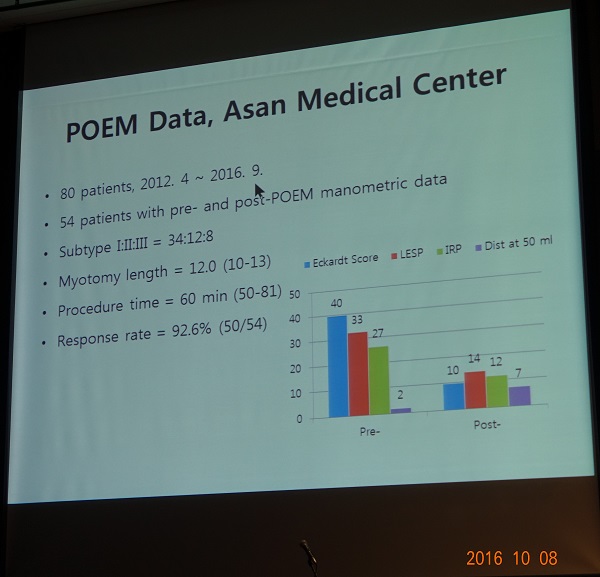
![]() 6. Endoscopic management of achalasia (МПяОЦЛъКДПј. СЄШЦПы)
6. Endoscopic management of achalasia (МПяОЦЛъКДПј. СЄШЦПы)
Pneumatic dilatationАњ Heller myotomyИІ КёБГЧб ПЌБИИІ СОЧеЧб ИоХИКаМЎ
СЄШЦПы БГМіДдРК МПяОЦЛъКДПјРЧ POEM МКРћРЛ РкЖћНКЗДАд КИПЉСжМЬНРДЯДй. Pioneer ДйПю И№НРРЬОњНРДЯДй. СИАцЧеДЯДй.
* ТќАэ: EndoTODAY Achalasia
![]() 7. Fluoroscopically-guided ballon dilation for achalasia (МПяОЦЛъКДПј. НХСіШЦ)
7. Fluoroscopically-guided ballon dilation for achalasia (МПяОЦЛъКДПј. НХСіШЦ)
ЧГМБШЎРхМњРК ГЛНУАцРИЗЮЕЕ НУЧрЧв Мі РжАэ fluoroscopyИІ РЬПыЧв МіЕЕ РжНРДЯДй.
FluoroscopyИІ РЬПыЧб double ballon dilatationРЬЖѓДТ ШяЙЬЗЮПю МњБтИІ МвАГЧи СжМЬНРДЯДй (Yi A. Abdom Imaging 2008).
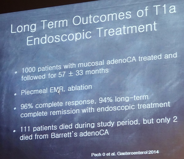
![]() 8. Barrett's treatment in 2016 (Shai Friedland, Stanford Universtiy, USA)
8. Barrett's treatment in 2016 (Shai Friedland, Stanford Universtiy, USA)
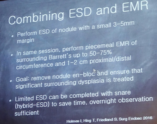
EMRРЧ ШПАњАЁ Рп РдСѕЕЧОю РжРИЙЧЗЮ ESDРЧ ШПАњИІ РдСѕЧЯБтДТ ОюЗЦНРДЯДй.
Combining ESD & EMR. ESDЗЮ noduleРЛ СІАХЧЯАэ СжКЏРК EMRЗЮ СІАХЧЯДТ РќЗЋ
DDW 2016ПЁ cryoablation АќЗУ УЪЗЯРЬ 3АГ ЙпЧЅЕЧОњНРДЯДй. ДыЛѓ СњШЏРК 2АГДТ Barrett, 1АГДТ squamous dysplasiaПДНРДЯДй.
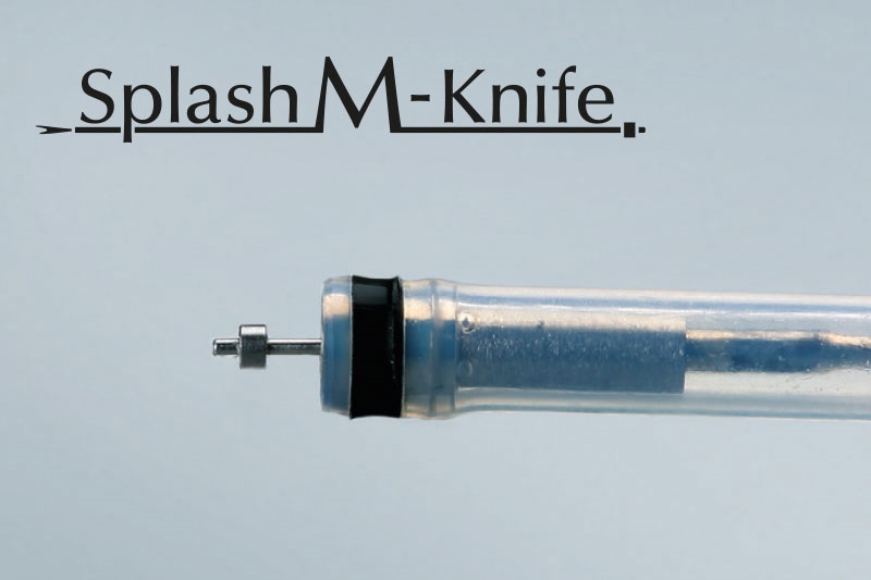
![]() 9. Esophageal stricture after ESD (Mitsuhiro Fujishiro, Japan)
9. Esophageal stricture after ESD (Mitsuhiro Fujishiro, Japan)
Splash M-knife: Submucosal injectionРЬ АЁДЩЧЯАэ hemostatic powerАЁ ССРН.
Circumferential ESD ШФ ИХПь ИЙРК ЧГМБ ШЎРхМњ(preventive balloon dilatation)РЛ ЧЯПДДј ПРЗЁЕШ ШЏРкИІ КИПЉСжОњНРДЯДй. УжБйПЁДТ Ию АЁСі ПЙЙц РќЗЋРЛ ЛчПыЧЯБт ЖЇЙЎПЁ НЩЧб strictureИІ КИРЬДТ ШЏРкДТ АХРЧ ОјНРДЯДй.
БтСИРЧ steroid injection РЬПмРЧ Ию АЁСі ЙцЙ§РЬ МвАГЕЧОњНРДЯДй. Fujishiro ЙкЛчРЧ АсЗаРК steroidПЭ shieldingРЛ ЧдВВ ЛчПыЧЯДТ АЭРЬ АЁРх ССРЛ АЭ ААДйДТ АЭРЬОњНРДЯДй. Triamcinolon injectionАњ PGA shieldingРЛ ЧдВВ ЛчПыЧЯДТ АцПь oral steroidДТ УГЙцЧЯСі ОЪДТДйАэ ЧеДЯДй.
(1) Biodegradable stent (Saito Y. Dig Dis Sci 2008) - commercially availableЧЯСі ОЪНРДЯДй.
(2) Cell sheet technology (Ohki. Gastroenterology 2012)
(3) Polyglycolic acid sheets and fibrin glue (Tokyo University)
(4) Triamcinolone injection and shielding with PGA and fibrin glue (Tokyo University)
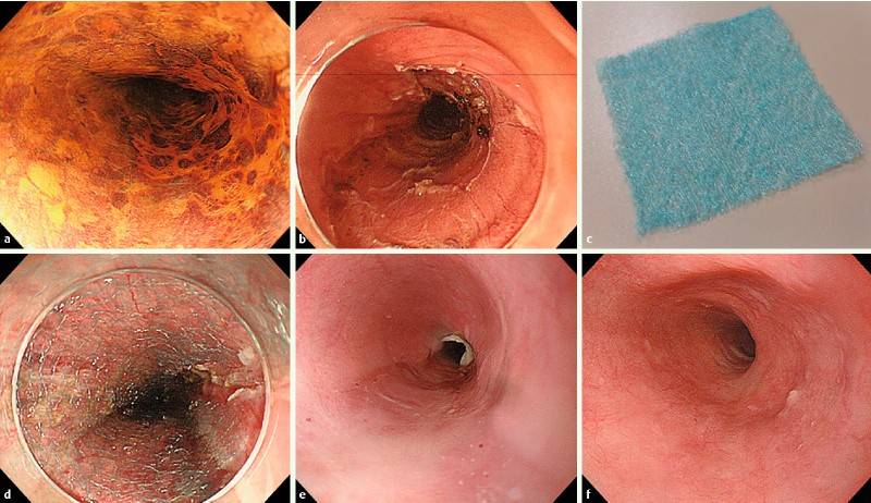
A representative case where postoperative stricture was prevented after wide-spreading ESD. Chromoendoscopy showed a semi-circumferential lesion in the esophagus (a), and the lesion was resected en bloc with endoscopic submucosal dissection (b). This resulted in endoscopic resection of over 3/4 the circumference of the esophagus (c). Triamcinolone was injected into the perimeter of the ESD defect (d), followed by shielding with PGA sheets and fibrin glue (e). Endoscopic follow-up 12 weeks later revealed no stricture (f). (Sakaguchi Y. Am J Gastroenterol 2016 )
* ТќАэ: EndoTODAY НФЕЕОЯ ГЛНУАцФЁЗс ШФ ЧљТј
![]() 12. Endoscopic gastrostomy and jejunostomy (МПяОЦЛъКДПј БшЕЕШЦ)
12. Endoscopic gastrostomy and jejunostomy (МПяОЦЛъКДПј БшЕЕШЦ)
ММ АЁСі ЙцЙ§РЬ РжНРДЯДй.
АњАХПЁДТ PEG НУМњ ДйРН ГЏКЮХЭ feedingРЛ НУРлЧЯПДРИГЊ УжБйПЁДТ PEG МіНУАЃ ШФКЮХЭ feedingРЛ НУРлЧЯДТ АцЧтРдДЯДй. PEG ШФ 4НУАЃКЮХЭ feedingРЛ ЧЯДТ АЭЕЕ ОШРќЧЯДйДТ ИоХИКаМЎРЬ РжОњНРДЯДй.
Wound infectionРЛ СйРЬБт РЇЧЯПЉ external bumperАЁ 1-2cmСЄЕЕ "free-float"ЧЯЕЕЗЯ РЏСіЧЯДТ АЭРЬ ССНРДЯДй.
Peristomal leakИІ ИЗБт РЇЧЯПЉ larger size tubeИІ РЬПыЧЯДТ АЭРК ЕЕПђРЬ ЕЧСі ОЪНРДЯДй. TubeИІ ЛЉАэ 24-48НУАЃ maturationРЛ БтДйИЎДТ АЭРЬ РЏПыЧеДЯДй. (РЬСиЧр comment: РЬ КЮКаРК АяЖѕЧеДЯДй. LeakИІ ЧиАсЧЯБт РЇЧЯПЉ PEG tubeИІ РсНУ ЛЉАэ ААРК РкИЎЗЮ ДйНУ Л№РдЧЯДТ ЙцЙ§РЬ РжНРДЯДй. КИХы 4НУАЃ СЄЕЕ ЛЉГѕРЛ Мі РжНРДЯДй. БзКИДй ПРЗЁ ЛЉГѕРИИщ ДйНУ ГжСі ИјЧЯДТ АцПьАЁ ЙпЛ§ЧеДЯДй. ТќАэ: EndoTODAY PEG leak)
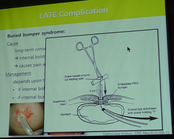
Buried bumper syndrome
Endoscopic jejunostomyРЧ ЙцЙ§РИЗЮДТ PEG with jejunal extension ЙцЙ§РЬ АЁРх ШчШї ЛчПыЕЫДЯДй.
* ТќАэ: EndoTODAY PEG
![]() 13. Radiologic gastrostomy and jejunostomy (Seung Kwon Kim, Washington University, USA)
13. Radiologic gastrostomy and jejunostomy (Seung Kwon Kim, Washington University, USA)
GastropexyАЁ ЧЪПфЧбСі ГэЖѕРЬ РжНРДЯДй. ПЌРкДТ gastropexyИІ ЛчПыЧЯАэ РжДйАэ ЧеДЯДй.
G tube feedingРЛ ЧЯДј ЛчЖїРЛ JejunostomyЗЮ КЏАцЧЯБтДТ ОюЗЦДйАэ ЧеДЯДй. УЙ gastrostomy tube Л№Рд НУ ЙцЧтРЬ fundusИІ ЧтЧЯДТ АцПьАЁ ИЙБт ЖЇЙЎРдДЯДй.
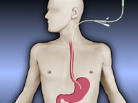
![]() 14. Percutaneous transesophageal gastrostomy (PTEG) (МПяОЦЛъКДПј НХСіШЦ)
14. Percutaneous transesophageal gastrostomy (PTEG) (МПяОЦЛъКДПј НХСіШЦ)
РЯКЛПЁДТ PTEG РќПы kitАЁ РжНРДЯДй.
НХСіШЦ МБЛ§ДдРК lidocaine hydrodissectionРЛ ХыЧи АцЗЮИІ ИИЕщОю СжОњНРДЯДй. Puncture-free balloonРЬ РжРИИщ ЦэЧЯСіИИ ПьИЎГЊЖѓПЁМДТ available ЧЯСі ОЪБт ЖЇЙЎРЬЖѓАэ ЧеДЯДй.
PTEG tubeИІ БГШЏЧЯДТ АЭРК НЌПю РЯРЬЖѓАэ ЧеДЯДй.
![]() [References]
[References]
1) 20160630 СІ2Тї SGI С§ДуШИ (АГВММКъЖѕНК КДПј)
© РЯПјГЛНУАцБГНЧ ЙйИЅГЛНУАцПЌБИМв РЬСиЧр. EndoTODAY Endoscopy Learning Center. Lee Jun Haeng.