EndoTODAY 내시경 교실
Beginner | ESA | Schedule | OPD
Seminars | Atlas | Recent | Links
![]() [Zenker diverticulum 젱커 개실] - 終
[Zenker diverticulum 젱커 개실] - 終
2020-4-25. 순천만내시경세미나 동영상 강의
2. 병태 생리
3. 역학
4. 증상
5. 내시경 소견
7. FAQs
8. References
![]() 1. Zenker 게실의 정의, 해부 및 역사
1. Zenker 게실의 정의, 해부 및 역사
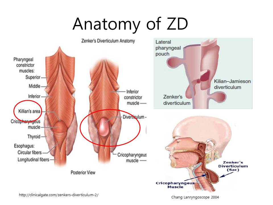
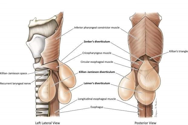
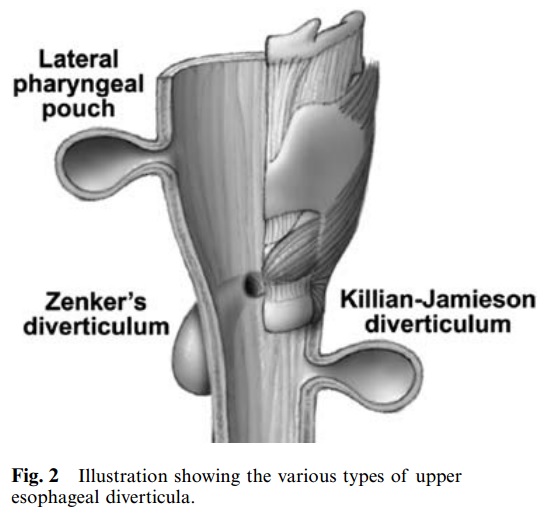
Cricopharyngeal muscle 상방에 위치하고 주머니(pouch)가 뒤쪽으로 향하는 게실이 Zenker diverticulum입니다. 옆으로 빠지는 것은 Killian-Jamieson diverticulum입니다.
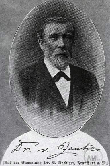
처음 기술한 사람은 Lundlow라는 분인데요 (Med Observ Inq 1769;3:85-101), Zenker라는 분이 1877년 체계적으로 정리한 모양입니다. 아래 사진이 Doctor Zenker입니다.
![]() 2. 병태생리
2. 병태생리
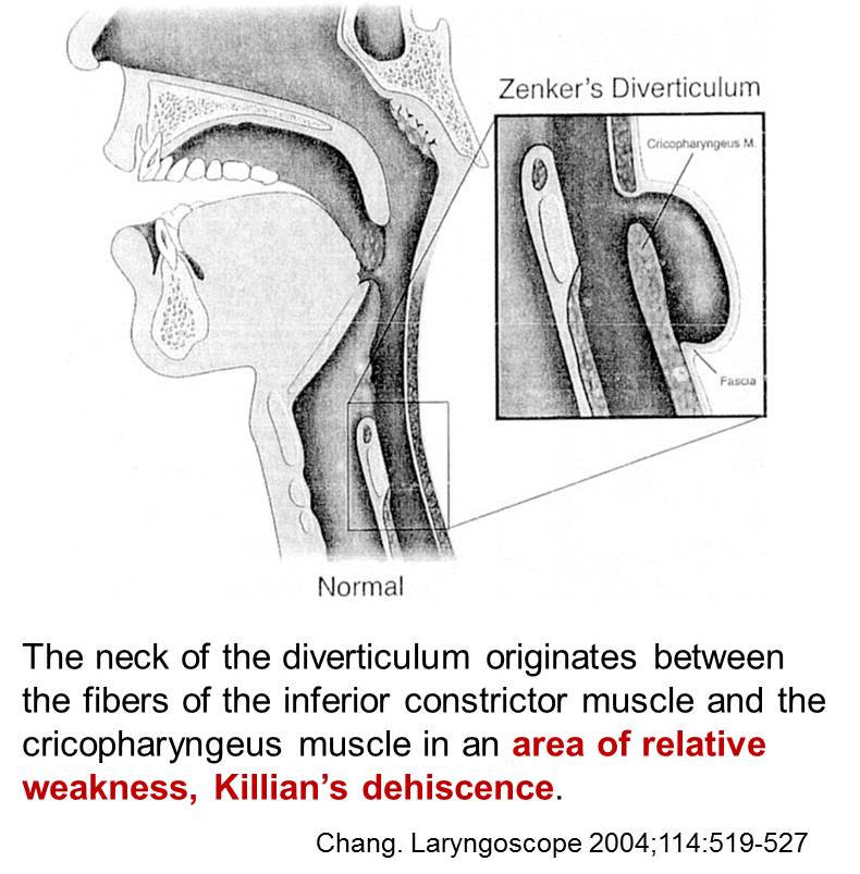
Zenker diverticulum occurs at an area of potential weakness in the inferior pharyngeal constrictor muscle referred to as the Killian dehiscence. It is located between the obliquely oriented fibers of the thyropharyngeal muscle and the horizontally oriented fibers of the cricopharyngeal muscle. Manometric examinations of patients with Zenker diverticulum have produced conflicting results, with some studies showing increased upper esophageal sphincter (UES) pressure and abnormal relaxation and others finding normal relaxation and low UES pressure. To date, no mechanism of pathogenesis has been generally accepted, although most recent studies confirm increased intrabolus pressure in patients with Zenker diverticulum.
Zenker diverticulum is associated with gastroesophageal reflux and hiatal hernia. Some studies have shown that as many as 94% of patients with pharyngeal pouches have concurrent hiatal hernias and gastroesophageal reflux. Increased UES pressure is also associated with gastroesophageal reflux, but whether the reflux, the increased UES pressure, or the Zenker diverticulum arises first in patients is controversial.
Pathologic studies of cricopharyngeal and hypopharyngeal muscle tissue resected from patients with Zenker diverticulum have shown that as many as 95% demonstrate abnormal histologic changes. These include atrophy, necrosis, hypertrophy, inflammation, and fibrosis. This muscle tissue also has been shown to have decreased anticholinesterase levels compared with those in normal tissue.
![]() 3. 역학
3. 역학
Frequency: In the US, fluoroscopic studies of the upper GI tract have shown that the prevalence of Zenker diverticulum is 0.01-0.1%. They are present in approximately 2% of patients with nonspecific dysphagia who are referred for fluoroscopy.
Sex: slightly more common in men than in women.
Age: seen most commonly in the elderly. More than 50% of affected patients present in the seventh or eighth decade of life.
Mortality/Morbidity: Weight loss occurs in approximately one third of patients; however, this is generally modest in severity. A more serious complication occurring in approximately 30% of patients is aspiration pneumonia. Squamous cell carcinoma developing within the diverticulum is a rare complication, occurring in 0.3-0.48% of patients; however, this has a high mortality rate. Other rare complications include ulceration, esophageal obstruction, hemorrhage, tracheodiverticular fistula, and perforation.
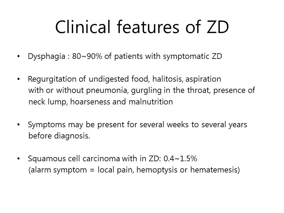
![]() 4. 증상 - 당연히 심할 때만 치료합니다.
4. 증상 - 당연히 심할 때만 치료합니다.
Transient dysphagia; early in the course
Pulmonary aspiration
Neck mass
Food regurgitation
Esophageal obstruction
Recurrent aspiration pneumonia
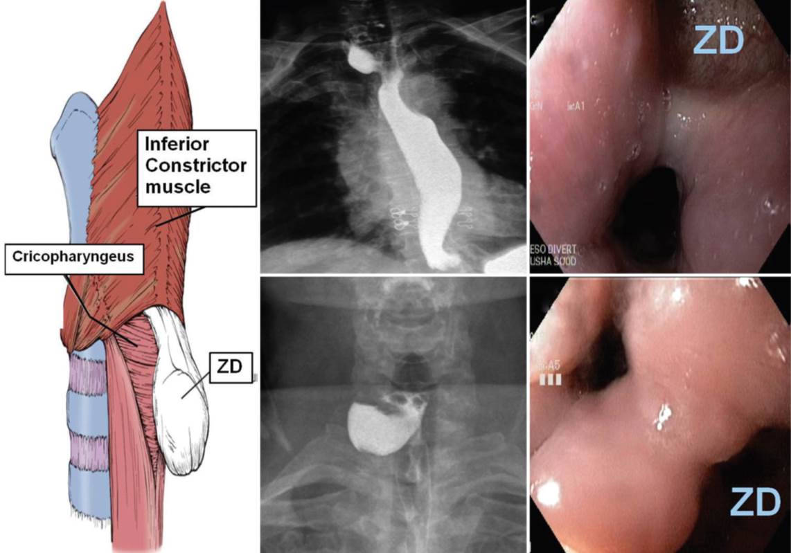
![]() 5. 내시경 소견
5. 내시경 소견
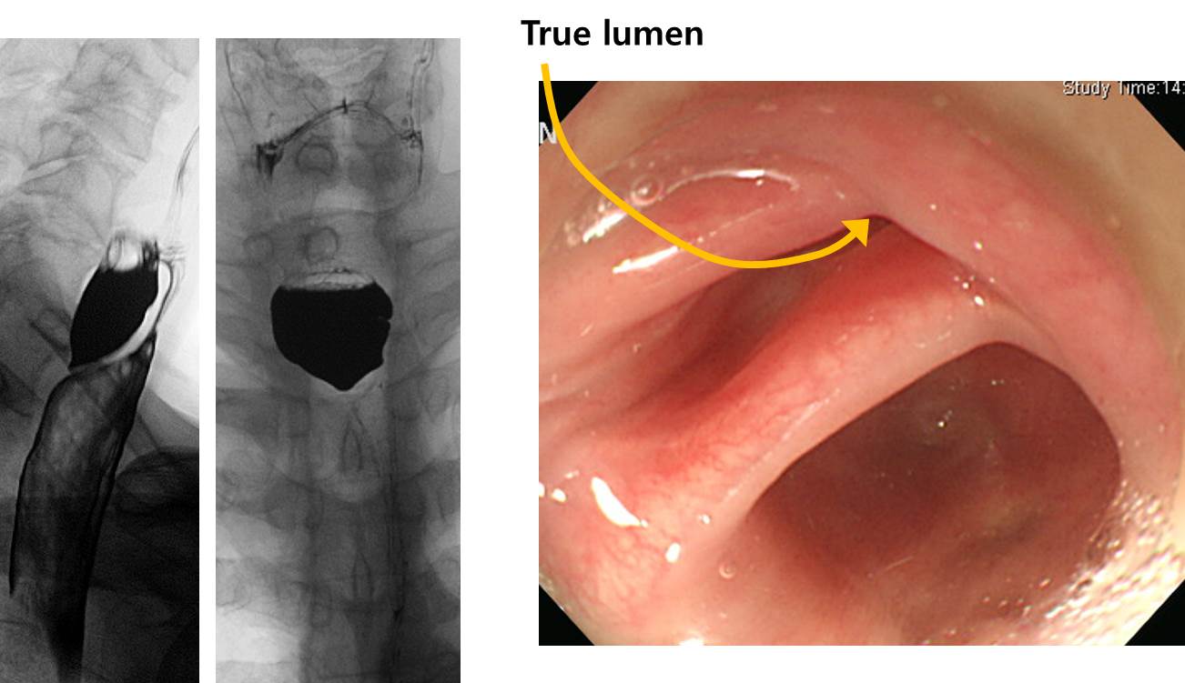
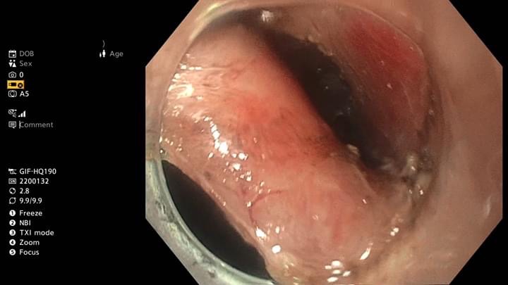
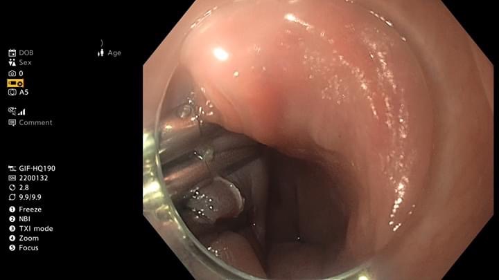
목의 통증으로 내시경 검사를 시행하여 상부식도의 erythematous swelling, stenosis 소견만 있었고 조직검사에는 염증 소견만 나왔고 neck CT에서 암 의심으로 의뢰되신 분입니다.
첫 내시경. 가는 내시경으로 바꾸어 통과할 수 있었음.
의뢰 후 내시경 재검에서 UES 직하방에 blind end를 이루는 pouch가 있었고, 이 pouch의 proximal 측면에 작은 opening이 관찰됨. 이 구멍을 통하여 식도 true lumen으로 들어갈 수 있었음.
상부식도 게실에 음식이 차 있고 true lumen으로 들어가는 길을 찾기 어려웠기 때문에 상부식도의 악성질환으로 오인되었던 것 같습니다. 고령 환자의 상부식도 질환에서는 반드시 diverticulum을 고려해야 합니다.
![]() 6. 치료
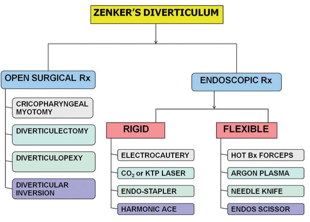
6. 치료
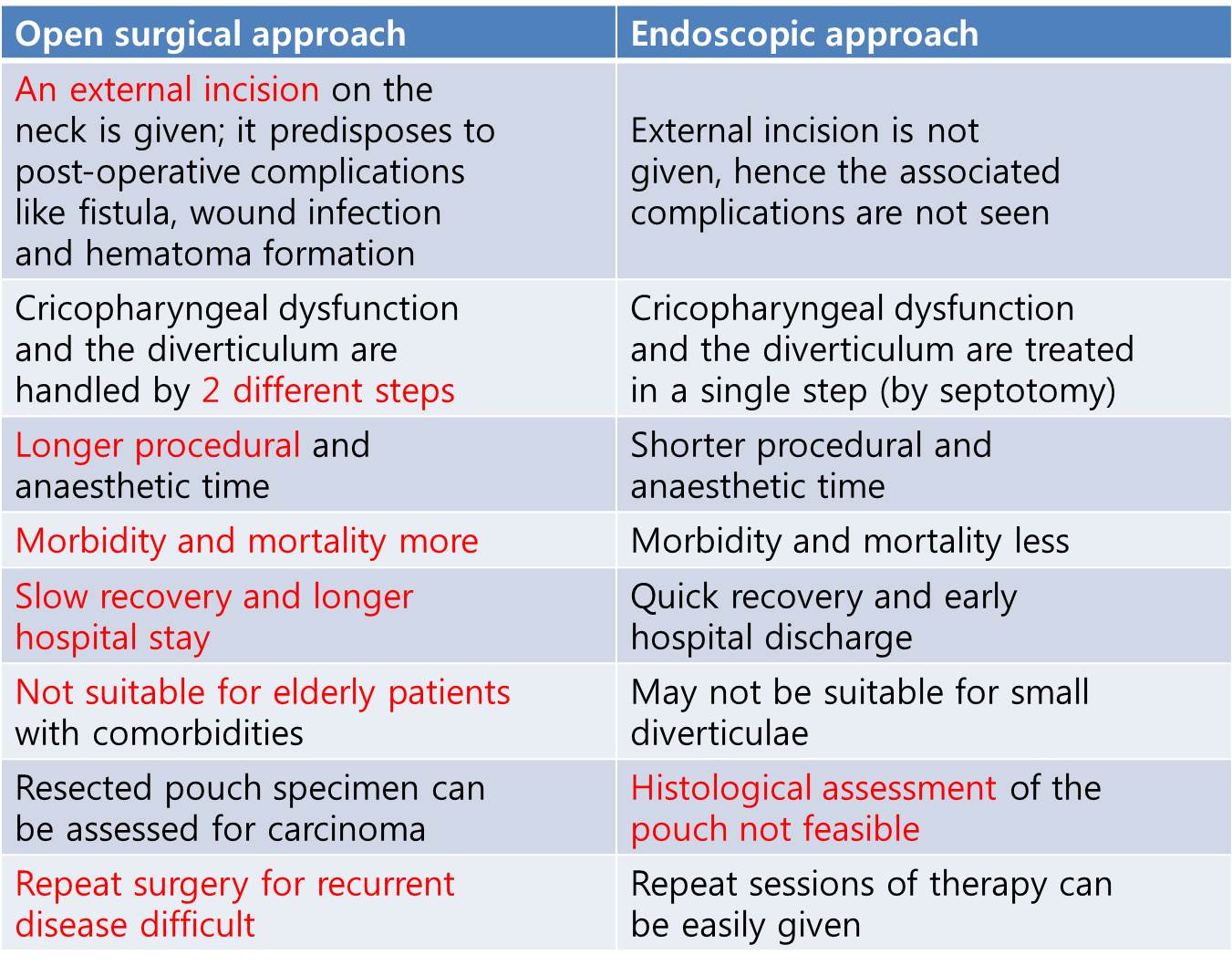
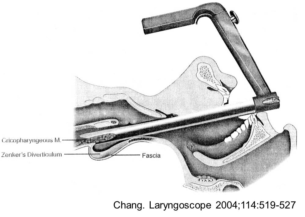
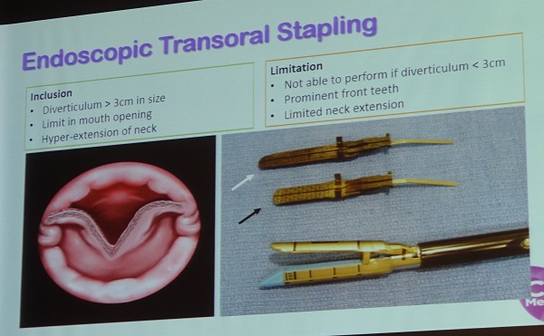
1) 외과적 절제술 (laparoscopic diverticulectomy)
2) Rigid esophagoscopy를 이용한 endoscopic diverticulostomy
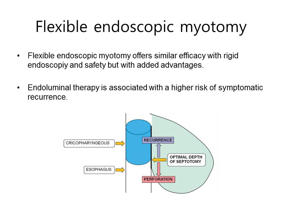
3) Flexible endoscopy를 이용한 endoscopic diverticulostomy
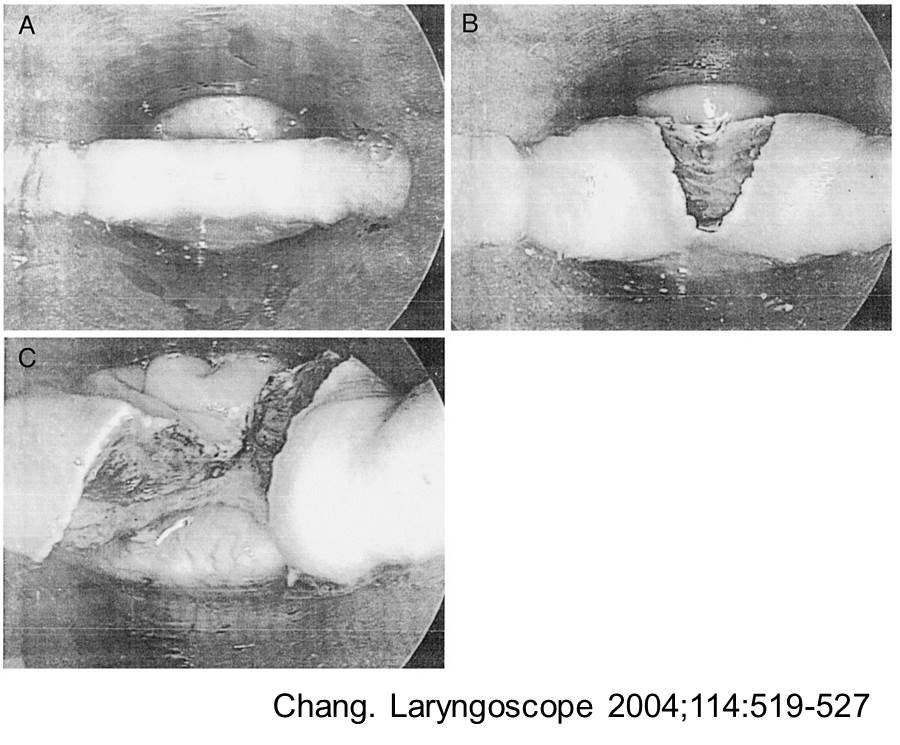
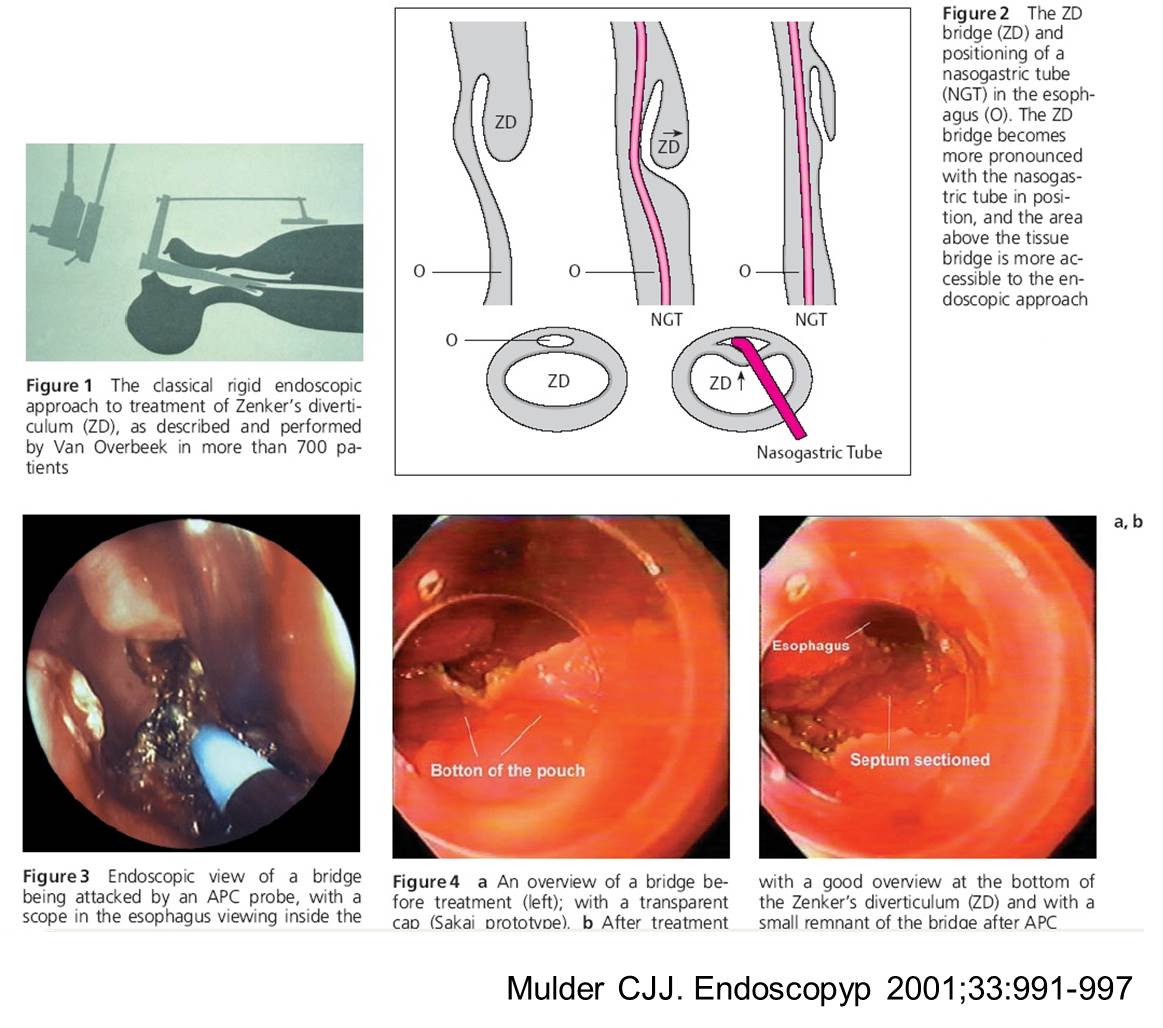
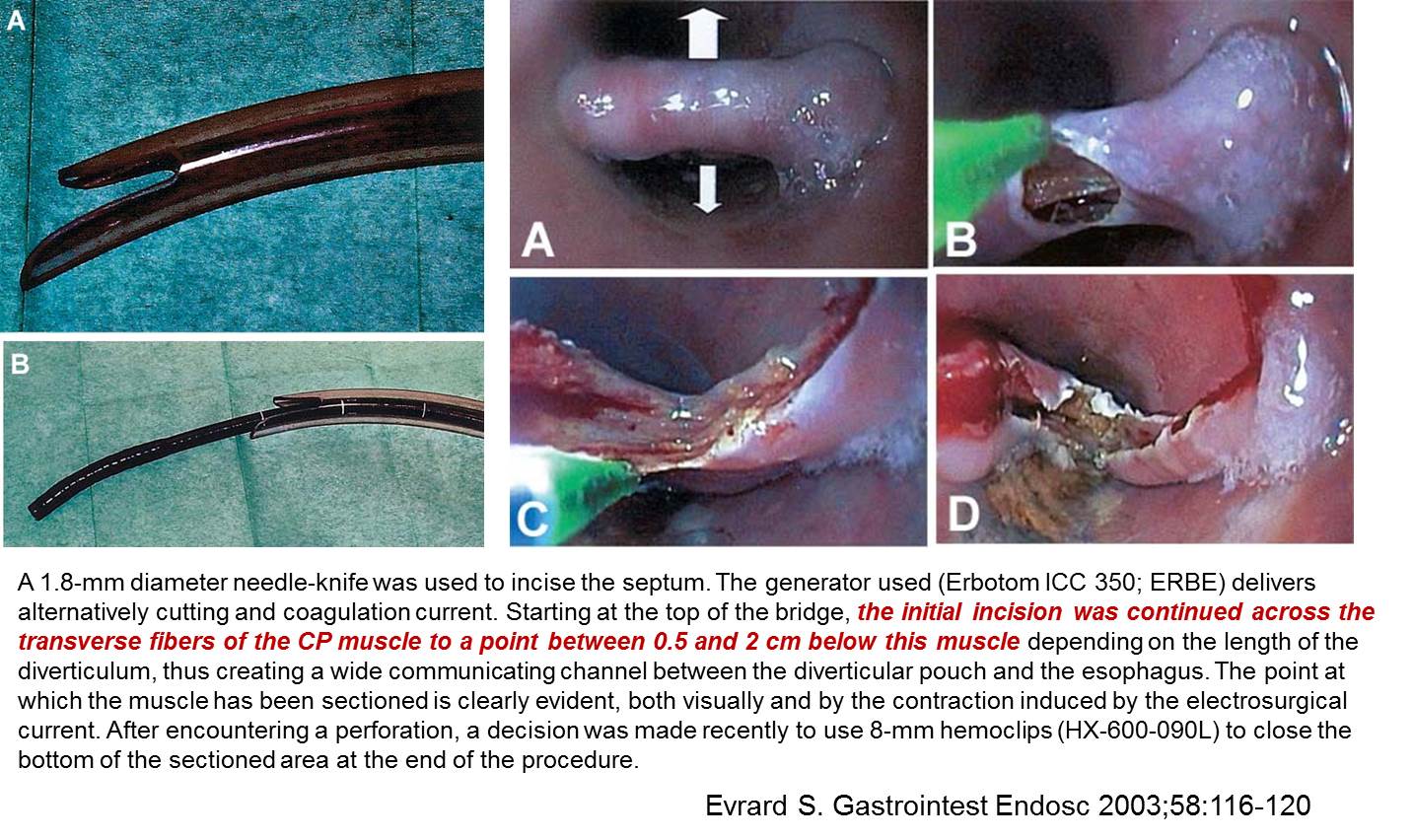
내시경으로 게실과 식도의 벽(septum)을 직접 자르는 방법입니다. Septum의 맨 기저부까지 거의 빵구나기 직전까지 자르는 것이 요점입니다. 처음 시도할 경우는 아래 그림과 같이 식도로 nasogastric tube를 넣어놓고 하는 것이 좋겠다는 글을 본적이 있는데 저는 nasogastric tube를 사용하지 않고 시행해도 좋다고 생각합니다.
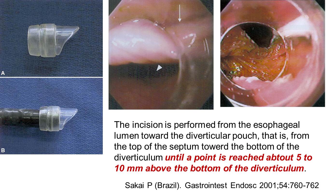
아래와 같은 Zenker diverticulostomy 전용 cap이 있으면 좋겠지만 구할 수 없습니다. 그냥 보통 cap을 이용하는 수밖에...
아래와 같은 Zenker diverticulostomy 전용 tube가 있으면 더없이 좋겠지만 구할 수 없습니다. 그냥 보통 cap을 이용하는 수밖에...
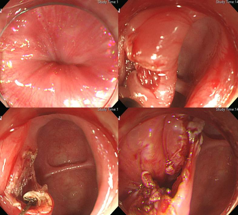
저도 많이 해보지 못했지만 한 예를 보여드리면 아래와 같습니다. 겁이 나서 septum을 다 자르지 못했는데도 환자의 증상은 현저히 좋아졌습니다. 당시 합병증으로 subcutaneous emphysema가 있었는데 이내 좋아졌습니다. 이 시술은 브라질에서 가장 많이 시행되었는데, 그들의 보고에 따르면 합병증의 빈도는 subcutaneous emphysema가 5%, 출혈이 3%라고 합니다 (Mulder CJ. Endoscopy 2001).
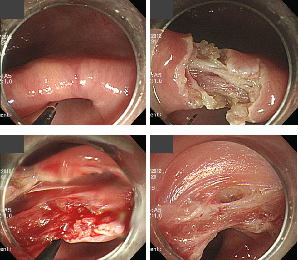
Zenker 게실을 내시경으로 자르는 데 가장 어려운 점을 내시경을 안정감있게 고정하기 어렵다는 것입니다. 오히려 중부식도 게실의 septum은 내시경으로 자르기가 쉽습니다. 안정감이 있기 때문입니다. 아래 사진은 중부식도 septum을 자르는 모습입니다. 막상 10분 정도 밖에 안 걸립니다.
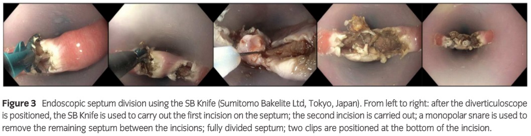
Digestive Endoscopy 2015년 11월호에 이탈리아에서 FB knife를 이용한 Zenker 게실 치료 논문을 발표하였습니다 (Battaglia G. DE 2015). 그냥 cutting만 하는 것이 보통인데 이탈리아 의사들은 두 번 cutting하고 그 사이를 snare로 잘라주었습니다. 그리고 맨 마지막에 clip을 해 주었네요. 지나치게 조심한 것 아닌가 싶습니다.
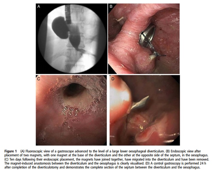
Zenker에서 사용할 수 있을지 의문이지만 2015년 11월 호 Gut지에 magnet를 이용한 식도게실 치료법이 소개되었습니다 (Bouchard S. Gut 2015). 게실의 벽을 중간에 두고 자석을 위치시키면 그로 인하여 fistula가 생깁니다. 그 이후 남은 septum을 내시경으로 자르는 방법입니다. 자석을 이용하지 않고 처음부터 Dual knife 같은 것을 이용하여 septum을 잘라도 충분할 것 같은데... 아무래도 내시경 술기에 자신이 없고 겁이 난다면 자석의 도움을 받을 수 있다고 생각합니다. 간단한 것을 괜히 복잡하게 하는 느낌도 들었습니다.
Endoscopy 2020년 4월호 E-Videos에 Zenker diverticulum septotomy 동영상이 실렸습니다 (Endoscopy 2020;52:308-309). 참고하시기 바랍니다.
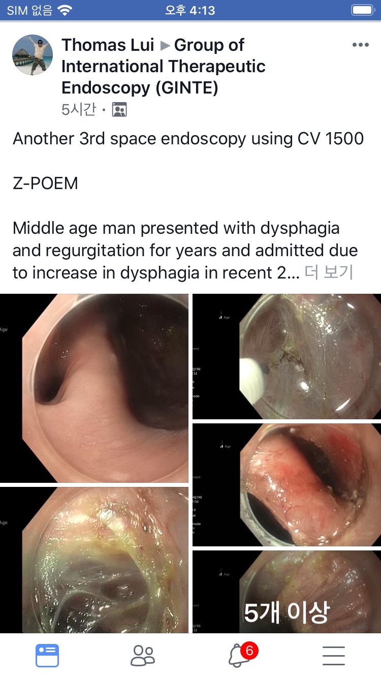
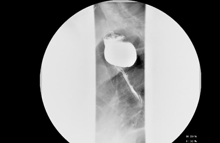
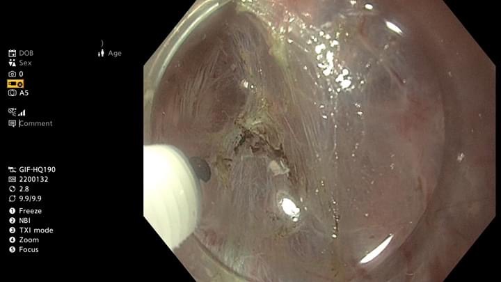
4) Z-POEM
Flexible endoscopy와 needle type knife를 이용한 Zenker diverticulum septotomy은 내시경을 안정적으로 유지하기 어려워 천공 위험이 있습니다. 최근에는 Achalasia에 사용되는 POEM을 Zenker 게실 치료에 적용하고 있습니다. Z-POEM이라 부릅니다. 저는 아직 시술 경험이 없어서 Facebook에 한 선생님이 소개한 내용을 옮깁니다.
![]() [FAQ]
[FAQ]
[2015-3-5. 애독자 질문]
항상 선생님의 EndoTODAY로 아침을 시작하고 있습니다. 저는 시골의 작은 병원 의사이지만, 다소 외진 곳이어서 다양한 증례를 접하게 됩니다. 항상 우문에 현답해 주시는 선생님께 많이 배우고 있습니다. 너무나 감사합니다.
질문은 식도 게실입니다. 특히, 식도 상부에서 관찰되는 이른바 Zenker's diverticulum 입니다. 이 환자분은 검진 내시경중에 식도 게실에 음식이 쌓여 있는 것이 발견되었습니다. 내시경 흡인으로 음식물을 다 끄집어냈습니다. 환자는 크게 불편해하지 않습니다. "가끔 뭐가 걸린 듯 해요"라고 합니다. 그래도, 입안에, 치아 사이에 음식이 끼여 있어도 거북한데, 식도안에 음식이 저만큼 고여있다니. 불편해 보입니다.
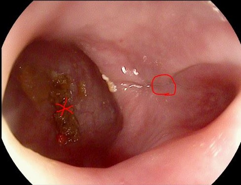
펠로우 시절에 구입한 [소화관 치료내시경 술기 아틀라스] (차재명, 정기욱 선생님 번역)을 보면, 제일 마지막이 '젠커게실의 내시경적 중격절개술' 입니다. 한다는 이야기는 들었지만, 본적은 없습니다. UpTodate 검색을 해보니 수술적인 방법과 내시경적 절제술에 대해 설명이 있지만, 구체적인 감이 잘 안옵니다. 선생님의 경험이나 의견을 바랍니다. 사진에 O 표시는 식도 lumen 이고 * 표시가 게실입니다.
[2015-3-5. 이준행 답변]
좋은 질문 감사합니다. 선생님께서 보내주신 사진에서는 septum의 endoscopic anatomy를 정확히 알기 어려워 치료계획을 확정하기 어렵습니다. Esophagography를 시행한 후 결정하셔도 좋겠습니다.
[2015-3-24. 애독자 의견]
어제 일요일 Meet the professor session에서 교수님을 오랜만에 뵙고 항상 열정적이셨던 강의를 다시 들을 수 있어서 좋았습니다. 오늘 메일은 다름이 아니라.. 몇 일전 엔도투데이 애독자가 보낸 Zenker diverticulum을 제가 나름대로 검토해 보니 사진으로 보나 위치정황으로 보나 Zenker가 아니고 Killian-Jamieson diverticulum인 것 같습니다. 내시경 사진을 보면, UES가 막 열리면서 UES직하방의 proximal cervical esophagus부위인 것 같습니다. (cf.젠커 게실은 UES직상부 또는 근접부위의 인두 점막의 게실돌출).
뒷쪽으로 빠진 것은 Zenker이고 옆으로 빠진 것은 Killian-Jamieson이라고 말씀해 주셨는데... 저널이나 해부학책을 살펴보면, UES 직상방 인지 아니면 직하방 인지가 더 중요하다고 언급되어 있습니다. 소위 경부식도괄약근 부위 또는 괄약근 직상부에서 뒷쪽으로 인두점막이 빠진 것이 Zenker이고, UES 직하방의 옆으로 식도점막이 빠진 것이 Killian-Jamieson diverticulum (proximal cervical diverticulum) 이라고 되어 있습니다. 즉, 중요한 포인트는 UES중심으로 게실위치가 인후두 쪽(즉, 엄밀히 말하면 인두점막의 게실돌출)인주 아니면 경부식도 근위부 쪽(경부 식도점막의 게실 돌출)인지 아닐까요? 왜냐하면, 이 두 게실은 증상발현이나 치료법에서 서로 많이 다르기 때문입니다 (Zenker가 훨씬 더 불편감 초래).
참고로 읽어본 저널 몇 개를 보내드립니다. 교수님께 검토를 부탁드립니다. 그럼, 큰 일교차에도 항상 건강하십시요.
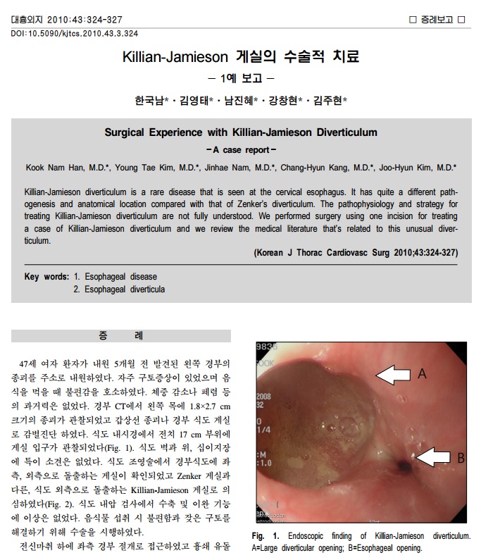
1) Killian-Jamieson 게실의 수술적 치료
2) Zenker's diverticula: pathophysiology, clinical presentation, and flexible endoscopic management.
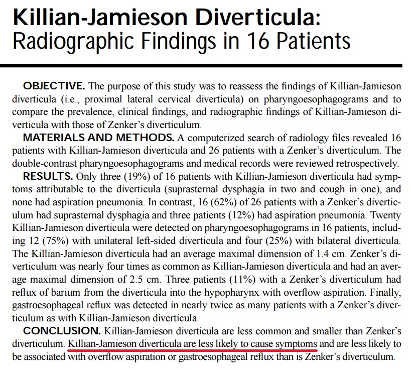
3) Killian-Jamieson Diverticula: Radiographic Findings in 16 Patients
[2015-3-24. 이준행 답변]
타당한 지적이라고 생각합니다. 일전에 Killian-Jamieson diverticulum 편에서 설명드린 바와 같이 두 게실의 가장 현저한 차이점은 Killian-Jamieson diverticulum는 cricopharyngeus의 아래 level, anterolateral aspect에 발생하고 Zenker’s diverticulum은 cricopharyngeus의 윗 level, posterior aspect에서 발생한다는 점입니다. 선생님의 지적과 저의 설명이 사실 같은 내용입니다.
일전의 애독자의 사진이 Zenker 게실이 아니라 K-J 게실인 것 같다는 선생님 말씀에 충분히 수긍합니다. 한 장의 사진만으로 구분이 쉽지 않지만... 앞으로 비슷한 환자를 보면 그냥 상부식도게실이라고 쓰지 않고 Zenker 쪽인지, K-J 쪽인지 구분하는 시도를 해 보겠습니다. 좋은 comment 감사드립니다.
![]() [References]
[References]
1) Pyriform sinus pouch/diverticulum
2) 식도 게실: Zenker diverticulum, Killian-Jamieson 게실, 중부식도 게실, 하부식도 게실
3) 위 게실
4) 십이지장 게실
5) Meckel 게실
6) 대장 게실과 게실염
7) Peroral endoscopic septotomy (POES) TOKYO LIVE 2022
8) 내시경 치료 동영상
© 일원내시경교실 바른내시경연구소 이준행. EndoTODAY Endoscopy Learning Center. Lee Jun Haeng.