EndoTODAY 내시경 교실
Beginner | ESA | Schedule | OPD
Seminars | Atlas | Recent | Links
![]() [Thursday Endoscopy Conference 20171109]
[Thursday Endoscopy Conference 20171109]
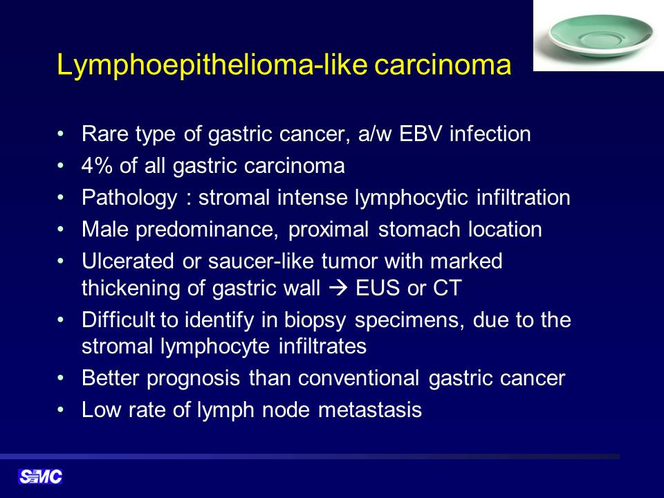
![]() 1. Lymphoepithelioma-like carcinoma
1. Lymphoepithelioma-like carcinoma
조직검사에서는 poorly differentiated adenocarcinoma였고 EUS에서 SM invasion이 뚜렷했습니다. 수술 후 최종 병리는 약간 의외로 lymphoepithelioma-like carcinoma였습니다.

tomach, subtotal gastrectomy:
. Early gastric carcinoma
1. Location : lower third, Center at proximal antrum and anterior wall
2. Gross type : EGC type IIc
3. Histologic type : lymphoepithelioma-like carcinoma
4. Histologic type by Lauren : mixed
5. Size : 1.8x1.4 cm
6. Depth of invasion : invades submucosa (sm3) (pT1b)
7. Resection margin: free from carcinoma, safety margin: proximal 1 cm, distal 8.7 cm
8. Lymph node metastasis : no metastasis in 34 regional lymph nodes (pN0)
9. Lymphatic invasion : not identified
10. Venous invasion : not identified
11. Perineural invasion : not identified
12. AJCC stage by 7th edition: pT1b N0
13. Epstein-Barr virus : positive
임상강사 백남영 선생님께서 정리한 내용입니다.
* 참고: EndoTODAY Lymphoepithelioma-like carcinoma
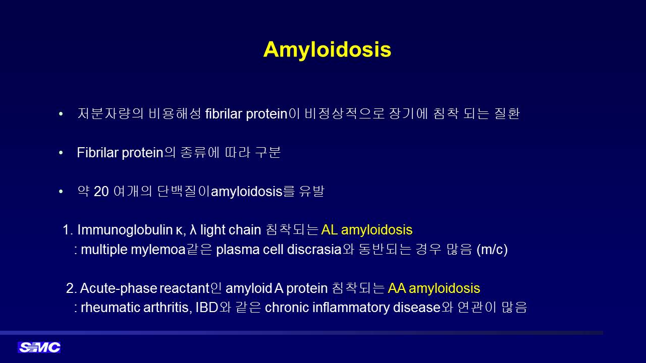
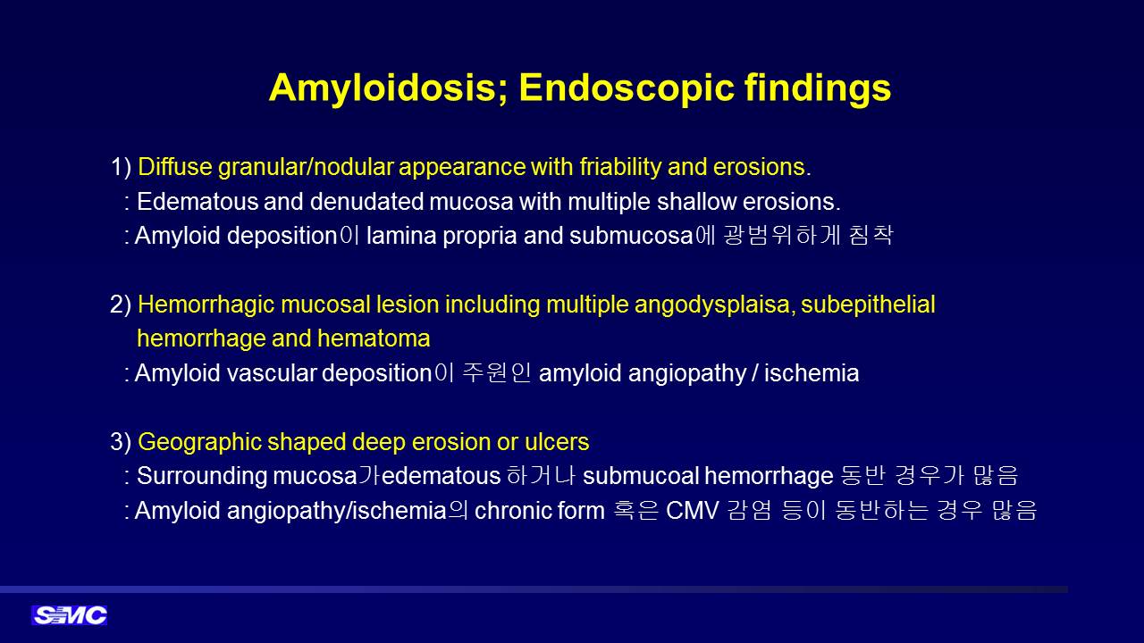
![]() 2. Isolated gastric amyloidosis
2. Isolated gastric amyloidosis
Postero-GC of high body, biopsy : Amyloidosis, Chronic gastritis, active, with inflamed granulation tissue, No H. pylori identified.
. Amyloid A: Negative
. Amyloid P: Positive
. Lambda : Positive
. Kappa : Focal positive
. TTR : Negative
전신 workup을 했으나 amyloidosis의 타 장기 침범의 증거는 없었습니다. 단독장기 침범의 amyloidosis는 예후가 좋은 것으로 되어 있어 경과관찰 하는 경우가 많지만, 이 환자는 매우 심하였고 단기 추적 내시경에서 조금 심해져 보이는 소견이 있어서 항암치료를 시작하였습니다. 아래는 일전에 임상강사 김동욱 선생님께서 정리해주신 내용입니다.
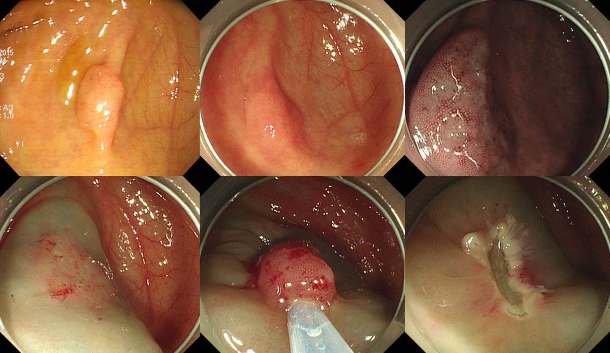
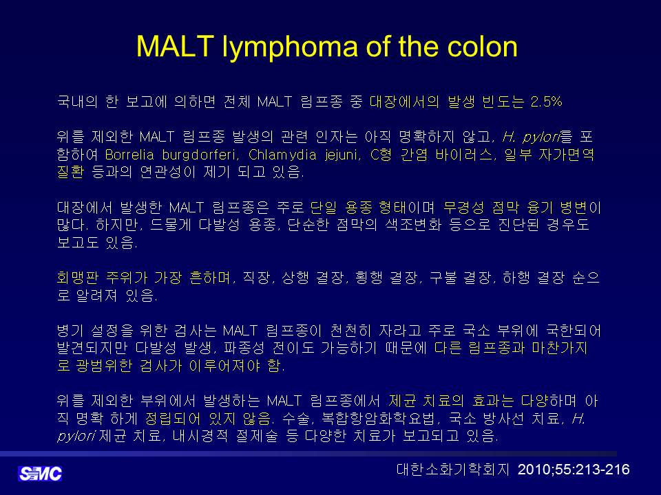
![]() 3. Cecal MALToma
3. Cecal MALToma
외부 내시경에서 cecum에 0.8cm의 flat elevated lesion으로 의뢰됨. 조직검사나 용종절제술은 시행되지 않은 상태였음.
병리결과 "dense infiltration of small lymphocytes with lymphoid follicle formation and multifocal lymphoepithelial lesions, suggestive of extranodal marginal zone lymphoa of MALT"였음. Staging workup에서 다른 부위 lymphoma는 없었음. 추가 치료 없이 6개월 후 추적내시경을 하였고 특이 소견이 없었음.
![]() 4. Lung cancer (adenocarcinoma) with anal metastasis
4. Lung cancer (adenocarcinoma) with anal metastasis
주소: anal pain, constipation
biopsy: rectal mucosa showing adenocarcinoma (M/D), EGFR: positive (1+, 1%), BFAR: not detected
Lung cancer with multiple metastasis가 있던 분으로 anal lesion에 대해서는 local RT를 추가하였습니다.
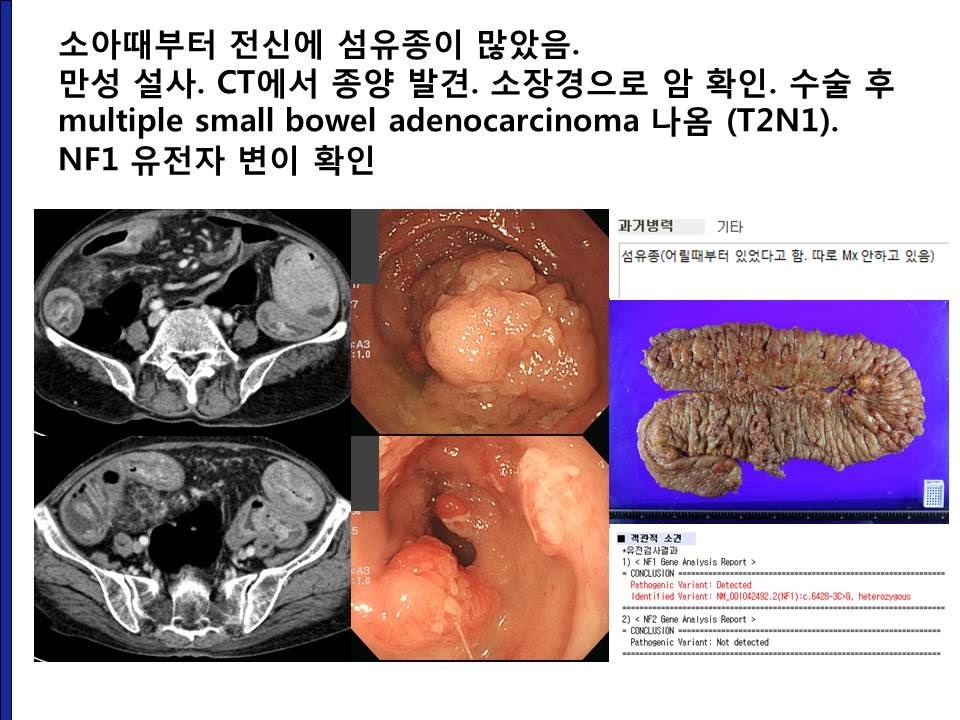
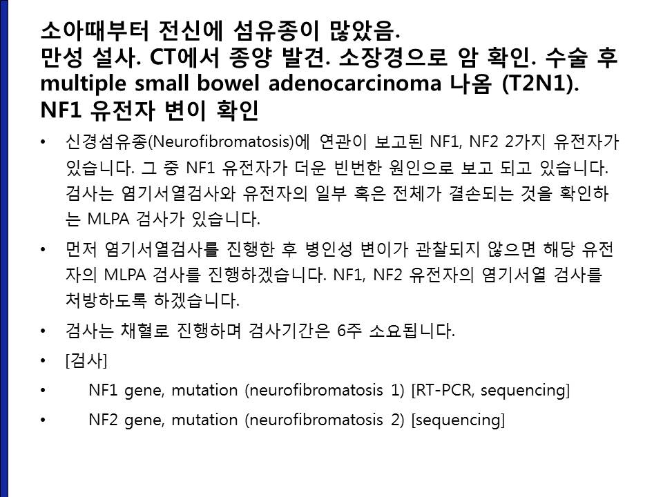
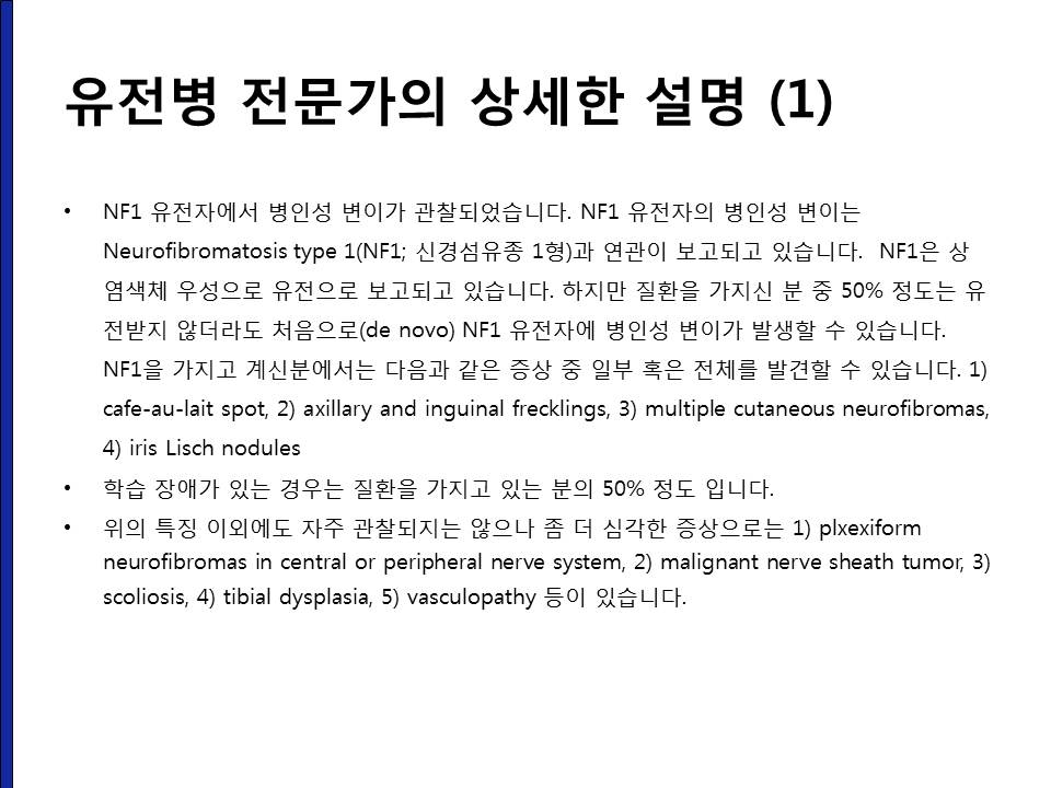
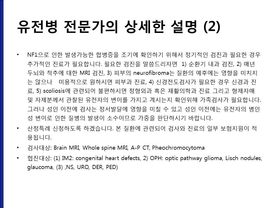
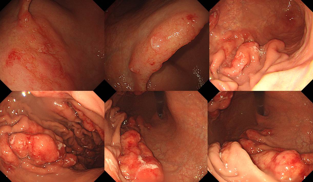
![]() 5. Neurofibromatosis with small bowel adenocarcinoma
5. Neurofibromatosis with small bowel adenocarcinoma
2015년 환자입니다. 50대 남성입니다. 설사로 workup 하여 드문 질환으로 확인되었기에 소개합니다.
![]() [References]
[References]
1) SMC Endoscopy Unit 삼성서울병원 내시경실
2) SMC Monday GI conference 삼성서울병원 일원내시경교실 월요점심소화기집담회
3) SMC Thursday endoscopy conference 삼성서울병원 일원내시경교실 목요점심내시경집담회
© 일원내시경교실 바른내시경연구소 이준행. EndoTODAY Endoscopy Learning Center. Lee Jun Haeng.