EndoTODAY 내시경 교실
Beginner | ESA | Schedule | OPD
Seminars | Atlas | Recent | Links
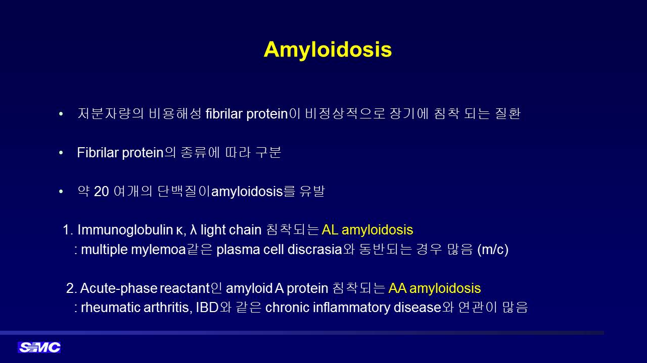
![]() [위/십이지장 아밀로이드증. Gastric and duodenal amyloidosis] - 終
[위/십이지장 아밀로이드증. Gastric and duodenal amyloidosis] - 終
표준 설명서 (환자용, 2023): 위장관 아밀로이드증은 예후가 매우 다양한 질환입니다. 위나 장에만 국한된 경우 치료없이 경과관찰을 합니다. 여러 장기 침범이 있는 경우 임파선암이나 다발성 골수종과 같은 혈액암에 준한 치료가 필요합니다. 증상없이 지내다가 부정맥 등 긴박한 상황이 발생하는 경우도 있습니다. 한마디로 향후 어떻게 될지 짐작하기 어려운 병입니다.
![]() 1. Gastric amyloidosis. 위 아밀로이도증
1. Gastric amyloidosis. 위 아밀로이도증
[증례 1]
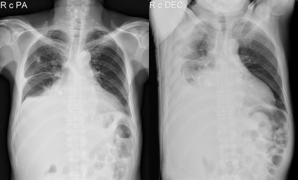
특이 병력이 없던 60세 여성으로 복부 팽만, 하지 부종, 호흡곤란으로 연고지 병원에 입원하여 검사를 받았고 위 조직검사에서 amyloidosis가 확인되었고 복부와 흉수가 있고 신장 침범이 의심된다는 소견으로 내원하였습니다. 입원하여 검사하던 중 수 일만에 cardiac arrest로 사망하였습니다. Amyloidosis의 예후는 예측할 수 없으며 언제든지 갑자기 돌아가실 수 있는 무서운 질환입니다. 특히 다장기 침범의 경우는 더욱 그러합니다.
[증례 2]
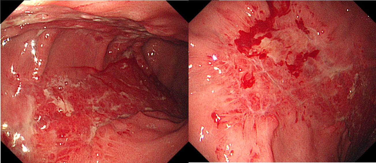
술을 많이 드시던 분으로 갑작스런 토혈로 응급실을 방문하여 내시경에서 이상한 모양의 병소가 발견되었습니다. 조직검사는 'Chronic gastritis, active, with amorphous deposit, consistent with amyloidosis. No H. pylori identified.'였습니다.
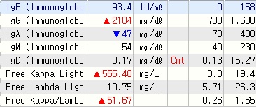
Albumin 4.2, globulin 3.6, A/G ration 1.2로 정상치를 약간 벗어나는 정도였으나, serum IgG 2104, free kappa light chain 555로 매우 높아져 있어 MM with AL amyloidosis로 진단되었음.
[증례 3]
70대 초반 여성으로 검진 내시경에서 위점막 이상소견 발견되어 조직검사를 시행하였고 amyloidosis로 진단되었습니다. 당시에는 stomach localized form으로 평가되었습니다.
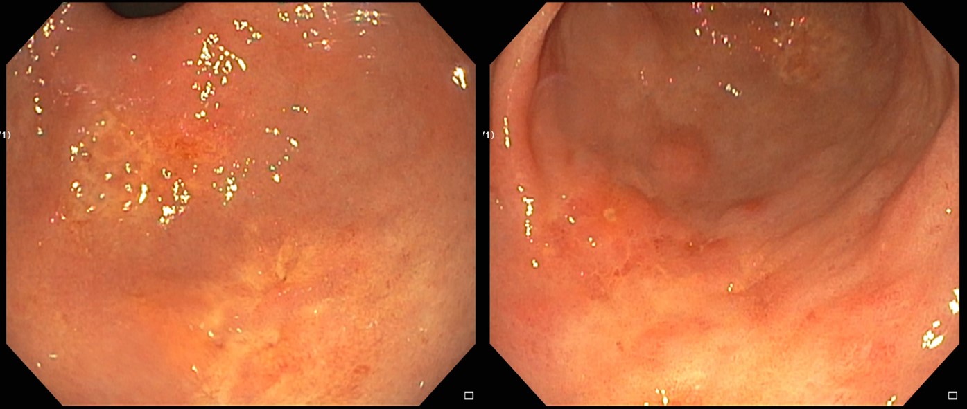
위조직검사, H&E
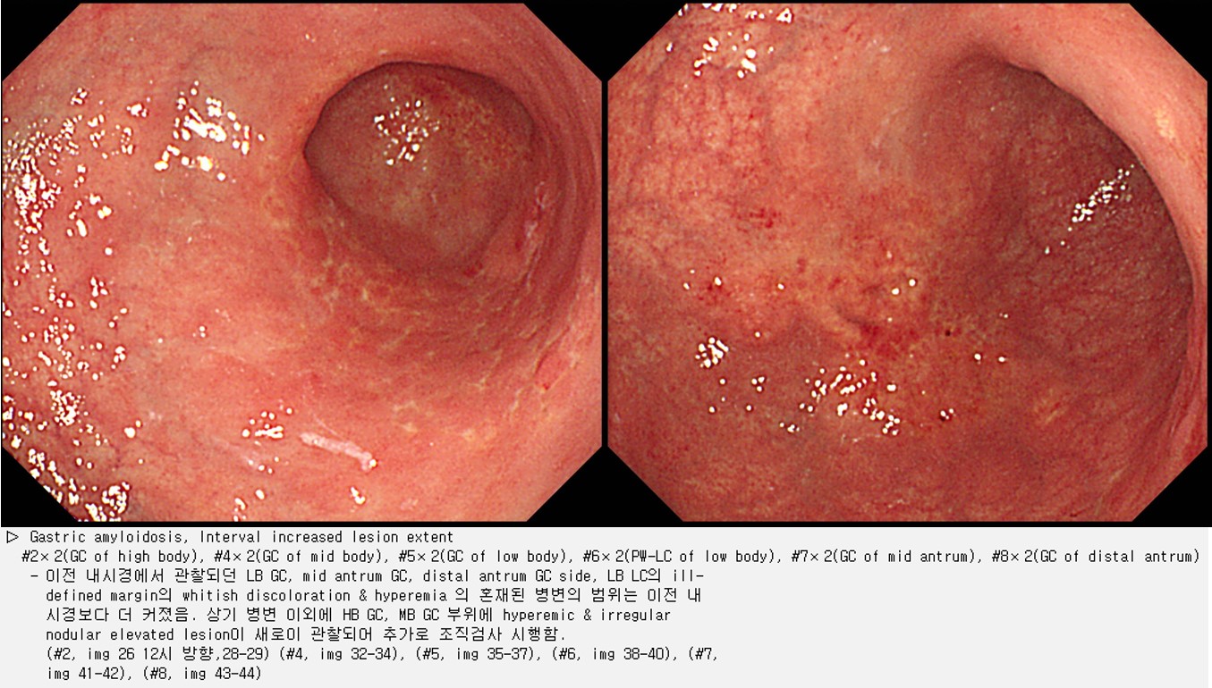
약 2년 후 weakness와 기립성 저혈압이 발생하여 다시 검사를 했습니다. 위 amyloidosis의 범위가 훨씬 넓어지고 표면 불규칙성도 심해졌습니다. 골수 조직검사와 심장 조직검사를 하였는데 모두 amyloid 침윤이 확인되었습니다. Systemic 치료를 시작하였습니다.
심장 조직검사, Congo red staining 후 일반 현미경으로 관찰하였을 때 Salmon color의 amorphous material이 잘 관찰되었습니다.
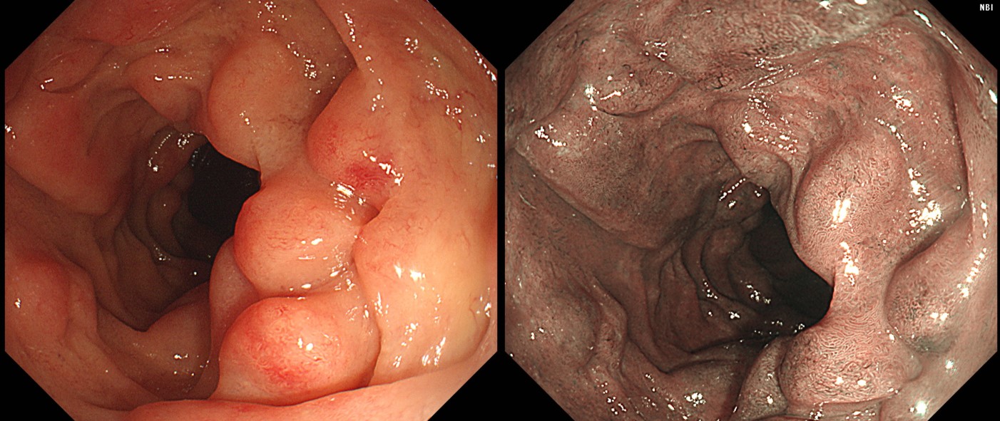
![]() 2. Duodenal amyloidosis 십이지장 아밀로이도증
2. Duodenal amyloidosis 십이지장 아밀로이도증
![]() [Brief review]
[Brief review]
2017년 6월 1일 목요집담회에서 임상강사 김동현 선생님께서 잘 정리해 주셨습니다.
![]() [Cases]
[Cases]
위 아밀로이도증. 보만 4형 진행성 위암과 감별진단에 유의해야 합니다.
베체병 환자에서 발생한 systemic amyloidosis
Localized gastric amyloidosis (2017-4-15. 순천만내시경세미나 순천플러스내과 안용환)
무증상 성인 건진에서 발견한 isolated gastric amyloidosis
Postero-GC of high body, biopsy : Amyloidosis, Chronic gastritis, active, with inflamed granulation tissue, No H. pylori identified.
. Amyloid A: Negative
. Amyloid P: Positive
. Lambda : Positive
. Kappa : Focal positive
. TTR : Negative
전신 workup을 했으나 amyloidosis의 타 장기 침범의 증거는 없었습니다. 단독장기 침범의 amyloidosis는 예후가 좋은 것으로 되어 있어 경과관찰 하는 경우가 많지만, 이 환자는 매우 심하였고 단기 추적 내시경에서 조금 심해져 보이는 소견이 있어서 항암치료를 시작하였습니다.
림프종 환자의 십이지장과 위에서 amyloidosis 확인됨.
유수현 선생님 정리
Localized asymptomatic gastric amyloisosis
Localized asymptomatic gastric amyloisosis
![]() [FAQ]
[FAQ]
[2017-11-5. 애독자 편지 (순천 플러스 내과 안용환 원장)]
교수님 안녕하셨습니까? 2017년 4월 순천만내시경세미나에서 발표했던 증례인 "localized gastric amyloidosis with kappa and lambda light chain co-expression"이 Clinical Endoscopy에 accept 되고 발표되어 이렇게 메일 보내드립니다 (Ahn. Clin Endosc 2017).
이번 증례를 준비하면서 localized gastric amyloidosis가 rare 한 질환이지만 내시경 검사에서 biopsy를 통해 확진되면 systemic 한 질환 여부를 확인하여 localized 형태로 확인 되면, EUS 또는 EMR/ESD 같은 invasive inspections을 수행하기 보다는 추적관찰을 하면서 질환의 변화를 잘 살피는 것이 중요하다는 내용을 전하고 싶었습니다.
"However, only 2% of patients with localized AL amyloidosis exhibited symptom progression, and overall survival was not different from that of the general population, except in cases involving the lungs. Localized AL amyloidosis involving the stomach was also associated with a good prognosis, and long-term outcomes were excellent. Therefore, if localized gastric AL amyloidosis is confirmed by histopathology, it is likely that invasive inspections, such as EUS, EMR, or ESD, are not necessary."
요즘은 아침에 교수님의 블로그를 읽는 것으로 하루를 시작하고 있습니다. 저는 계속 읽어와서 그런지 EndoToday Update 블로그로 바뀌고 나서 읽기가 더 편해진 것 같습니다. ^^
날씨도 추워지고, 소화기내과 의사들이 바빠지는 시기^^ 인 것 같습니다. 건강하시고, 좋은 자리에서 또 뵙도록 하겠습니다. 감사합니다.
[2017-11-7. 이준행 답변]
안용환 선생님. 증례 잘 보았습니다 (Ahn. Clin Endosc 2017). 지난 순천만세미나에서 논의되었던 내용이 더욱 보강되어 잘 정리된 것 같습니다. Discussion 참 잘 쓰셨습니다.
개업가에서도 증례에 대하여 관심을 갖고 최선의 치료법을 고민하고, 간혹 학회에서 발표도 하시는 선생님의 활동은 많은 후배들에게 모범이 된다고 생각합니다. 우리 나라에 선생님 같은 분들이 좀 더 많아지기를 바랍니다.
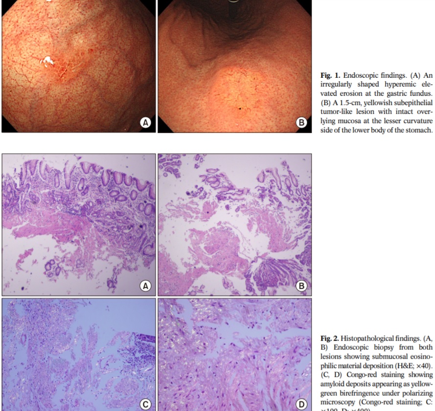
Endoscopic findings. A round lesion approximately 20 mm in diameter, with central depression and a pale-colored base on the lesser curvature of the mid-gastric body.
Histopathologic findings. (A) Congo red staining shows amorphous amyloid proteins (Congo red, ×200). (B) Apple-green birefringence is shown using polarized light microscopy after Congo red staining (Congo red, ×200).
Immunohistochemical staining shows the presence of plasma cells with co-expression of kappa (A) and lambda (B) light chains in the mucosal layer.
일전에 한 번 말씀드린 바 있으나, 전신 침범 없이 한두 장기에 국한된 localized form의 amyloidosis를 가끔 경험합니다. 대부분 예후는 좋다고 생각됩니다. 관련 문헌을 소개합니다.
"Localized deposition of amyloid may occur in individual organs, in the absence of systemic involvement. The reason for localized deposition is unknown, but it is hypothesized that deposits result from local synthesis of amyloid protein, rather than the deposition of light chains produced elsewhere. We identified 20 cases of localized amyloidosis at our institution between 1993 and 2003. There were 11 males and nine females in the group. The mean age at the time of diagnosis was 65.5 years. Organs involved included skin, soft tissues, oropharynx, larynx, lung, bladder, colon, conjunctiva, and lymph node. In six of nine patients typed, the amyloid light chain was lambda. In those patients where follow-up was available (mean 7.6 years), none developed systemic disease. Localized amyloidosis occurs in a variety of organ systems. Evolution into systemic amyloidosis was not seen in our series of patients, supporting the hypothesis of local production of amyloid protein in these cases." (Biewend ML. Amyloid 2006)
![]() [References]
[References]
© 일원내시경교실 바른내시경연구소 이준행. EndoTODAY Endoscopy Learning Center. Lee Jun Haeng.