 EndoTODAY 내시경 교실
Beginner | ESA | Schedule | OPD
Seminars | Atlas | Recent | Links
![]() [EsoTODAY 036 - Esophageal neuroendocrine carcinoma. 식도 신경내분비암]
[EsoTODAY 036 - Esophageal neuroendocrine carcinoma. 식도 신경내분비암]
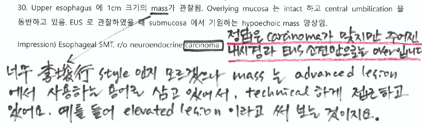
![]() [증례 1]
[증례 1]
내년도에 저희 팀에서 fellow를 하실 선생님들과 함께 내시경 description exercise 문제를 풀고 있습니다. 말하자면 내시경 선행학습니다. 그 중 한 증례를 소개합니다.
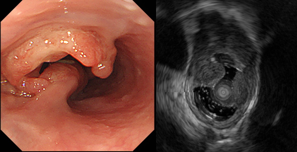
소견: 상부식도 5시 방향에 1cm 크기의 정상 점막으로 덮인 molar tooth shape의 elevated lesion이 있습니다. EUS에서 2nd lay에서 origin하는 mixed hyperechoic lesion입니다.
진단: Esophageal SET (granular cell tumor, more likely)
[이준행 comment]
식도 SET (SMT라고 불러도 좋습니다)입니다. 어금니 모양이므로 대부분 granular cell tumor라고 답하는 문제입니다. 정답은 아니지만 내시경으로는 그렇게밖에 할 수 없습니다. 조직검사가 꼭 필요한 증례입니다.
정답은 large cell neuroendocrine carcinoma였습니다. 내시경과 EUS 소견만으로 정확한 진단을 맞출 수 없는 것은 당연한 일입니다. 조직검사도 neuroendocrine tumor 인 것까지만 알려주고, neuroendocrine carcinoma인 것은 ESD 후 알게 되었습니다. 일반적인 granular cell tumor와 다른 점을 짚어본다면... 색조 정도를 말할 수 있겠습니다. Granular cell tumor는 좀 더 노랗게 비쳐 보이니까요. 한 선생님께서 neuroendocrine carcinoma를 생각하셨습니다. 정답을 제시한 점은 좋기는 한지만, 일반적인 것을 먼저 떠올리는 것이 자연스러울 것 같습니다.
조직검사에서 neuroendocrine tumor로 나와 ESD를 하여 large cell neuroendocrine carcinoma with submucosal and lymphatic invasion으로 나와 수술을 권했습니다.

1. Name of Procedure: ESD
2. Site of Tumor: Esophagus
3. Diagnosis: Large cell neuroendocrine carcinoma
4. WHO classification(2010): Neuroendocrine carcinoma
5. Multiplicity: Single
6. Size: 0.9x0.8 cm
7. Extent: Mucosa and submucosa
8. Grading: Mitotic Count: >20/10 HPF. Ki-67 labeling index: G3>20%
9. Immunohistochemical Stains - Synaptophysin : Positive, Chromogranin A: Positive, CD56: Positive, Ki-67: Positive (60%), PHH-3: Positive (198/10 HPFs)
10. Lymphovascular invasion: Present
11. Perineural invasion: Not identified
12. Resection Margins: Involved by tumor with cautery artifacts
ESD 병리. 점막층은 비교적 intact 한데 그 아래로 homogenous한 신경내분비세포가 넓게 보임.
ESD 병리. 뚜렷한 endolymphatic emboli가 관찰되었음.
수술 결과에서는 아래와 같이 lymph node metastasis가 있었습니다. 역시 수술을 보내기를 잘 했다고 생각하였습니다. 상부식도 병소인지라 환자께서 수술 후 고생을 많이 하셨습니다.
Esophagus and upper stomach, Ivor Lewis operation:
; "LRLN", 0/2; "L9", 0/1; "L10", 0/1; "RD", 0/1; "5", 0/1; "7", 0/2; "8u", 0/1)
Status post endoscopic submucosal dissection (D13-7695)
No residual tumor
1) tumor size: cannot be determined (no residual tumor)
2) depth of invasion: cannot be determined (no residual tumor)
3) endolymphatic tumor emboli: not identified
4) perineural invasion: not identified
5) resection margins: free from carcinoma, safety margin: proximal, 1.5 cm ; distal, 18 cm ;
6) metastasis to 1 out of 25 regional lymph nodes (1/25: "LC omentum", 0/0; "G1", 0/3; "G2", 0/3; "G3", 0/8; "RRLN (right recurrent laryngeal nerve)", 1/2
7) treatment effect: not applicable
수술 병리 (림프절). 림프절에 신경내분비암의 침윤이 있었음.
![]() [증례 2]
[증례 2]
Dysphagia and chest pain
Composite squamous and pooly differentiated neuroendocrine carcinoma
자료 정리: 2020년 임상강사 2년차 이동규
![]() [References]
[References]
1) EsoTODAY - Esophageal diseases
2) SmallTODAY - Small bowel diseases
3) ColonTODAY - Colorectal diseases
4) Dr. Sinn's LiverTODAY - Liver diseases
© 일원내시경교실 바른내시경연구소 이준행. EndoTODAY Endoscopy Learning Center. Lee Jun Haeng. (2017-12-24)
