EndoTODAY 내시경 교실
Beginner | ESA | Schedule | OPD
Seminars | Atlas | Recent | Links
![]() [Traction ESD. 견인 ESD] - 終
[Traction ESD. 견인 ESD] - 終
![]() 1. Introduction to traction ESD (출처: 천기누설)
1. Introduction to traction ESD (출처: 천기누설)
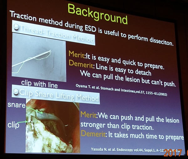
ESD는 single hand surgery입니다. Working channel을 통하여 ESD knife를 내밀어 병소를 절제하기 때문입니다. 일본에서는 두 개의 내시경을 삽입하여 시술하는 의사도 있다는 이야기를 듣기도 했지만 한 환자에게 두 개의 내시경을 삽입하는 것은 간단한 일이 아닙니다. Two hand surgery를 흉내내기 위하여 여러 시도가 있었지만 최근에는 traction method가 유용하게 사용되고 있습니다.
위각이나 위체하부 소만 병소는 ESD가 쉽지 않습니다. Submucosal dissection 과정에서 submucosal layer가 잘 노출되지 않는 경우가 많기 때문입니다. 이럴 때는 traction ESD가 딱입니다. Circumferential cutting 후 실을 연결한 clip을 병소의 proximal에 장착하고 실을 살살 당기면 submucosal layer가 아주 잘 보이게 되므로 시술이 쉬워집니다. 맨 마지막 사진을 보시면 수거한 specimen의 distal end에 clip이 장착되어 있고 실이 연결된 모습니다. 병리 결과는 아래와 같았습니다. [2019-7-14]
ESD: Early gastric carcinoma
1. Location : angle, lesser curvature
2. Gross type : EGC type IIc
3. Histologic type : tubular adenocarcinoma, moderately differentiated (70%) >> signet ring cell carcinoma (30%)
4. Histologic type by Lauren : intestinal
5. Size of carcinoma : (1) longest diameter, 10 mm (2) vertical diameter, 3 mm
6. Depth of invasion : invades mucosa (lamina propria) (pT1a)
7. Resection margin : free from carcinoma(N), safety margin : distal 8 mm, proximal 17 mm, anterior 8 mm, posterior 12 mm, deep 500 ㎛
8. Lymphatic invasion : not identified(N)
9. Venous invasion : not identified(N)
10. Perineural invasion : not identified(N)
11. Microscopic ulcer : absent
12. Histologic heterogeneity: present
위 증례와 거의 비슷한 경우입니다. Traction ESD를 시행하여 비교적 빠르고 안전하게 시술할 수 있었습니다.
ESD: Early gastric carcinoma
1. Location : high body, greater curvature
2. Gross type : EGC type IIa
3. Histologic type : tubular adenocarcinoma, moderately differentiated
4. Histologic type by Lauren : intestinal
5. Size of carcinoma : (1) longest diameter, 22 mm (2) vertical diameter, 14 mm
6. Depth of invasion : invades mucosa (muscularis mucosa) (pT1a)
7. Resection margin : free from carcinoma(N)
8. Lymphatic invasion : not identified(N)
9. Venous invasion : not identified(N)
10. Perineural invasion : not identified(N)
11. Pre-existing adenoma : none
12. Microscopic ulcer : absent
13. Histologic heterogeneity: absent
14. Associated finding: Gastritis cystica superficialis
위각부 선종 내시경 치료 후 재발로 시행한 ESD였습니다. Dual knife로 circumferential cutting을 하였고 IT-2 knife로 submucosal dissection을 시도하였으나 병소의 위치와 submucosal fibrosis로 인하여 cutting을 위한 점막하층 노출이 어려웠습니다. 치실과 clip으로 traction을 하였고 점막하층이 충분히 노출되어 훨씬 용이하게 submucosal dissection을 할 수 있었습니다. 최종 병리결과는 well differentiated tubular adenocarcinoma, 22mm, MM invasion, lateral margin (-), L/V (-/-) 로 나왔습니다. 다소 고생스러웠지만 one piece resection을 위하여 노력한 보람이 있다는 생각이 들었습니다.
![]() 2. [일본소화기내시경학회 2017 심포지엄] Traction method for ESD
2. [일본소화기내시경학회 2017 심포지엄] Traction method for ESD
발표 1. Clip-with-line method로 pharyngeal cancer를 치료할 수 있으므로 이비인후과 의사들의 Da Vinci를 이용한 로봇 수술을 일부 대체할 수 있습니다.
발표 2. Superficial pharyngeal cancer를 ESD로 치료하면서 치료효과는 좋아졌으나 시술시간이 길어지면 laryngeal edema가 문제가 되었습니다. Double scope ESD법 으로는 통상의 내시경을, 코로는 가는 내시경을 삽입하여 시술하는 방법)을 적용하여 치료시간을 단축시켜 효과적으로 치료할 수 있습니다. 평균 시술시간을 103분(conventional ESD)에서 64분(double scoep ESD)으로 줄일 수 있었다고 합니다 (2시간 이상 소요된 경우: cESD 43%, dsESD 16%).
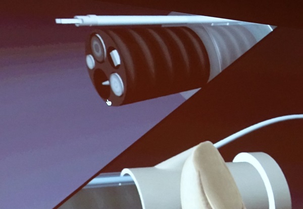
발표 3. Novel overtube with traction channel. (1) Overtube에 traction catheter를 넣을 수 있는 channel을 만들었고 (2) 회전 기능을 부여하였음 (참고; two channel endoscope에서는 회전이 불가능) . 식도 ESD 과정에서 부분적으로 절제된 병소를 잡은 후 적당히 돌려서 점막하층을 최대한 노출시킬 수 있었습니다.
발표 4. Tread traction method with Mohican strategy로 식도 ESD에 도움을 받았습니다.
발표 5. CONNETCT-G study: 위 ESD에서 dental floss clip을 사용한 ESD 법과 conventional ESD의 유용성을 비교한 다기관 전향적 연구였습니다. Dental floss clip ESD (DFC ESD) 법으로 fundus 병소의 ESD도 상당부분 가능하다고 합니다. 흥미로운 것은 primary end point가 ESD procedure time이었다는 것입니다.^^ 결과에서 procedure time은 큰 차이가 없었는데 천공률은 의미있는 차이가 있었습니다 (conventional ESD 2.2% (7/316), DFC ESD 0.3% (1/319)). Upper/middle third, greater curvature의 병소에 대해서는 DFC ESD를 적용하면 좋겠다는 연자의 의견이 있었습니다.
발표 6. Clip and snare method using pre-looping technique.
발표 7. S-O clip method. 두 clip이 연결된 형태였습니다. 하나는 부분적으로 절제된 병소에 부착시키고 다른 하나의 clip (spring이 있음)은 nylon loop를 통하고 또 다른 clip을 이용하여 정상 위벽에 부착시킵니다. ESD 과정에서 지속적으로 병소를 들어서 점막하층을 노출시키는 효과가 있습니다. S-O clip을 부착시키는데 4-5분 정도 걸리지만, 전체적인 ESD speed를 25% 정도 향상시킬 수 있다고 합니다.
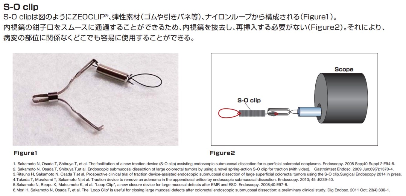
두 clip 사이에 용수철이 위치하여 지속적으로 당겨줄 수 있습니다. (참고자료 PDF)
발표 8. Clip Snare Lifting ESD
발표 9. 십이지장 ESD에서 tread traction method와 clip snare lifting method의 비교하였는데 CSL에서는 dividing이나 peeling 등이 일어날 수 있기 때문에 십이지장에서는 tread traction이 더 좋다고 합니다.
발표 10. Simplified magnetic-anchor-guided ESD (MAG ESD) (Matsuzaki I. Gastrointest Endosc 2017)
Magnetic anchor-guided endoscopic submucosal dissection for a laterally spreading tumor. A, Endoscopic image of the lesion in the cecum. B, The magnet attached to a hemoclip with thread. C, The external magnet with a flexible arm. D, After partial dissection, the mucosal edge before magnetic anchor is attached. E, Direct visualization of the submucosal layer by traction using magnetic anchor. F, Endoscopic view after endoscopic submucosal dissection, showing a large artificial ulcer.
발표 11. Ring shape tread countertraction ESD. 두 clip을 실로 연결하여 하나는 병소에 하다는 반대편 벽에 고정시키는 방법이었습니다. 발표 7 (S-O ring)과 비슷한 idea인 것 같습니다.
발표 12. S-O clip(2016년에 상용화 되었다고 합니다)의 traction 방법을 변경시킨 방법을 적용한 연구였습니다. 과거에는 oral side에 클립을 장착하였는데 이번에는 anal side에 클립을 장착하였습니다. Circumferential cutting이 끝난 직후 S-O clip을 장착하면 시술이 쉬워진다고 합니다.
발표 13. Traction assisted C-EDS (TAC ESD)에 대한 randomized controlled trial이었습니다. (오사카의 Uedo 선생님 팀)
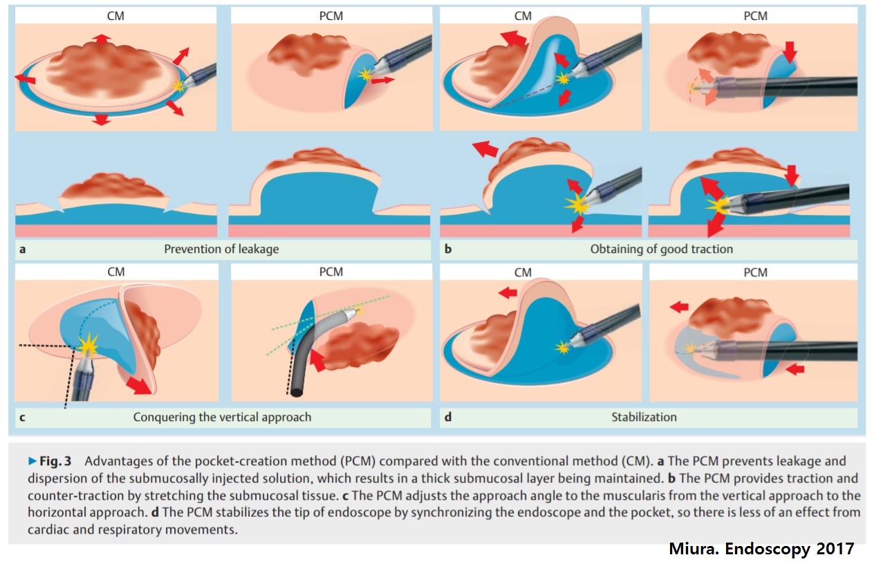
발표 14. Pocket creation metod (PCM). Conventional ESD에서는 flap을 만들지만, PCM에서는 flap을 만들지 않는 일종의 traction method입니다. 다만 long tapered hood (Short ST hood, Fujifilm)을 이용하여야 효과적입니다. 연자는 Fujifilm 내시경과 Flush knife (Fujifilum)와 Clutch cutter (Fujifilm)을 사용하였습니다. Submucosal fibrosis가 있어도 효과적으로 치료할 수 있다고 합니다. PCM은 POEM과 유사하므로 서구에서 ESD를 배우는 방법으로 좋을 것 같다는 제안이 있었습니다.
[이준행 혼잣말] Traction ESD라는 제목의 2시간 30분짜리 독립 세션이었습니다. 각 병원에서 서로의 방법을 약간씩 변형하여 나름대로 새로운 방법을 개발하고 있다는 점에서 일본 내시경계의 저력이 느껴졌습니다.
![]() 3. 대장내시경에서 traction을 적용하기 위한 여러 방법
3. 대장내시경에서 traction을 적용하기 위한 여러 방법
Double balloon endoluminal platform (Endosc Int Open 2020;8(10):E1273)
![]() 4. EndoGEL ESD hands-on training에서 사용하고 있는 traction method
4. EndoGEL ESD hands-on training에서 사용하고 있는 traction method
EndoGEL ESD hands-on training에서는 중력을 이용할 수 없으므로 (1) cap을 적용하거나 (2) traction-assisted ESD가 필요합니다. 회사에서는 cap을 적용한 방법을 추천하고 있으나 저는 traction ESD 방법을 개발하여 사용하고 있습니다.
A small flap was made by the partial submucosal dissection
This is a home-made traction device.
A small metal clip, which I bought at the Office Depot, was linked to the weight with a dental floss.
The traction device was applied the small flap. Now, you can see the submucoal layer with parallel fibers.
The submucosal layer can be dissected by the right to left movement of the Dual knife tip. No submucosal injection is required for the EndoGEL ESD training.
The traction was adjusted to make a better view.
We are using a stand for a more realistic traction. I need to find a good pully for this purpose.
Koken simulator에서 EndoGEL ESD를 하기 위한 platform을 개량하였습니다. (2020-4-14)
종이 box에 EndoGEL을 위치시킨 후 전벽, 후벽, 천장, 바닥과 같이 여러 방향으로 돌려가며 ESD 연습을 할 수 있습니다. Traction을 창의적으로 적용해야 합니다.
EndoGEL ESD hands-on training을 위한 작전 계획. 자체 제작한 traction 추를 이용한 needle type knife ESD를 시행하기로 하였습니다.
EndoGEL ESD recent setting (2020년 6월)
![]() [References]
[References]
1) Traction ESD (VOD. Abe. IDEN 2021)
2) 상부 ESD 에서 견인(traction)의 응용 윤영훈. 2022-10-4. KSGE webinar. log-in 要
3) 하부 ESD 에서 견인(traction)의 응용 양동훈. 2022-10-4. KSGE webinar. log-in 要
4) [2026 HUG 교육위원회 자료] Endoscopic submucosal excavation with internal traction 부산대 김수진
© 일원내시경교실 바른내시경연구소 이준행. EndoTODAY Endoscopy Learning Center. Lee Jun Haeng.