 EndoTODAY 내시경 교실
Beginner | ESA | Schedule | OPD
Seminars | Atlas | Recent | Links
![]() [GERD TODAY 032. Esophageal parakeratosis] - 終
[GERD TODAY 032. Esophageal parakeratosis] - 終
[2019-11-19. 애독자 질문]
BMI 30 정도인 젊은 분 검진 위내시경입니다.
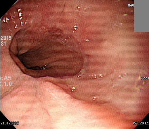
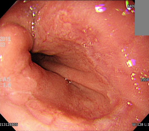
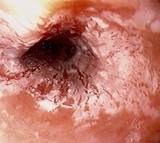
EGJ 1시 방향 erosion이 단순한 reflux esophagitis (LA-A)로 보기엔 뭔가 옆으로 infiltrative해 보이고 색조도 불균질하며 조직검사를 했습니다. 결과는 아래와 같았습니다
Tissue from gastro-esophageal junction, endoscopic biopsy ;
1. Esophageal mucosa, with 1) xanthoma, focal, 2) chronic active inflammation, 3) reactive basal cell hyperplasia, 4) parakeratosis, 5) some eosinophils infiltration ( 10 - 15 / 40 HPF)
2. Gastric mucosa, with 1) chronic inflammation, 2) regenerative glands
Note : The possibility of reflux esophagitis with focal xanthoma, reactive basal cell hyperplasia and parakeratosis is suggested. With treatment of inflammation, follow up biopsy is recommended.GERD에서 eosinophil infiltration은 흔한 소견이지만 parakeratosis가 좀 낯선 용어였습니다. Uptodate에서 esophageal parakeratosis 로 나온 사진은 사뭇 다른 양상이고 text로 나온 관련 내용은 아래와 같습니다.
Parakeratosis - Esophageal parakeratosis appears endoscopically as whitish, membranous, linear plaques that do not stain (turn brown) when sprayed with Lugol's solution. Biopsies reveal epithelial acanthosis, basal hyperplasia, and a dense, compact layer of parakeratosis, often featuring cytoplasmic eosinophilia and pyknotic nuclei, covered by an outer layer of nonnucleated squamous cells.
The clinical significance of esophageal parakeratosis is unclear; associations with esophageal and head and neck carcinoma have been reported, but the areas of parakeratosis themselves have not been shown to give rise to neoplasia. As an example, a prospective study of 400 patients with newly diagnosed squamous cell cancers of the head and neck found that close to 40 percent of patients had esophageal parakeratosis but none of the cancers clearly arose from such areas. Another report described an association with submucosal fibrosis of the oral cavity, particularly in smokers and those who chewed betel nuts. Because of the association with squamous cell cancer, we suggest careful evaluation of the esophagus and head and neck.
EGJ에서 parakeratosis가 상당히 심심찮게 나오는 소견인지, 또 parakeratosis의 임상적 의미 그리고 비교적 젊은 사람한테서 이런 소견이 나왔을 때 향후 f/u이나 cancer risk등에 대해서는 어느 정도 설명해주는게 좋을지 교수님의 고견을 여쭤봅니다.
[2019-11-19. 이준행 답변]
일전에 본 병원에서도 식도의 focal lesion처럼 보였던 parakeratosis를 경험한 바 있지만 (Dig Endosc 2012) 흔한 소견은 아닙니다.
내시경에서는 white plaque처럼 보이는데 전암성 병소로 생각되지는 않습니다. 따라서 암발생 가능성을 설명할 필요는 없겠고 나중에 추적검사 정도 한번 해 보시면 될 것 같습니다.
보내주신 증례 사진은 젊은 비만 환자에서 흔한 hiatal hernia와 이로 인한 SCJ blurring과 하부식도 squamous epithelial hyperplasia입니다. 1시 방향의 불규칙한 점막이 약간 걱정되기는 하지만 조직검사에서 나온 바와 같이 염증성 변화입니다. Definite한 mucosal break는 없어 보입니다 (12시 30분 방향의 가는 선 모양이 다소 애매하기는 하지만 longitudinal erosion은 아닌 것 같습니다).
조직검사에서 parakeratosis라고 주신 것은 정말 parakeratosis인지 acanthotic squamous epithelium의 hyperplasia인지 병리과 선생님과 다시 한번 상의해 보면 좋겠습니다. 왜냐하면 내시경 소견은 전혀 parakeratosis 같지 않거든요.
여하튼 결론은 비만 환자의 hiatal hernia와 그로 인한 EG junction 주변의 염증 소견이며 parakeratosis라는 병리소견이 보인 것은 다소 의외이며 임상적 의의는 거의 없을 것 같습니다. 체중 감량이 중요할 것 같습니다.
![]() [References]
[References]
3) EndoTODAY 위식도역류질환에 대한 흔한 질문
![]() © 일원내시경교실 바른내시경연구소 이준행. EndoTODAY Endoscopy Learning Center. Lee Jun Haeng. (since 1999-8-23)
© 일원내시경교실 바른내시경연구소 이준행. EndoTODAY Endoscopy Learning Center. Lee Jun Haeng. (since 1999-8-23)