 EndoTODAY 내시경 교실
Beginner | ESA | Schedule | OPD
Seminars | Atlas | Recent | Links
![]() [일원내시경교실 목요점심집담회 2016-3-31]
[일원내시경교실 목요점심집담회 2016-3-31]
![]() 1. Pathology of the ischemic colitis
1. Pathology of the ischemic colitis
목요집담회에서 ischemic colitis 증례가 있었습니다. 이에 대한 병리 소견을 김순영 선생님께서 정리하셨습니다.
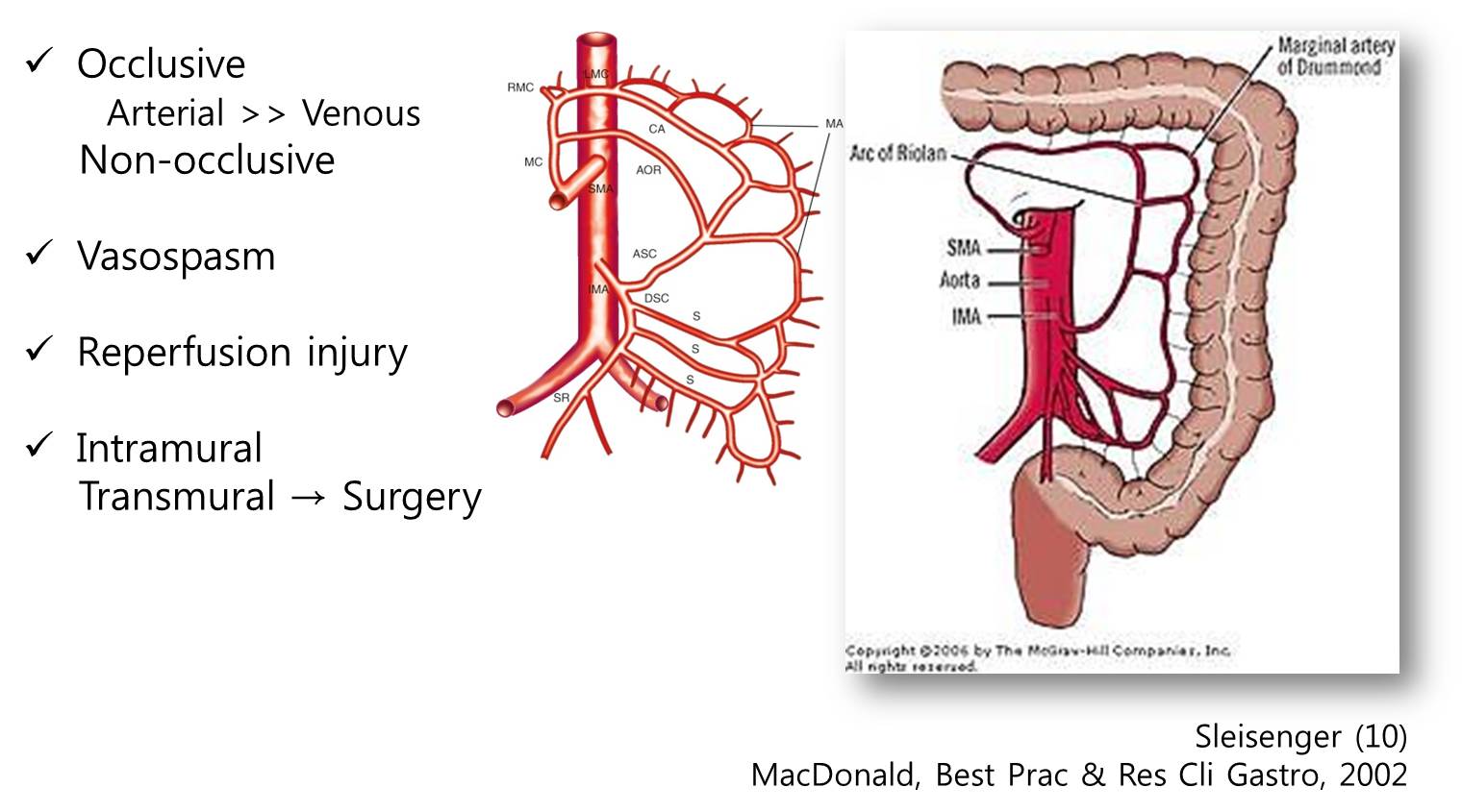

Ischemic colitis (H&E). Necrosis of superficial crypts with viable crypt bases is present (arrows). The changes are pauciinflammatory and characteristic hyalinization of the lamina propria with congestion and red cell extravasation (arrowheads) is seen. There is edema of the submucosa
![]() 증례 2. Herpetic esophagitis (s/p total gastrectomy due to gastric cancer)
증례 2. Herpetic esophagitis (s/p total gastrectomy due to gastric cancer)
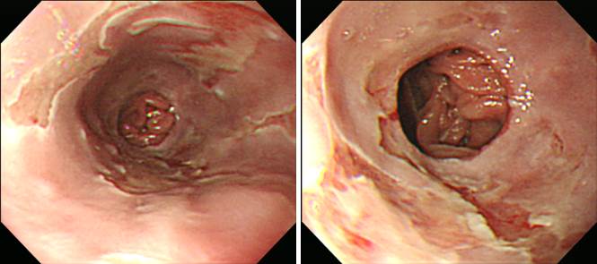
Sleisenger 책에서 옮깁니다. "The endoscopic appearance is characterized by diffuse friability; ulceration; and exudates, mostly in the distal esophagus. Classically, the earliest esophageal lesions are rounded 1- to 3-mm vesicles in the mid to distal esophagus, the centers of which slough to form discrete circumscribed ulcers with raised edges. These lesions can also be appreciated radiographically."
* 참조: EndoTODAY 바이러스 식도염
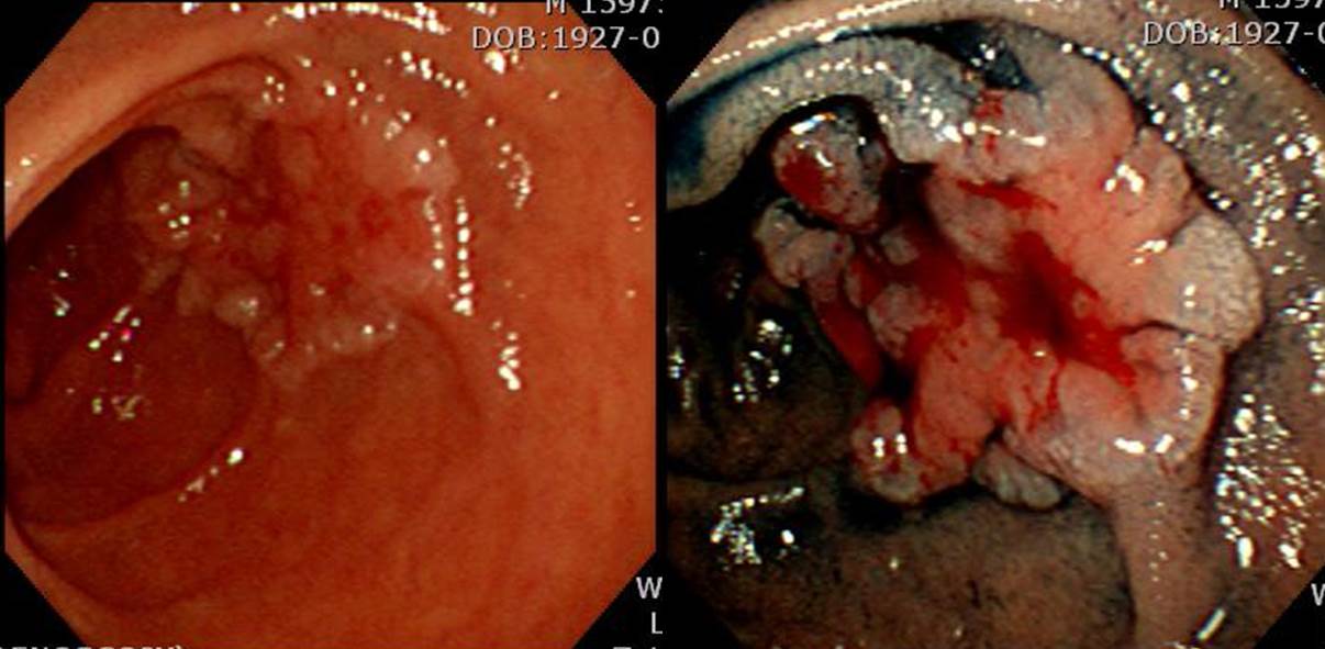
![]() 증례 3. Atypical regenerating glands
증례 3. Atypical regenerating glands

첫 조직검사가 H. pylori (+) gastritis, active with ulcer and atypical regenerating glands로 나왔지만 제균치료와 궤양치료 후 호전되었고 장기 경과관찰에서도 문제가 없었습니다. Atypical regenerating glands는 간혹 암일 수도 있지만 다수는 이와 같이 양성 질환으로 나옵니다. 여하튼 주의해야 합니다.
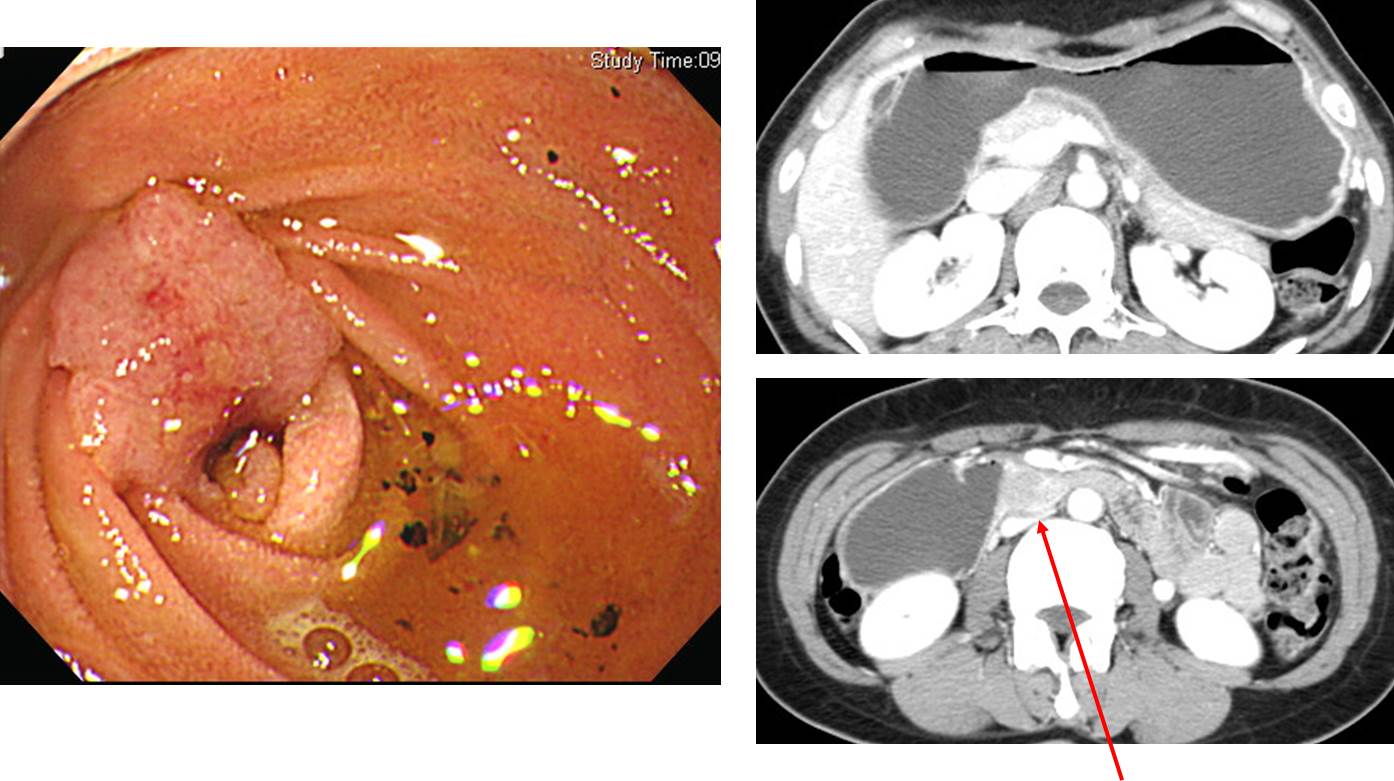
![]() 증례 4. Duodenal adenocarcinoma
증례 4. Duodenal adenocarcinoma

외부 내시경에서 "duodenal 3rd part ulcer and stricture (biopsy: atypical glands)" 소견으로 의뢰된 분으로 내시경 재검에서 M/D adenocarcinoma로 수술한 증례입니다. 내시경 소견과 수술 병리를 소개합니다.
내시경 소견: Duodenal 2nd to 3rd portion부위에 lumen을 encircling하는 mass 관찰되었으며 소아용 대장 내시경을 이용하여 진입 시도하였으나 mass의 위치가 angulation 직후이고 lumen이 좁아져있어 scope이 통과되지 못했습니다. 위내시경으로 바꾸어 try했으나 최대한 삽입해도 mass의 입구까지 밖에 도달하지 못하여, 다시 소아용 대장내시경에 cap을 장착후 try했으나 역시 진입불가능하여 최대한 forcep을 밀어넣어 조직검사를 시행했습니다.
DUODENAL CANCER
1. Type of specimen : PPPD
2. Histopathologic Diagnosis : Adenocarcinoma, moderately differentiated
(1) Tumor site : duodenal
(2) Tumor size : 4.5x4 cm
(3) T3 : Tumor invades through the muscularis propria into the subserosa
(4) Involvement of pancreas: absent
(5) N1 : Regional lymph node metastasis (2/16: "peri-SMA tissue" for frozen section-1, 0/1; "LN8,12", 0/6; "periCBD&peripancreatic LN", 1/3; periduodenal, 1/2; peripancreatic, 0/4)
(6) M0 : No distant metastasis
(7) Negative (pancreas, common bile duct, retropancreatic) resection margins
상부위장관 내시경으로 도달할 수 있는 부위에 위치한 십이지장암 몇 증례를 소개합니다.
초고령으로 수술을 못하시고 1년 조금 넘어 obstruction 증상 발생
Partial resection으로 치료하였고 점막에 국한된 0.6cm 암이었습니다. 최근 같아서는 내시경 절제술로 치료할 수 있다고 생각됩니다.
M/D adenocarcinoma, 3.5 x 3 cm, extension to subserosa, metastasis to 3 out of 10 regional lymph nodes
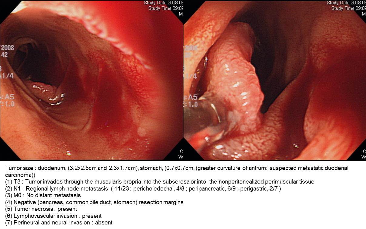
Duodenal cancer (M/D adenocarcinoma, 4 x 2 cm, extension to periduodenal soft tissue and pancreas, LN 1/34) + stomach cancer
Obstruction (+), hepatic metastasis (+)
Poorly differentiated carcinoma
Mucinous adenocarcinoma, 5 x 5 x 3 cm, directly invades other organs (pancreas), involvement of vessel (SMA and SMV: op. record without histologic evaluation), regional lymph node metastasis (13/16)
O & C due to SMA, pancrease, stomach metastasis
Bowel habit change로 대장내시경에서 우측 대장암이 발견되었고 우연히 시행한 위내시경에서 duodenal adenocarcinoma도 함께 진단되었음. Whipple 수술과 right hemicolectomy를 동시에 시행하였음.
Duodenal papillary adenocarcinoma
Duodenal signet ring cell carcinoma
Depressed type FAP-associated early duodenal carcinoma
© 일원내시경교실 바른내시경연구소 이준행