 EndoTODAY ГЛНУАц БГНЧ
Beginner | ESA | Schedule | OPD
Seminars | Atlas | Recent | Links
![]() [РЯПјГЛНУАцБГНЧ ИёПфСЁНЩС§ДуШИ 2016-4-7]
[РЯПјГЛНУАцБГНЧ ИёПфСЁНЩС§ДуШИ 2016-4-7]
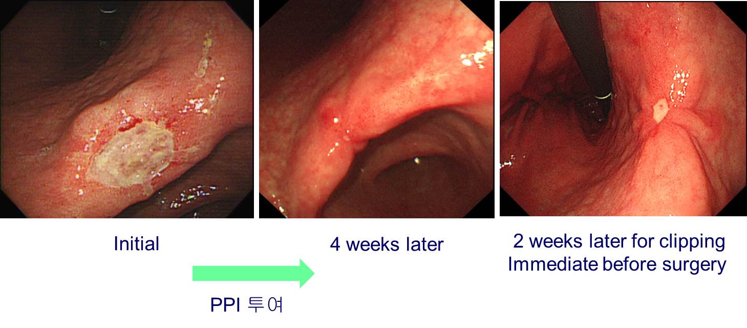
![]() 1. Change of EGC morphology probably by PPI
1. Change of EGC morphology probably by PPI
РЇАЂРЧ ЧдИєЧќ КДМвПЁМ СЖСїАЫЛчЧЯПЉ atypical cellРЬ РжДйАэ РЧЗкЕЧОњНРДЯДй. АЫЛч СїШФКЮХЭ PPIИІ ЕхНХ АЭ ААНРДЯДй. Ор 4Сж ШФ ГЛНУАцПЁМ БЫОчЧќ КДМвДТ АХРЧ ОЦЙАОю РжОњНРДЯДй. ПмКЮ СЖСїАЫЛч РчЦЧЕЖАњ КЛ КДПјРЧ ГЛНУАц СЖСїАЫЛч РчАЫПЁМ И№ЕЮ ОЯРИЗЮ ГЊПЭ МіМњРЛ ЧЯПДНРДЯДй. КЛ КДПјПЁМДТ PPIИІ ЕхИЎСі ОЪОвАэ Ор 2Сж ШФ МіМњЧЯПДНРДЯДй. МіМњ РќГЏ clippingРЛ РЇЧЯПЉ ГЛНУАц РчАЫРЛ ЧЯПДНРДЯДй. 3ЙјРЧ ГЛНУАц ЛчСјРЛ Рп КИНУБт ЙйЖјДЯДй. СЖБтРЇОЯРК natural historyПЁ РЧЧЯПЉ И№ОчРЬ КЏЧЯБтЕЕ ЧЯСіИИ PPI Ею ОрСІПЁ РЧЧб ПЕЧтЕЕ ЛѓДчЧеДЯДй.
Early gastric carcinoma
1. Location : lower third, Center at body and lesser curvature
2. Gross type : EGC type IIc and IIa
3. Histologic type : tubular adenocarcinoma, moderately differentiated
4. Histologic type by Lauren : intestinal
5. Size : 3.9x2.5 cm
6. Depth of invasion : invades mucosa (muscularis mucosae) (pT1a)
7. Resection margin: free from carcinoma, safety margin: proximal 3.5 cm, distal 6.3 cm
8. Lymph node metastasis : no metastasis in 65 regional lymph nodes (pN0)
9. Lymphatic invasion : not identified
10. Venous invasion : not identified
11. Perineural invasion : not identified
12. AJCC stage by 7th edition: pT1a N0
РЇОЯРЧ РкПЌЛчИІ КИПЉСи АњАХ СѕЗЪИІ МвАГЧеДЯДй.
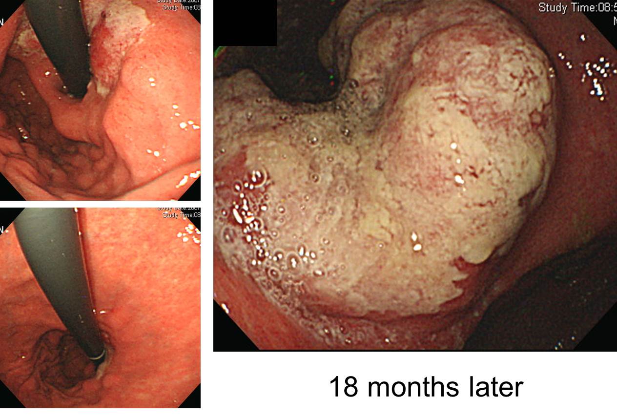
70Ды ГВРкРдДЯДй. CardiaИІ involveЧЯДТ РЇОЯРИЗЮ МіМњРЛ БЧЧЯПДРИГЊ МіМњЧЯСі ОЪАэ АцАњАќТћРЛ ПјЧЯНУОю follow-up lossАЁ ЕЧОњДйАЁ 1До РќКЮХЭ dysphagiaАЁ РжОю РЧЗкЕШ ШЏРкРдДЯДй. 18 АГПљ Рќ ЛчСјРЛ КИИщ КДМвРЧ distal marginРК upper bodyПЭ mid-bodyРЧ АцАш СЄЕЕПДРИГЊ УжБй ГЛНУАцПЁМДТ cardia involveАЁ РжОњАэ (ЛчСјЛ§ЗЋ), angleБюСі КДМвАЁ ГаОюСГНРДЯДй. КёБГРћ КќИЅ МгЕЕРЧ progressionРЛ КИРЮ СјЧрМКРЇОЯРЬОњДј АЭРИЗЮ ЦЧДмЧЯПДНРДЯДй.
АњАХПЁДТ АэЗЩРЧ ШЏРкЕщПЁАд МіМњРЛ БЧЧЯДТ АЭРК ИХПь ОюЗЦАэ РЇЧшЧб РЯЗЮ АЃСжЕЧОњНРДЯДй. БзЗЏГЊ УжБйПЁДТ АэЗЩРЧ ШЏРкЖѓАэ ЧЯДѕЖѓЕЕ performanceАЁ ССРК АцПьДТ РћБиРћРЮ ФЁЗсИІ БЧЧЯДТ АЭРЬ КИХыРдДЯДй. МіМњПЁ ЕћИЅ РЇЧшМКРК НЧСІ ПЌЗЩКИДйДТ cardiopulmonary functionПЁ РЧЧЯПЉ АсСЄЕЧДТ АЭРИЗЮ Л§АЂЕЧБт ЖЇЙЎРдДЯДй. МіМњРЬ АЁДЩЧб СјЧрМКРЇОЯ ШЏРкПЁМ УЪАэЗЩ ШЏРкЖѓАэ ЧЯДѕЖѓЕЕ СпГт ШЏРкПЁ КёЧЯПЉ МіМњПЁ ЕћИЅ morbidityПЭ mortalityАЁ РЏРЧЧб ТїРЬАЁ ОјДйДТ АЭРК ПЉЗЏ КИАэПЁМ ЙнКЙРћРИЗЮ ШЎРЮЕЧАэ РжНРДЯДй.
ОюЖВ МБЛ§ДдРК РЬЗБ ИЛОИРЛ ЧЯМЬНРДЯДй. "ГЊРЬДТ ДмСі М§РкРЯ ЛгРЬДй."
![]() 2. PylorusПЁ ДъОЦРжДТ СјЧрМК РЇОЯ
2. PylorusПЁ ДъОЦРжДТ СјЧрМК РЇОЯ

Advanced gastric carcinoma
1. Location : [1] lower third, [2] duodenum, Center at antrum and lesser curvature
2. Gross type : Borrmann type 2
3. Histologic type : tubular adenocarcinoma, poorly differentiated
4. Histologic type by Lauren : diffuse
5. Size : 4x3.1 cm
6. Depth of invasion : invades muscularis propria (pT2)
7. Resection margin: free from carcinoma
8. Lymph node metastasis : metastasis to 5 out of 30 regional lymph nodes (pN2) (5/30: "1", 0/4; "3", 1/10; "4", 1/2; "4sb", 0/1; "5", 1/1; "6", 1/4;
"8a", 0/3; "7", 0/2; "9", 0/0; "11p", 0/2; "12a", 1/1)
9. Lymphatic invasion : present
10. Venous invasion : not identified
11. Perineural invasion : not identified
12. Peritoneal cytology : negative
13. AJCC stage by 7th edition: pT2 N2
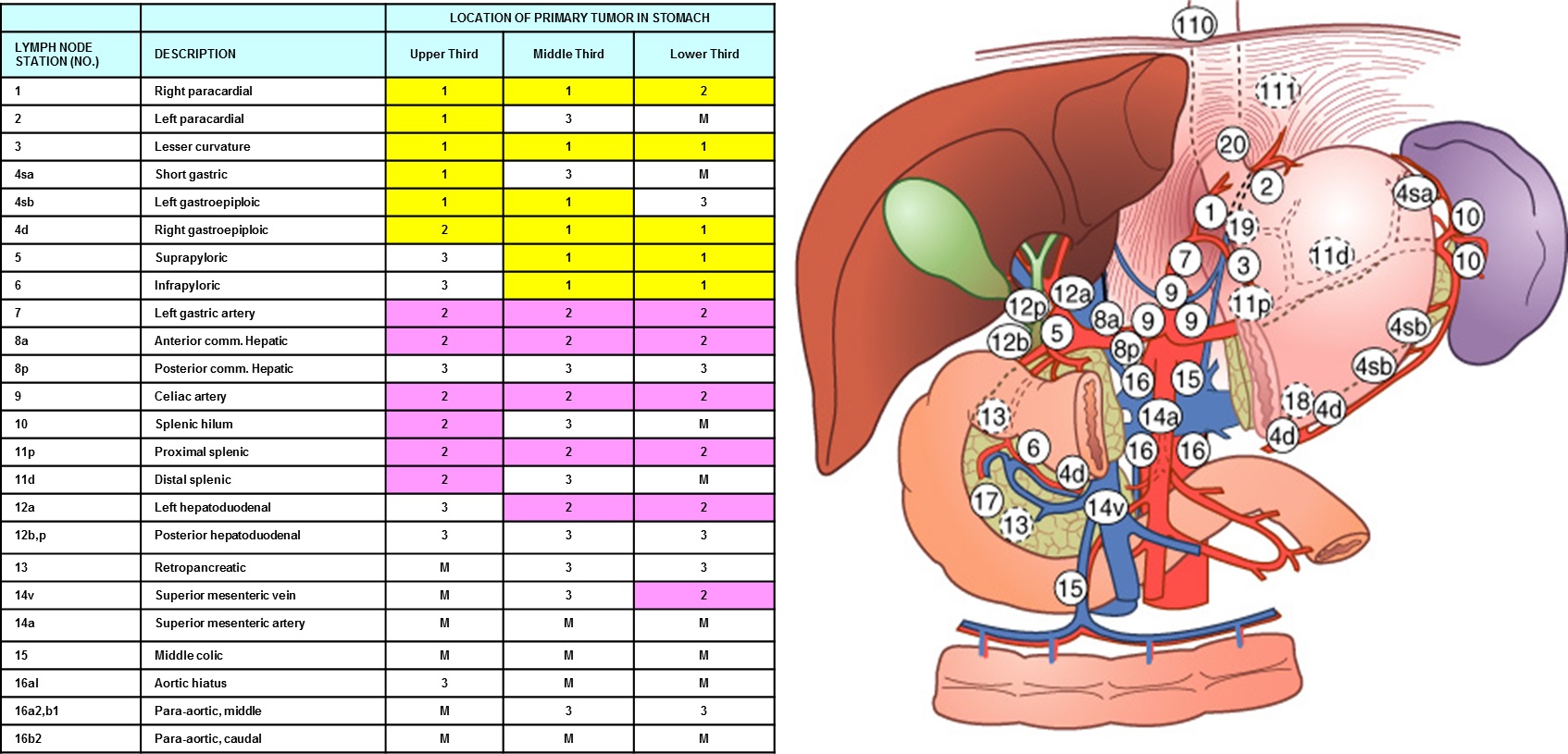
Л§АЂКИДй ИВЧСР§ РќРЬАЁ ЧіРњЧпНРДЯДй. 3Йј, 4Йј, 5Йј, 6Йј, 12aЙјПЁ АЂАЂ РќРЬЕЧОю РжОњНРДЯДй. ОЦЗЁ lymph node stationРЛ ТќСЖЧЯНУБт ЙйЖјДЯДй.
РЇ ИВЧССОРК АЃШЄ Borrmann type IIПЭ БИКаРЬ ОюЗЦНРДЯДй. РЬ ШЏРкДТ gastric biopsyПЁМЕЕ diffuse large B cell lymphomaАЁ ГЊПдНРДЯДй.
* ТќАэ: EndoTODAY diffuse large B cell lymphoma
Stomach, endoscopic submucosal dissection:
2014Гт СѕЗЪРдДЯДй. LUQ pain, hematochezia, weight loss 3~4kg/10days ЕюРИЗЮ CTИІ НУЧрЧЯАэ "colon(splenic flexure) wall thickening with omentum invasion РжОю r/o colon ca, r/o metastatic cancer" МвАпРИЗЮ РЧЗкЕЧОњНРДЯДй. ДыРхГЛНУАцАњ CT ЦЧЕЖРЛ ЧЯПДНРДЯДй. CT ЦЧЕЖРЬ ИХПь ШЧИЂЧпНРДЯДй.
CT ЦЧЕЖ: Left sideРЧ gastrocolic ligamentИІ ЕћЖѓМ irregular enhancing massАЁ РжРН. АцАшАЁ СССі ОЪОЦ ХЉБтРЧ УјСЄПЁ СІЧбРЬ РжРИГЊ 6 cm РЬЛѓРЧ extentИІ КИРг. РЬ КДКЏРК ЛѓЙцРИЗЮ stomachРЧ body greater curvature sideПЭ ЧЯЙцРИЗЮДТ transverse colonРЧ upper wallБюСі extensionЧЯАэ РжРН. InvolvementЕШ bowel wall thickeningРЬ РжРИГЊ КёБГРћ layeringРЬ РЏСіЕЧАэ РжРН. БзЗЏГЊ transverse colonПЁМДТ focalЧЯАд layeringРЬ РЏСіЕЧСі ОЪДТ КЮКаРЬ РЧНЩЕЪ. Left side mesorectumПЁ Ор 3.3 cm sizeРЧ enhancing massАЁ РжАэ ПЊНУ АцАшАЁ СССі ОЪРИИч СжКЏРИЗЮ infiltrationРЛ ЕПЙнЧЯАэ РжРН. RectumРЧ left side wallАњ abuttingЧЯАэ РжРИИч РЮСЂЧб rectal wallПЁ wall thickeningРЬ РжАэ ПЊНУ layeringРЬ РЏСіЕЧАэ РжРН. Pelvic cavityПЁ МвЗЎРЧ fluid collectionРЬ РжРН. UterusПЁ IUD insertion stateРг. КЙА ГЛ РЧЙЬРжАд ФПСј lymph node КИРЬСі ОЪРН. Бз Пм liverПЭ spleen, pancreas, both kidneysПЁ ЦЏРЬМвАп ОјРН. GBПЁ ЦЏРЬМвАп ОјАэ biliary tree dilatation ОјРН. ScanПЁ ЦїЧдЕШ basal lungАњ boneПЁ ЦЏРЬМвАп ОјРН.
Transvaginal biopsyИІ НУЧрЧЯПДАэ ДйРНРЧ АсАњПДНРДЯДй. Inflamed granulation tissue with abscess and dense fibrosis. Bacterial colony present, consistent with actinomycosis
Treatment: IV penicillin G
2017Гт 3Пљ ГЛНУАцЧаШИ БГРАРкЗсАЁ КЙКЮ ЙцМББеСѕ (actinomycosis) РЬОњНРДЯДй.
1) SMC Endoscopy Unit ЛяМКМПяКДПј ГЛНУАцНЧ
2) SMC Monday GI conference ЛяМКМПяКДПј РЯПјГЛНУАцБГНЧ ПљПфСЁНЩМвШБтС§ДуШИ
3) SMC Thursday endoscopy conference ЛяМКМПяКДПј РЯПјГЛНУАцБГНЧ ИёПфСЁНЩГЛНУАцС§ДуШИ
© EndoTODAY Endoscopy Learninng Center. Jun Haeng Lee.
3. 8ГтРќ hilar mass (diffuse large B cell lymphoma)ЗЮ ЧзОЯФЁЗс ЙоДј ШЏРкРЧ gastric recurrence
4. 60 Ды ПЉ. EGC. ESD НУЕЕЧЯПДРИГЊ БэОюМ МіМњРЛ ЧЯПДНРДЯДй.
Early gastric carcinoma
1. Location : antrum, lesser curvature
2. Gross type : EGC type IIa & IIc
3. Histologic type : tubular adenocarcinoma, moderately differentiated
4. Histologic type by Lauren : intestinal
5. Size of carcinoma : (1) longest diameter, 20 mm (2) vertical diameter, 17 mm
6. Depth of invasion : invades submucosa, (depth of sm invasion : 3000 Ї) (pT1b)
7. Resection margin : involved deep resection margin by carcinoma, safety margin : distal 8 mm, proximal 6 mm, anterior 6 mm, posterior 6 mm, deep 0 mm
8. Lymphatic invasion : present
9. Venous invasion : not identified(N)
10. Perineural invasion : not identified(N)
11. Microscopic ulcer : absent
12. Histologic heterogeneity: absent
5. 40 Ды ПЉ. Actinomycosis (КЙКЮ ЙцМББеСѕ)
CONCLUSION: Inflammatory lesion such as actinomycosis R/O Malignancy such as transverse colon cancer with peritoneal seeding.
RECOMMENDATION: Transrectal biopsy for mesorectal mass.
[References]