 EndoTODAY 내시경 교실
Beginner | ESA | Schedule | OPD
Seminars | Atlas | Recent | Links
![]() [Thursday Endoscopy Conference 20170608]
[Thursday Endoscopy Conference 20170608]
2017-6-8
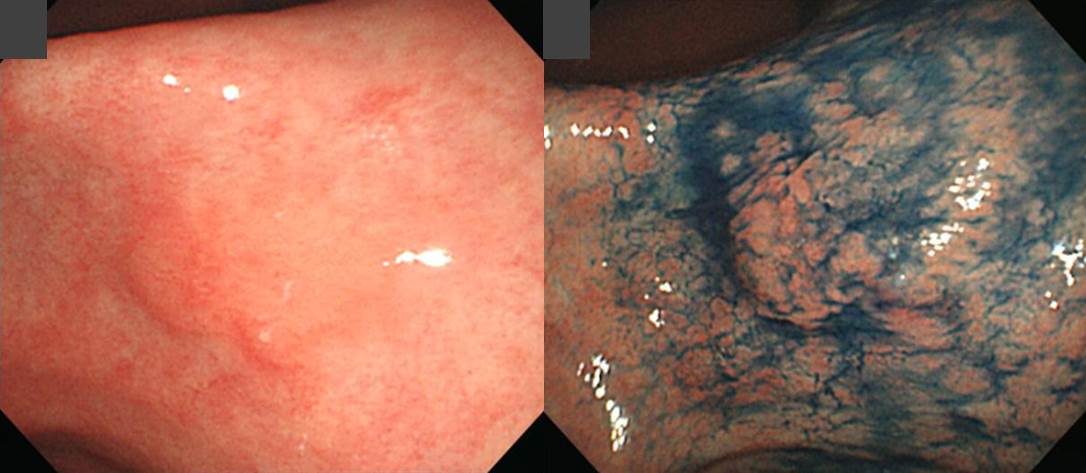
![]() 1. Delayed bleeding after gastric ESD
1. Delayed bleeding after gastric ESD
화요일에 시술하고 토요일에 출혈한 환자입니다 (4일째 출혈). 토혈이나 검은 변이 있으면 인근 병원 방문을 권했습니다. 인근 의료기관을 방문하였는데 조처가 다소 미흡하지 않았나 생각됩니다. 다행스럽게 환자 상태는 stable 했습니다. 앞으로 좀 더 자세히 설명드려야겠다는 생각을 하였습니다.
![]() 2. Adenoma with HGD
2. Adenoma with HGD
72세 여성 검진 내시경
ESD: Tubular adenoma with high grade dysplasia:
1) size: 1.8x1.2 cm
2) gross type: flat
3) negative resection margins
![]() 3. Metachronous gastric cancer (deep SM invasion, lymphatic positive)
3. Metachronous gastric cancer (deep SM invasion, lymphatic positive)
과거 조기위암 내시경치료를 받은 여성입니다. 치료부에 가깝지만 다소 떨어진 위치 이소성 위암이 발견되어 ESD를 하였는데 수술이 필요한 결과가 나왔습니다.
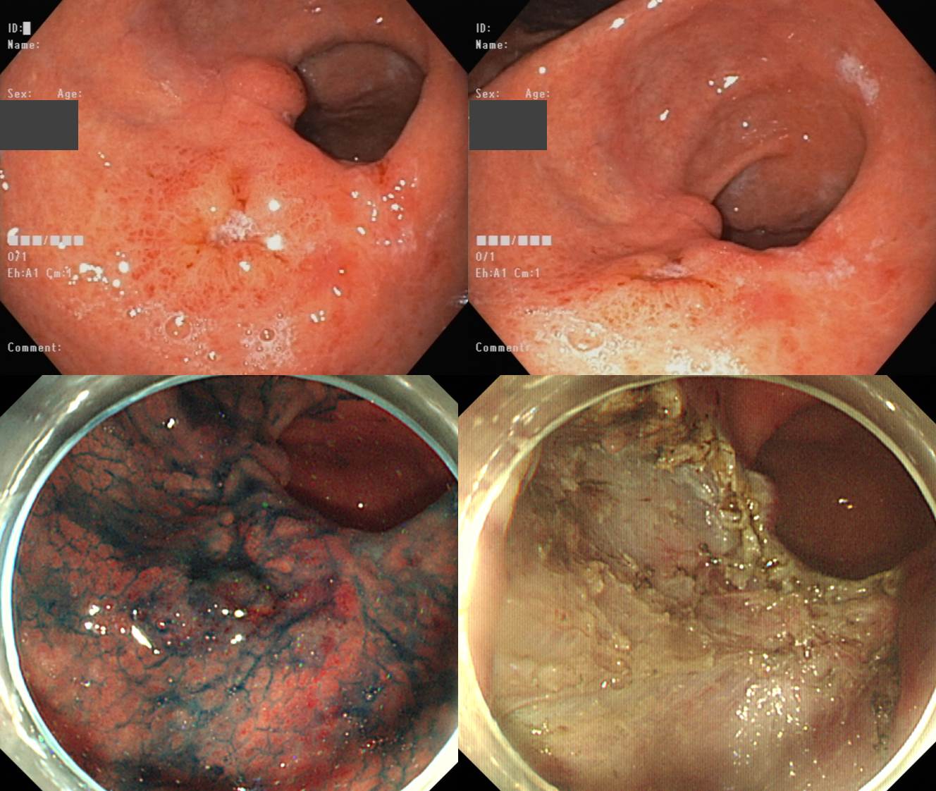
Stomach, endoscopic submucosal dissection:
Early gastric carcinoma
1. Location : mid antrum
2. Gross type : EGC type IIc
3. Histologic type : tubular adenocarcinoma, moderately differentiated
4. Histologic type by Lauren : intestinal
5. Size of carcinoma : (1) longest diameter, 13 mm (2) vertical diameter, 12 mm
6. Depth of invasion : invades submucosa, (depth of sm invasion : 1000 ㎛) (pT1b)
7. Resection margin : involved deep resection margin by carcinoma with cauterized artifacts safety margin : distal 15 mm, proximal 13 mm, anterior 16 mm, posterior 14 mm, deep 0 mm (sm only)
8. Lymphatic invasion : present (+++)
9. Venous invasion : suspicious
10. Perineural invasion : not identified(N)
11. Microscopic ulcer : absent
12. Histologic heterogeneity: absent
![]() 4. Early gastric cancer
4. Early gastric cancer
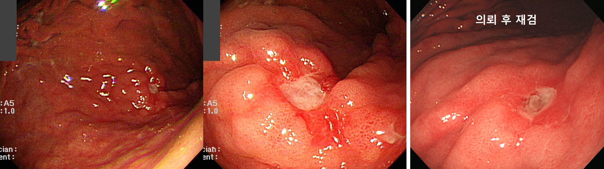
Stomach, subtotal gastrectomy:
Early gastric carcinoma
1. Location : middle third, Center at body and posterior wall
2. Gross type : EGC type IIc
3. Histologic type : tubular adenocarcinoma, poorly differentiated
4. Histologic type by Lauren : intestinal
5. Size : 3x1.5 cm
6. Depth of invasion : invades mucosa (muscularis mucosa) (pT1a)
7. Resection margin: free from carcinoma
8. Lymph node metastasis : no metastasis in 37 regional lymph nodes (pN0)
9. Lymphatic invasion : not identified
10. Venous invasion : not identified
11. Perineural invasion : not identified
12. Peritoneal cytology : negative
![]() 5. Early colon cancer
5. Early colon cancer
검진 대장내시경에서 발견된 병소로 ESD를 시행하였습니다.
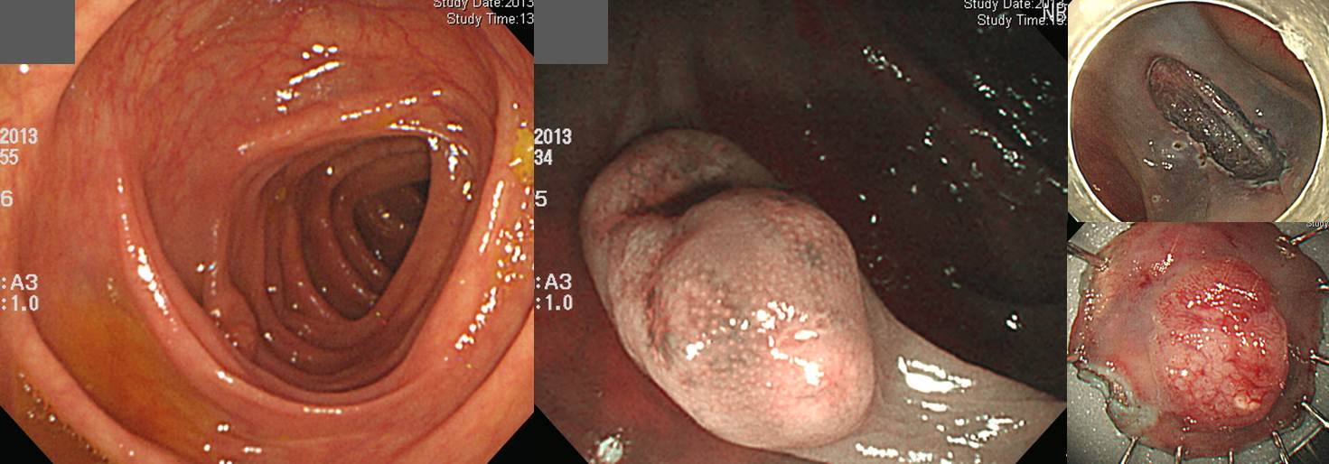
Ascending colon, endoscopic submucosal dissection:
Adenocarcinoma, well differentiated :
1) size of carcinoma: (1) longest diameter, 12 mm (2) vertical diameter, 7 mm
2) invasion into submucosa: present (depth of submucosal invasion: 2200 ㎛)
3) lymphovascular invasion: not identified(N)
4) perineural invasion: not identified(N)
5) tumor budding: not identified(N)
6) resection margins: free from carcinoma(N) (0.02 cm apart from deep resection margin)
Deep SM invasion으로 right hemocolectomy 시행하였고 no residual tumor가 나왔습니다.
![]() [References]
[References]
1) SMC Endoscopy Unit 삼성서울병원 내시경실
2) SMC Monday GI conference 삼성서울병원 일원내시경교실 월요점심소화기집담회
3) SMC Thursday endoscopy conference 삼성서울병원 일원내시경교실 목요점심내시경집담회
© 일원내시경교실 바른내시경연구소 이준행. EndoTODAY Endoscopy Learning Center. Lee Jun Haeng.