 EndoTODAY 내시경 교실
Beginner | ESA | Schedule | OPD
Seminars | Atlas | Recent | Links
![]() [Thursday Endoscopy Conference 20170615]
[Thursday Endoscopy Conference 20170615]
![]() 1. ESD for EGC
1. ESD for EGC
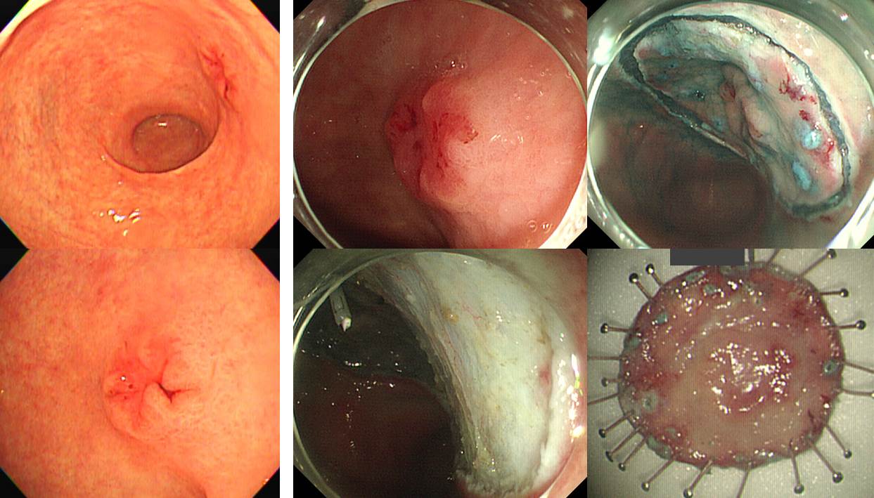
Stomach, endoscopic submucosal dissection:
Early gastric carcinoma
1. Location : mid antrum, postero-lesser curvature
2. Gross type : EGC type IIa+IIc
3. Histologic type : tubular adenocarcinoma, moderately differentiated
4. Histologic type by Lauren : intestinal
5. Size of carcinoma : (1) longest diameter, 16mm (2) vertical diameter, 14mm
6. Depth of invasion : invades mucosa (muscularis mucosa) (pT1a)
7. Resection margin : free from carcinoma(N), safety margin : distal 9 mm, proximal 13 mm, anterior 18 mm, posterior 10 mm
8. Lymphatic invasion : not identified(N)
9. Venous invasion : not identified(N)
10. Perineural invasion : not identified(N)
11. Microscopic ulcer : absent
12. Histologic heterogeneity: absent

Stomach, endoscopic submucosal dissection:
Pyloric gland adenoma with high grade dysplasia
1. Location : mid body, greater curvature
2. Gross type : elevated
3. Size of adenoma : (1) longest diameter, 20 mm (2) vertical diameter, 16 mm
4. Resection margin : negative resection margins(N)
![]() 3. Cardia EGC in the background of Barrett's esophagus
3. Cardia EGC in the background of Barrett's esophagus
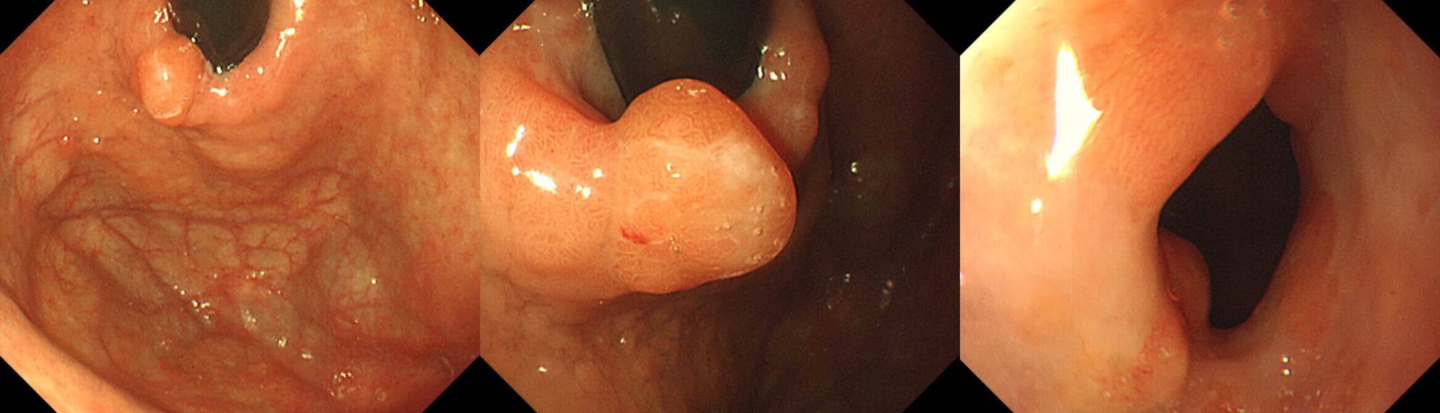
Early gastric carcinoma in the background of Barrett's esophagus ;
1. Location : upper third, Center at cardia (Siewert II) and lesser curvature
2. Gross type : EGC type I
3. Histologic type : tubular adenocarcinoma, moderately differentiated
4. Histologic type by Lauren : intestinal
5. Size : 3.5x2.2 cm
6. Depth of invasion : invades mucosa (lamina propria) (pT1a)
7. Resection margin: free from carcinoma, safety margin: proximal 1 cm, distal 17.2 cm
8. Lymph node metastasis : no metastasis in 21 regional lymph nodes (pN0), (0/21: "3,5", 0/3; "4,6", 0/3; "2", 0/2; "5", 0/0; "6", 0/3; "7", 0/3; "9", 0/2; "8a", 0/2; "11p", 0/0; "4sb", 0/0; "1", 0/1; "12a", 0/2)
9. Lymphatic invasion : not identified
10. Venous invasion : not identified
11. Perineural invasion : not identified
12. Peritoneal cytology : negative
13. AJCC stage by 7th edition: pT1a N0
내시경 육안소견에서 바렛이라고 보지 않았는데 병리에서 그렇게 이야기하면 바렛암인가요, 아닌가요? 늘 고민입니다. 진단기준은 바렛암인데.... 제가 본 22번째 환자입니다.
![]() 4. Adenoma with HGD
4. Adenoma with HGD
조직검사는 atypical glands with regenerative change였으나 내시경 육안소견이 종양성으로 판단되어 ESD 시행

Tubular adenoma with high grade dysplasia
1. Location : distal antrum
2. Gross type : depressed
3. Size of adenoma : (1) longest diameter, 8 mm (2) vertical diameter, 8 mm
4. Resection margin : negative resection margins(N)
![]() [References]
[References]
1) SMC Endoscopy Unit 삼성서울병원 내시경실
2) SMC Monday GI conference 삼성서울병원 일원내시경교실 월요점심소화기집담회
3) SMC Thursday endoscopy conference 삼성서울병원 일원내시경교실 목요점심내시경집담회
© 일원내시경교실 바른내시경연구소 이준행. EndoTODAY Endoscopy Learning Center. Lee Jun Haeng.