EndoTODAY 내시경 교실
Beginner | ESA | Schedule | OPD
Seminars | Atlas | Recent | Links
![]() [Thursday Endoscopy Conference 20170914]
[Thursday Endoscopy Conference 20170914]
![]() 1. FD 환자에서 우연히 발견된 자궁 clear cell carcinoma
1. FD 환자에서 우연히 발견된 자궁 clear cell carcinoma
자궁 근종의 가능성도 고려하였으나 크기가 커서 수술
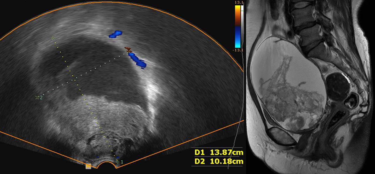
Ovary and salpinx, right oophorectomy, bilateral salpingectomy, total omentectomy, appendectomy and total hysterectomy with pelvic lymph node dissection and paraaortic LN dissection:
Ovary, right: clear cell carcinoma, grade III
1) Tumor size: 11x10 cm
2) Ovarian surface involvement: absent
3) Involvement of uterine serosa
4) No involvement of Bilateral salpinges, Omentum, appendix and left ovary
5) Lymphovascular invasion without D2-40 immunohistochemistry: Negative
6) No metastasis in all 14 regional lymph nodes
7) Hemorrhagic corpus luteum, left ovary
Myometrium: Leiomyomas (5.5x5 cm)
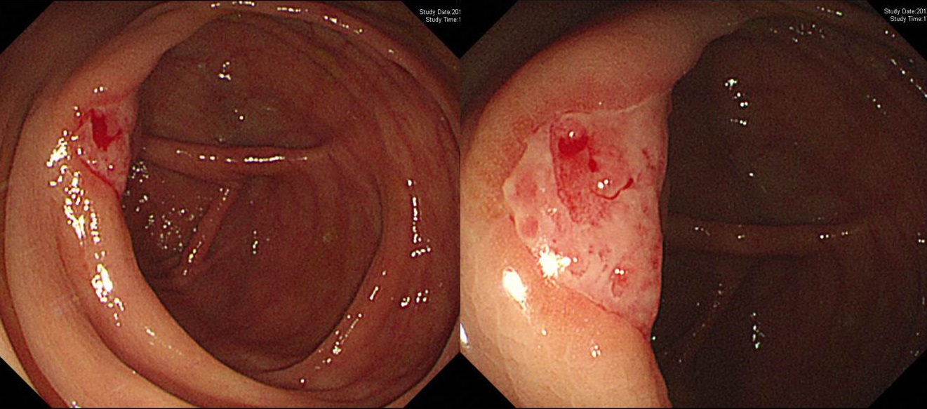
![]() 2. 류마티스 관절염 환자의 베체 장염
2. 류마티스 관절염 환자의 베체 장염
![]() 3. Paraesophageal hernia in Marfan syndrome
3. Paraesophageal hernia in Marfan syndrome
Ghent1 nosology (an internationally accepted diagnostic criterion for this syndrome.) involves major and minor criteria for the diagnosis of Marfan syndrome and includes seven fields: skeletal, cardiovascular, ocular, pulmonary, skin and integument, dura matter, and genetic analysis. The gastrointestinal involvement has not been mentioned because of its rare association. (sciencedirect.com)
Marfan 증후군 환자 paraesophageal hiatal hernia (mixsed type)
위 Marfan 증후군 환자의 6 년 전 내시경 사진
아래는 Marfan 환자 중 hiatal hernia가 심하다 못해 gastric volvulus가 된 증례보고입니다.
Figure 1. (A) Gastroesophageal junction (small white arrow) and fundus (large white arrow). (B) Pylorus (white arrow) beyond which the endoscope was not navigable. (sciencedirect.com)
Figure 2. (A) CECT coronal image shows malrotated stomach to be in the right hemithorax (long white arrow) and large intestine in the left thoracic cavity (white broken arrow). Multiple air-fluid levels are seen with maintained contrast enhancement of the wall of the herniated bowel loops. Omental herniation is also seen (small white arrow). (B) CECT axial image shows omental herniation in between anteriorly displaced inferior vena cava and posteriorly placed aorta (white arrows). (C) CECT axial image shows bilateral meningoceles at the sacral level (black arrows). The sacral pedicles were thinned out with dural ectasia. (D) CECT axial image shows dilatation of the sinus of valsalva (white arrow). CECT = contrast-enhanced computed tomography. (sciencedirect.com)
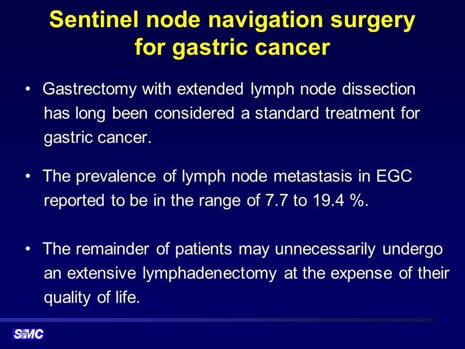
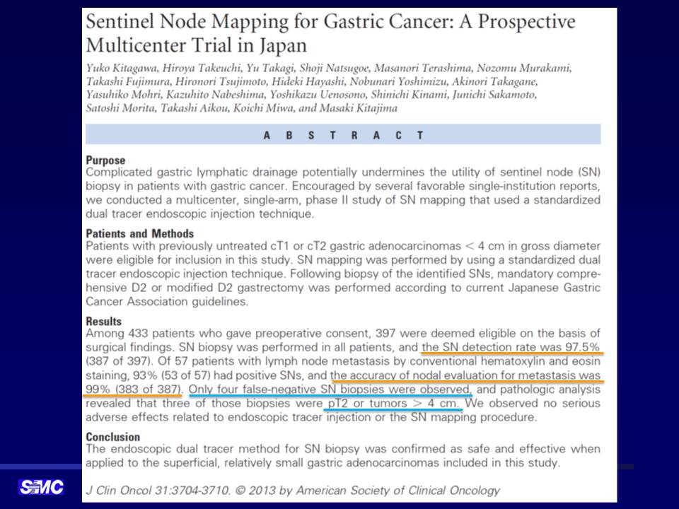
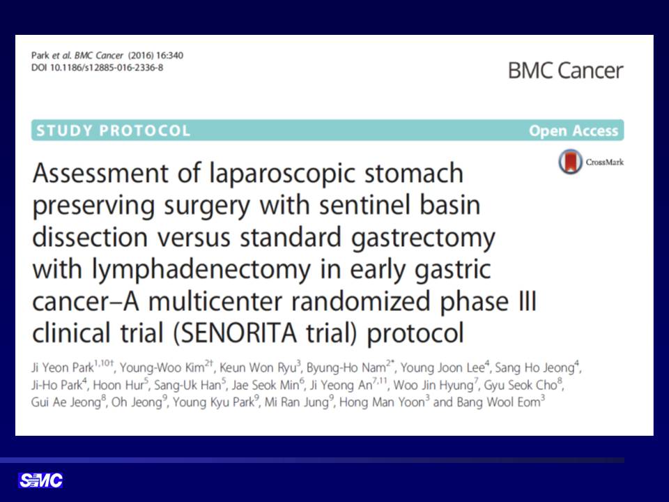
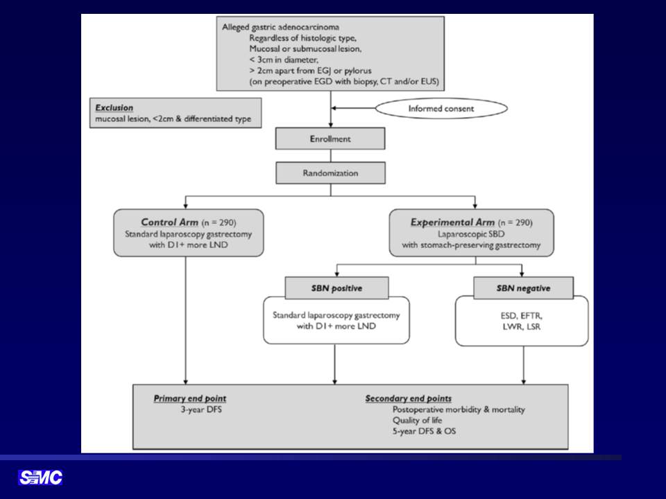
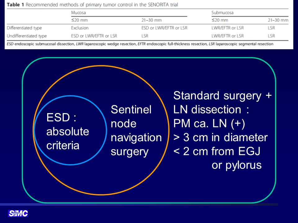
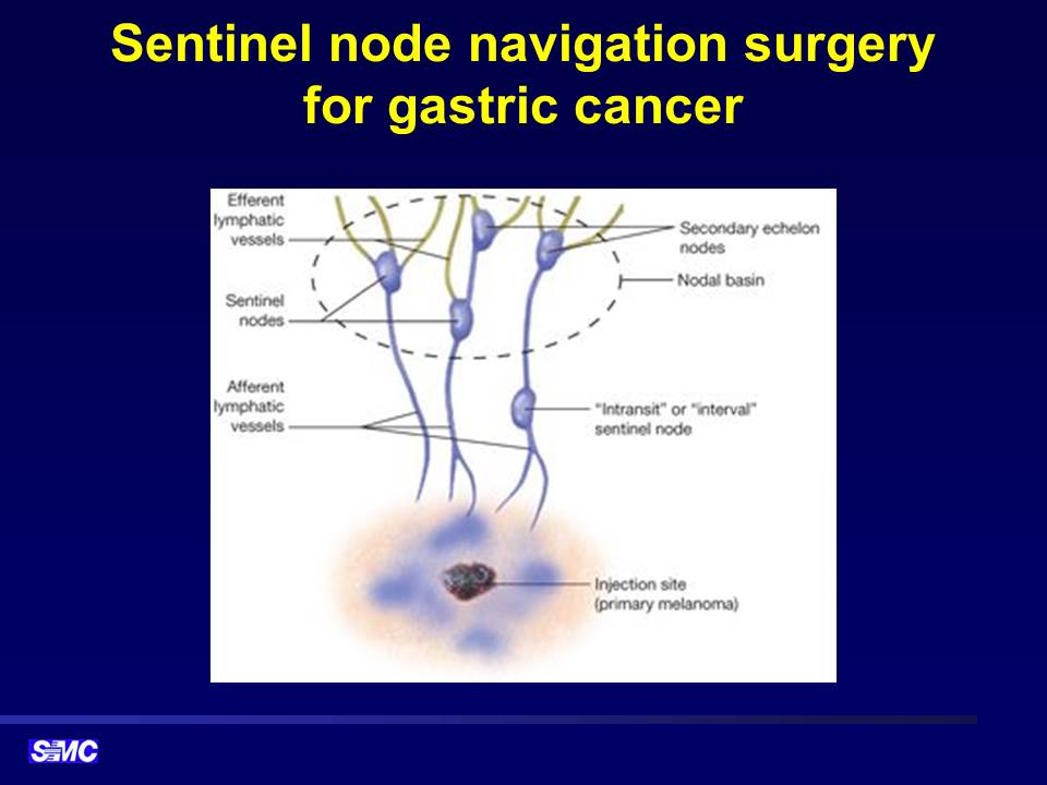
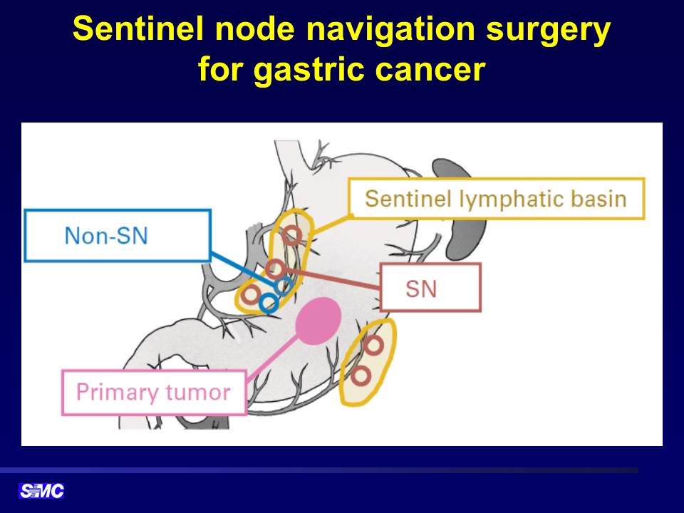
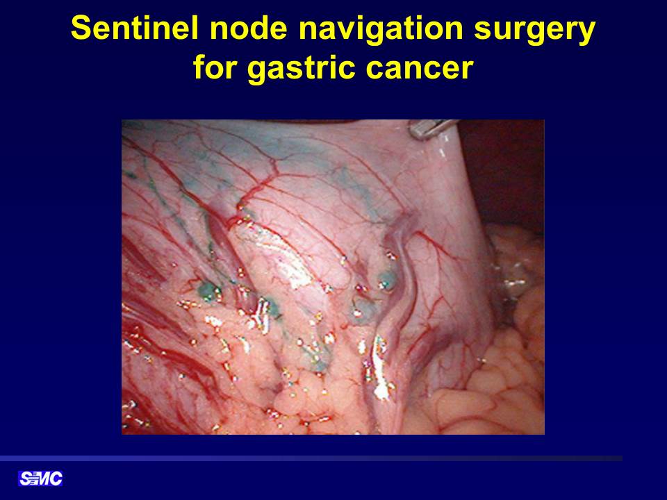
![]() 4. Sentinel node navigation surgery for gastric cancer
4. Sentinel node navigation surgery for gastric cancer
![]() [References]
[References]
1) SMC Endoscopy Unit 삼성서울병원 내시경실
2) SMC Monday GI conference 삼성서울병원 일원내시경교실 월요점심소화기집담회
3) SMC Thursday endoscopy conference 삼성서울병원 일원내시경교실 목요점심내시경집담회
© 일원내시경교실 바른내시경연구소 이준행. EndoTODAY Endoscopy Learning Center. Lee Jun Haeng.