EndoTODAY 내시경 교실
Beginner | ESA | Schedule | OPD
Seminars | Atlas | Recent | Links
![]() [One point lesson 3 - 식도암]
[One point lesson 3 - 식도암]
EndoTODAY 식도암의 진단과 치료를 참고하시기 바랍니다.
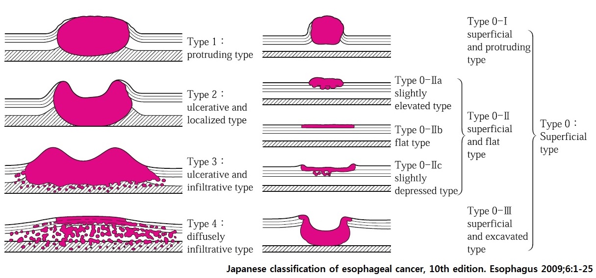
식도암은 위암만큼 내시경 분류가 유용하지 않습니다. 내시경 결과지에는 superficial esophageal cancer 혹은 advanced esophageal cancer라고 간단히 쓰는 경우가 많습니다. 그러나 식도암에서도 위암처럼 내시경 분류를 적용하면 여러 측면에서 유용할 것 같습니다. 2009년 발표된 Japanese classification of esophageal cancer (10th ed)에서 제시된 방법을 따릅시다 (Esophagus 2009:6:1-25). 위암의 분류와 매우 유사하기 때문에 외우기는 쉽습니다.
[표재성 식도암]
| 표재성 식도암의 내시경 소견 |
|
1) 점막의 혼탁과 조잡
2) 혈관망의 변화 혹은 소실 (점막하 혈관상의 소실) 3) 점막 발적 4) 융기성 병변 (융기가 높을 수록 심달도는 깊어짐) 5) 함몰성 병변 6) 식도의 비정상적인 연동운동 (고유근층으로의 침범및 종괴형성) |
M3 표재성 식도암
넓은 M2 표재성 식도암
[진행성 식도암]
내시경 진단은 Advanced esophageal cancer, 4 cm, type 2로 쓰실 것을 권합니다. Type 3 같다구요? 상관없습니다.
Invasive squamous cell carcinoma, moderately differentiated, esophagus:
1) tumor size: 4x2 cm
2) extension to perimuscular adventitia
3) endolymphatic tumor emboli: not identified
4) perineural invasion: present
5) involvement of radial margin
6) negative resection margins (proximal, 3 cm ; distal, 10 cm)
7) metastasis to 3 out of 62 regional lymph nodes
내시경 진단은 Advanced esophageal cancer, 3 cm, type 3로 쓰실 것을 권합니다. Type 2 같다구요? 상관없습니다.
Esophagus and upper stomach, Ivor Lewis operation:
Invasive squamous cell carcinoma, moderately differentiated;
1) tumor size: 3.5x1.8cm
2) extension to perimuscular adventitia
3) endolymphatic tumor emboli: absent
4) perineural invasion: absent
5) negative resection margins (proximal, 5.8cm ; distal, 8.2cm)
6) no metastasis in 59 regional lymph nodes
내시경 진단: Advanced esophageal cancer, 3 cm, type 3
Esophagus and upper stomach, Ivor Lewis operation : Invasive squamous cell carcinoma, moderately differentiated, middle thoracic esophagus :
1) invasive carcinoma size: 3.5x2.5 cm
2) extension to periesophageal soft tissue (adventitia)
3) endolymphatic tumor emboli: present
4) perineural invasion: not identified
5) resection margins: free from carcinoma, but very close to circumferential resection margin. safety margin: proximal, 4.5 cm ; distal, 8.5 cm ; circumferential (adventitial) margin(deep), 50 ㎛
6) metastasis to 1 out of 29 regional lymph nodes without extracapsular extension, (size of the largest metastasis: 0.4 cm) (1/29 : "7(Subcarinal)", 1/10; "Gastric lymph node 1", 0/2; "Gastric lymph node 3", 0/10; "Gastric lymph node 4", 0; "Lt Recurrent Laryngeal Nerve lymph node", 0/4; "Rt Recurrent Laryngeal Nerve lymph node", 0/3)
7) treatment effect: no prior treatment
[식도 melanoma]
일견 advanced esophageal cancer, 9cm, type 3로 쓰기 쉬울 것 같습니다. 그러나 검은 색 plaque와 mass 사이사이의 검은 색에 주의해주시기 바랍니다.
Esophagus and upper stomach, Ivor Lewis operation:
Malignant melanoma:
1) tumor size: 9x9 cm
2) extension to proper muscle layer
3) depth of invasion: 1.5 cm
4) endolymphatic tumor emboli: present
5) necrosis: about 10 %
6) growing with pushing border
7) mitosis: 9/10 HPFs
8) negative mucosal resection margins, but lymphatic emboli present in "proximal resection margin"
9) metastasis to 9 out of 25 regional lymph nodes
* 참고: EndoTODAY 식도 흑색종
© 일원내시경교실 바른내시경연구소 이준행. EndoTODAY Endoscopy Learning Center. Lee Jun Haeng.