EndoTODAY 내시경 교실
Beginner | ESA | Schedule | OPD
Seminars | Atlas | Recent | Links
![]() [StomachTODAY 038. Gastric Interted Hamartomatous Polyp. GIHP. 위 속말림 과오종성 용종]
[StomachTODAY 038. Gastric Interted Hamartomatous Polyp. GIHP. 위 속말림 과오종성 용종]

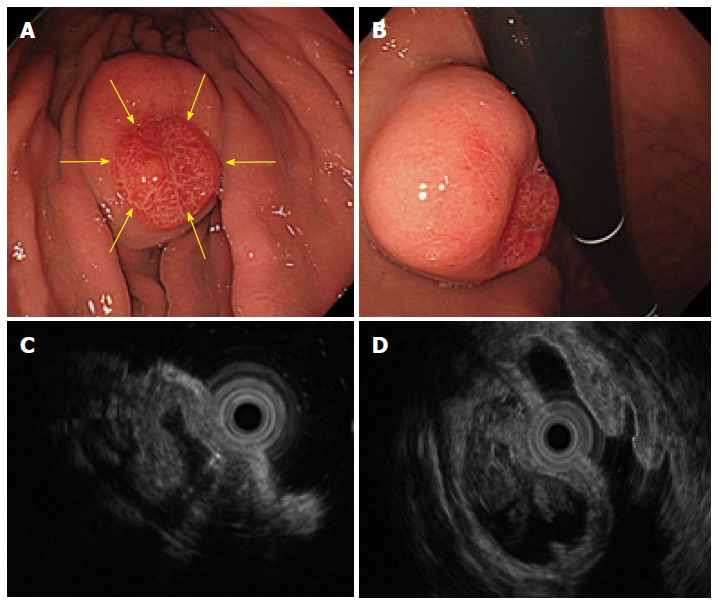
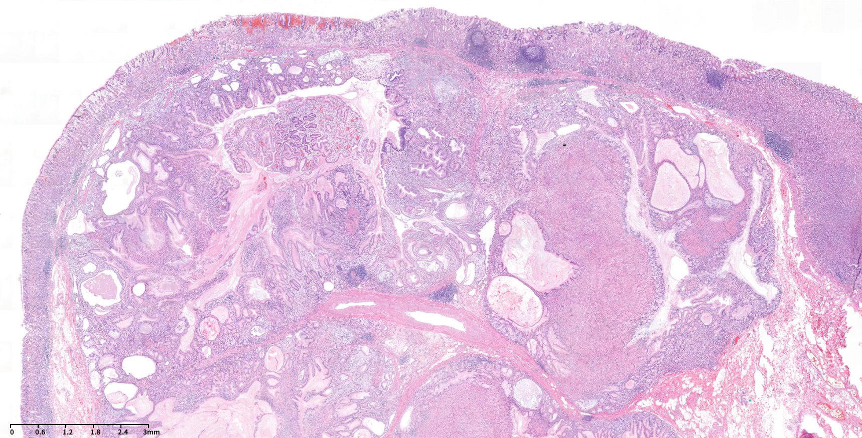
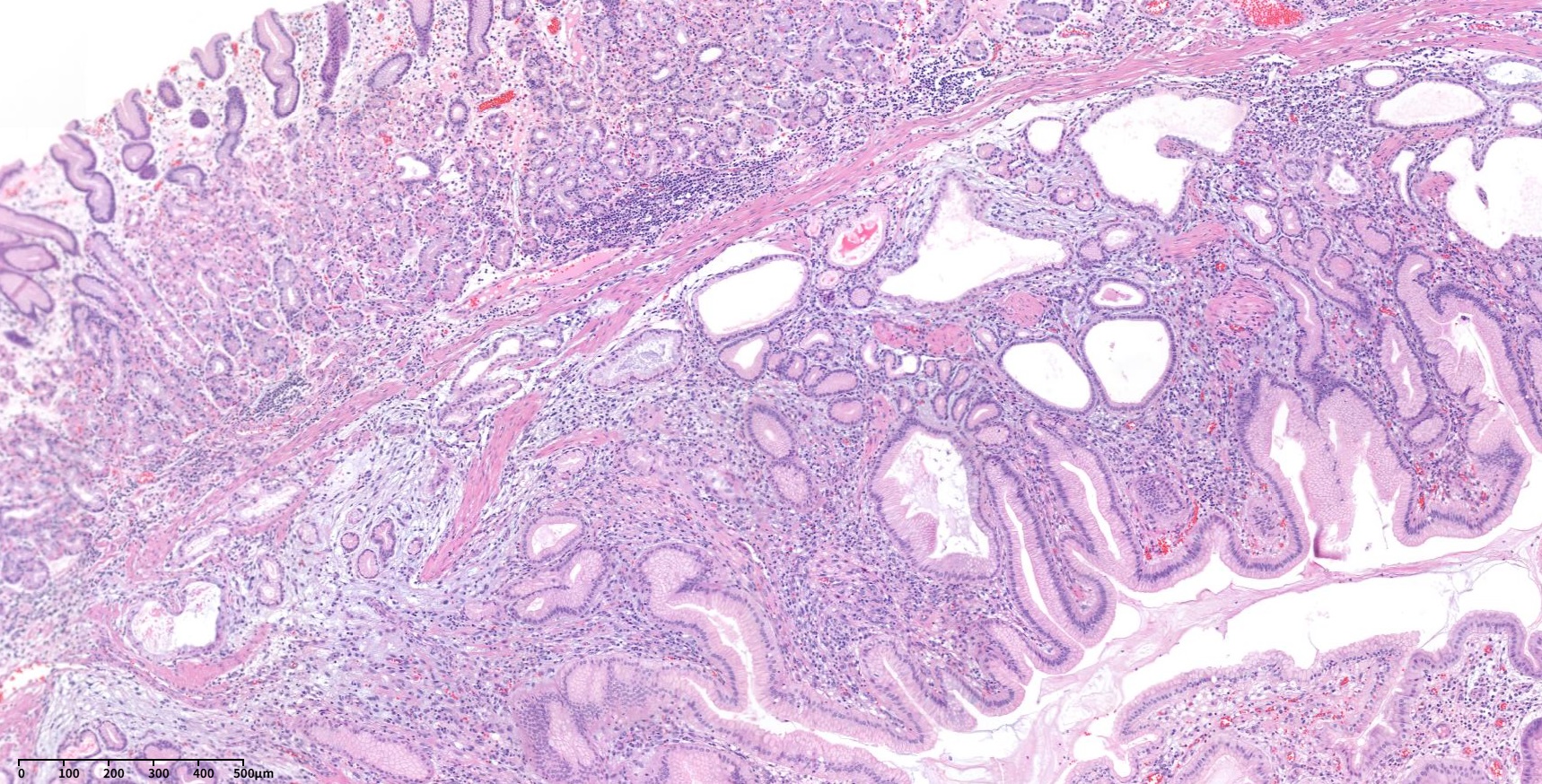
60세 남성이 건강검진 내시경에서 우연히 발견된 위저부의 3cm크기의 umbilicated ulcer를 동반한 점막하종양 소견으로 의뢰되었다. 내시경 검사 당시 조직검사 부위 중 한 곳에서 white thick mucinous material이 삐져나오는 소견이 보였고 (사진 1시 방향) 찐득한 느낌의 젤리같기도 했지만 포셉으로 잡히지는 않는다는 소견이 기술되어 있었다. 외부 조직검사 결과는 stromal cell proliferation in lamina propria였고 악성 GIST (gastrointestinal stromal tumor) 가능성이 고려되었다. 의뢰 후 외부 슬라이드 재판독한 소견은 Spindle cell tumor. differential diagnosis: GIST이었다. 내시경 재검을 하였고 육안 소견은 비슷했으며 조직검사 재검은 Chronic gastritis, active, with erosion, No definite submucosal tissue included로 보고되었으며 DOG-1과 C-KIT (CD 117)에 대한 면역형광염색은 음성이었다. CT를 시행하였으며 위저부에 약 3 cm의 endophytic mass가 있으며 central umbilication이 있었으며 정상 mucosa로 덮여있어 점막하종양의 가능성이 있는 것으로 판독되었다. 비록 조직검사에서 확인되지는 않았으나 spindle cell proliferation이 확인된 바 있고 내시경 육안소견에서 중앙의 표면이 불규칙한 함몰부가 있었으므로 GIST의 가능성을 고려하여 외과 의뢰하여 쐐기절제술을 하였다. 최종 병리 결과는 Herniated gastric mucosa with marked smooth muscle proliferation in submucosa, consistent with hamartomatous inverted polyp (1.7x1.6 cm)이었으며 desmin 면역형광염색은 양성이었다. 위 속말림 과오종성 용종 (gastric inverted hamartomatous polyp, GIHP)은 위점막하층에서 위샘이 증식되고 낭성 확장을 보이는 것을 특징으로 하는 질환이다. 병리학적으로 평활근 증식을 보일 수 있다. 일반적인 과오종성 용종이 외성장(exophytic growth)을 보이는 것에 비하여 GIHP는 내성장(endophytic growth)을 보인다. 내시경 소견에서 표면으로부터 우유같은 뮤신이 배출될 수 있다. 초음파 내시경 소견은 이소성 췌장 조직과 구분이 어렵다. 약 20%의 악성화 가능성이 있으므로 2cm 이상에서는 절제술을 권한다.
[2020-2-19. 애독자 질문]
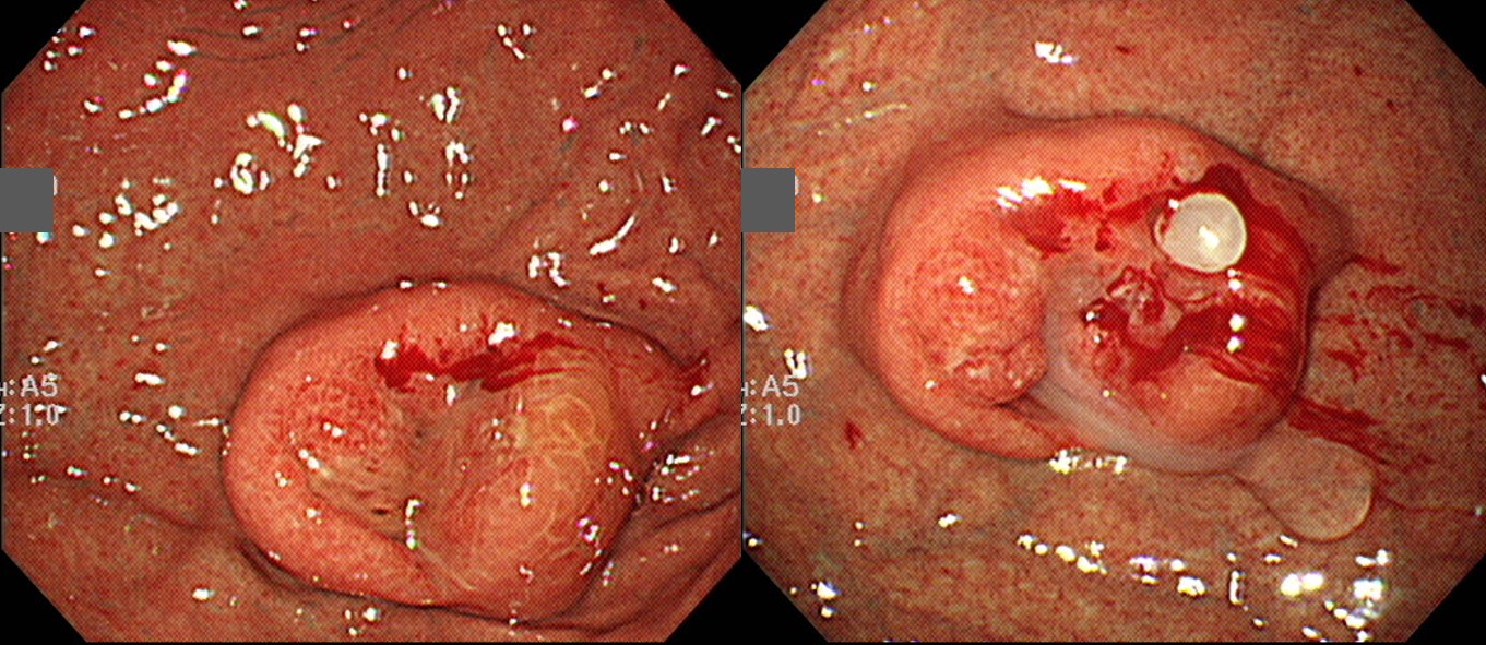
GC/fundus에 Gastric SET (3.0cm) with umblicated ulcer가 있어 bite on bite로 조직검사를 했습니다. 희한하게도 조직검사 부위 중 한 곳에서 white thick mucinous material이 삐져나오더군요 (우측 사진 1시 방향). 이게 뭐지~ 하고 포셉으로 찝어보기도 했었는데 찐득한 느낌의 젤리같기도 했지만 포셉으로 잡히지는 않았습니다.
Stomach, fundus, greater curvature, endoscopic biopsy ; Erosion with chronic atrophic gastritis, active, with
1) polypoid foveolar hyperplasia
2) cystically dilated glands
3) regenerative glands
4) lymphoid cells infiltration containing lymphoid follicles
5) stromal cells proliferation in lamina propria.
** Microscopic findings by Sydney system ;
1. Neutrophils : marked
2. Mononuclear cells : marked
3. Atrophy : mild
4. Intestinal metaplasia : absent
[ Note ] The finding is suspicious for mucosal polypoid lesion including hyperplastic polyp. Stromal cells are proliferated, but benign-looking and suspicious for reactive change, so it is difficult to diagnose gastro-intestinal stromal tumor lesion. And definite malignant lesion is not noted.의뢰 후 슬라이드 재판독한 결과는 "Stomach, fundus, greater curvature, biopsy : Spindle cell tumor. Differential diagnosis: Gastrointestinal stromal tumor"였습니다.
수술 후 최종병리 결과는 다음과 같았습니다.
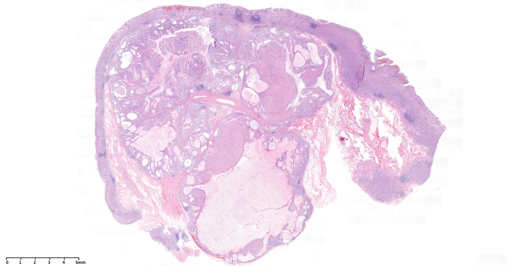
Stomach, wedge resection: Herniated gastric mucosa with marked smooth muscle proliferation in submucosa, consistent with hamartomatous inverted polyp (1.7x1.6 cm)
문헌을 찾아보았습니다.
Gastric Inverted Hamartomatous Polyps Clin Med Insights Gastroenterol 2016
GIHPs are a distinct entity characterized by submucosal growth of hypertrophic glands with cystic dilatation. They are distinct from the other types of hamartomatous polyps, which have an exophytic configuration contrary to the endophytic nature of these polyps. On endoscopic examination, these are reported as solitary submucosal masses. On endoscopy, extrusion of milky mucinous material from the surface of the lesion and calcifications from the biopsy site may provide a clue to diagnosis. On histology, there is cystic proliferation of glands, which may be accompanied by smooth muscle proliferation, and formation of ectopic duct-like structures has also been reported. In addition, fibroblastic and neural proliferation may also be seen with glandular elements. Diagnosis of GIHP is difficult without pathologic examination and may mimic ectopic pancreas on endoscopy and endosonography. Certain features have been suggested on endoscopic ultrasound imaging, such as hyperechoic lesions with hypoechoic spots, which might be suggestive of GIHPs. En bloc removal is recommended in lesions >2 cm due to the associated malignant risk (up to 20% risk of malignancy). Though it is rare, Hirasaki et al have reported a case of GHIP associated with signet ring cell carcinoma.
문헌에서 설명하고 있는 내시경 소견 및 조직 결과가 제 증례와 매우 흡사해서 신기했는데요, 뒤에 붙어있는 내용들을 보면 malignant risk가 상당히 되는 듯 싶습니다. 이 증례의 경우 향후 계획을 어떻게 잡는것이 최선의 선택일지 궁금합니다.
[2020-3-4. 이준행 답변]
저도 매우 드물게 경험하는 종류입니다. 몇 문헌을 보더라도 milky material이 특징으로 되어 있는 것 같습니다.
일반적인 용종에 비하여 다소 투명하고 pale해 보이는 점막이고 EUS에서 cystic nature가 확인되었습니다.
병리 보고서에서 용어를 조금 다르게 사용하고 있으나 비슷한 nature라고 생각되는 SMT를 경험한 적이 있습니다.
Wedge resection: herniated gastric mucosa in the submucosa with cystic dilatation
Herniated gastric mucosa in the submucosa
Wedge resection: Inverted hyperplastic polyp with focal inflammatory myofibroblastic tumor-like stroma (1.3x1cm)
내시경 절제술이 쉽지 않은 종류입니다만 간혹 내시경으로 절제한 증례 보고가 있습니다 (대한소화기학회지 2016:67:98-102).
이와 같은 상황에서 암이 동반된 경우는 단 한례 본적이 있습니다. Wedge resection을 하였는데 hamartomatous inverted polyp with mucosal adenocarcinoma였습니다.
GIST에서 low risk, intermediate risk에서 수년 후 전이하는 예가 있는 것은 resected specimen을 보더라도 병리학적으로 암인지 아닌지 명확히 구분할 수 없기 때문입니다. 그런데, hamartomatous inverted polyp에서 암이 발생하면 보통의 위선암입니다. 병리학적으로 암인지 아닌지 명확히 구분할 수 있는 종류입니다. 이 증례처럼 wedge resection 하였고 최종 병리에서 암이 없으면 암위험이나 전이 위험을 고려할 필요가 없을 것 같습니다. 상황 종료라고 보아도 무난할 것 같습니다.
참고: EndoTODAY EGC arising from herniated gastric mucosa in the submucosa
![]() [References]
[References]
1) EsoTODAY - 십이지장질환 증례토의
2) SmallTODAY - 소장질환 증례토의
3) ColonTODAY - 대장질환 증례토의
4) Dr. Sinn's LiverTODAY - 간질환 증례토의
© 일원내시경교실 바른내시경연구소 이준행. EndoTODAY Endoscopy Learning Center. Lee Jun Haeng (2020-3-4. Update: 2024-12-13)