EndoTODAY 내시경 교실
Beginner | ESA | Schedule | OPD
Seminars | Atlas | Recent | Links
![]() [Thursday Endoscopy Conference 20170323]
[Thursday Endoscopy Conference 20170323]
![]() 1. EVL due to cirrhosis realated with porphyria (erythropoietic protoporphyria ?)
1. EVL due to cirrhosis realated with porphyria (erythropoietic protoporphyria ?)
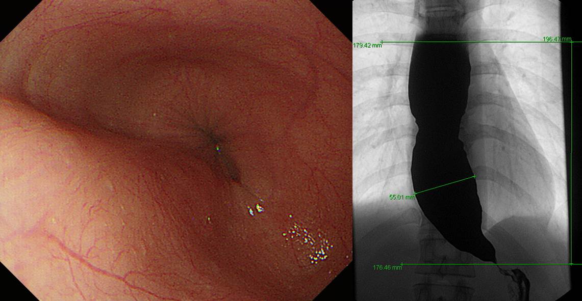
![]() 2. Achalasia
2. Achalasia
POEM 시행 후 증상이 호전되었습니다.
* 참고: EndoTODAY 식도이완불능증과 POEM
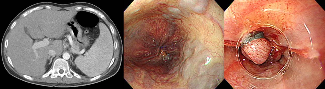
![]() 3. Periampullary cancer (most likely duodenal cancer)
3. Periampullary cancer (most likely duodenal cancer)
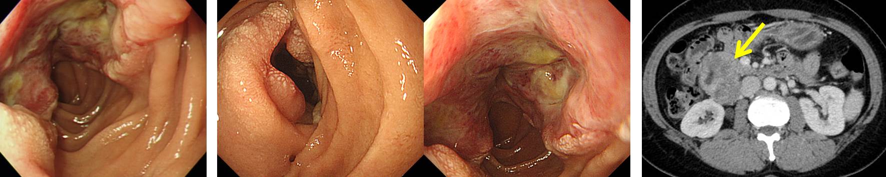
![]() 4. Borrmann type IV with small ulcer
4. Borrmann type IV with small ulcer
위체상부 전벽의 작은 궤양형 위암(poorly differentiated adenocarcinoma)으로 의뢰된 50대 여성입니다. 환자의 성별과 나이, 세포형, 병소의 위치, 주변 점막이 약간 두꺼운 점을 고려하여 보만 4형 진형성 위암의 가능성을 고려하였습니다. 내시경 검사 의뢰시 "작은 함몰형 위암으로 오셨습니다. 그러나 주변 위벽이나 fold가 두꺼워진 부위는 없는지 잘 살펴봐 주세요."라고 부탁하였습니다. 아니나 다를까 보만 4형 진행성 위암으로 나왔습니다.

Stomach, total gastrectomy:
Advanced gastric carcinoma1. Location : [1] upper third, [2] middle third, Center at body and anterior wall
2. Gross type : Borrmann type 4
3. Histologic type : tubular adenocarcinoma, poorly (poorly cohesive) differentiated
4. Histologic type by Lauren : diffuse
5. Size : about 9.5x7 cm
6. Depth of invasion : invades serosa (pT4a)
7. Resection margin: free from carcinoma
8. Lymph node metastasis : metastasis to 0 out of 28 regional lymph nodes (pN0)
9. Lymphatic invasion : present
10. Venous invasion : not identified
11. Perineural invasion : present
수술 결과를 보고 검사를 하셨던 fellow 선생님께서 "교수님. 역시 졌습니다."라는 제목의 메일을 보내오셨습니다.^^ "안녕하세요. 저번에 메일주셨던 그 환자분입니다! 모처럼 만난 보만 4형 진행성 위암이었습니다. 위주름의 변화가 그리 심하지 않다고 생각했는데... 수술결과는 10cm 크기로 나왔습니다. 사진을 다시 보니, 약간 의심스러운 fold가 더 멀리까지 보이는 것 같습니다."
제가 답장을 보냈습니다. "보만 4형 진행성 위암은 늘 어렵습니다. (1) 여성, (2) 위체부, (3) 전형적인 EGC가 아닌 경계가 명확하지 않은 작은 함몰형 병소, (4) P/D 또는 SRC에서는 보만 4형 진행성 위암인데 작은 함몰형 병소로만 관찰되는 경우를 의심해야 합니다. 이 환자는 4가지 모두 해당하였습니다. 내시경 소견으로 병소의 크기와 깊이를 짐작하는 것도 좋지만 임상상도 잊지 말아주세요."
* 참고: EndoTODAY 작은 함몰형 병소를 동반한 보만 4형 진행성 위암
![]() 5. Classification of capsule endoscopy findings (P0-2)
5. Classification of capsule endoscopy findings (P0-2)
Highly relevant P2 lesion (임상적응에 부합하는 결과를 얻음) - Angioectasia, Dieulafoy’s lesion, varices, presence of active bleeding, ulcerations, multiple (≥3) erosions, and diverticula
Less relevant P1 lesion (임상적응에 부합하지 않는 우연한 소장질환을 진단) - Red spots, visible submucosal veins, and erosions (<3)
Absent P0 lesion (소장질환의 소견을 진단하지 못함)
![]() 6. 2016년 2월 25일 목요내시경집담회에서 소개된 Capsule 내시경 관련 내용 리뷰
6. 2016년 2월 25일 목요내시경집담회에서 소개된 Capsule 내시경 관련 내용 리뷰
나이에 따른 차이
Active small bowel bleeding
Inflammatory lesions
NSAID induded enteropathy
Vascular lesions
Neoplastic lesions
Meckel's diverticulum
![]() [References]
[References]
1) SMC Endoscopy Unit 삼성서울병원 내시경실
2) SMC Monday GI conference 삼성서울병원 일원내시경교실 월요점심소화기집담회
3) SMC Thursday endoscopy conference 삼성서울병원 일원내시경교실 목요점심내시경집담회
© 일원내시경교실 바른내시경연구소 이준행. EndoTODAY Endoscopy Learning Center. Lee Jun Haeng.