 EndoTODAY 내시경 교실
Beginner | ESA | Schedule | OPD
Seminars | Atlas | Recent | Links
![]() [Thursday Endoscopy Conference 20170413]
[Thursday Endoscopy Conference 20170413]
![]() 1. Pill esophagitis
1. Pill esophagitis
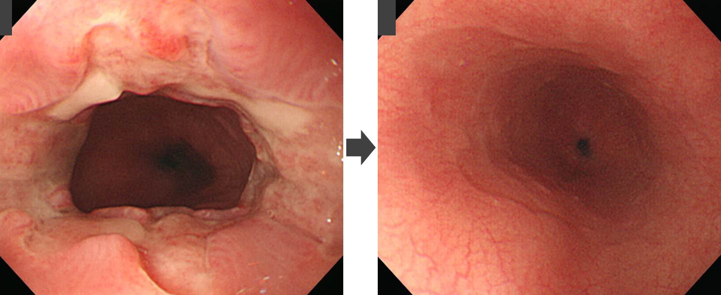
Urticaria 피부과 약 드셨던 분입니다. 38cm 하부식도 병소입니다.
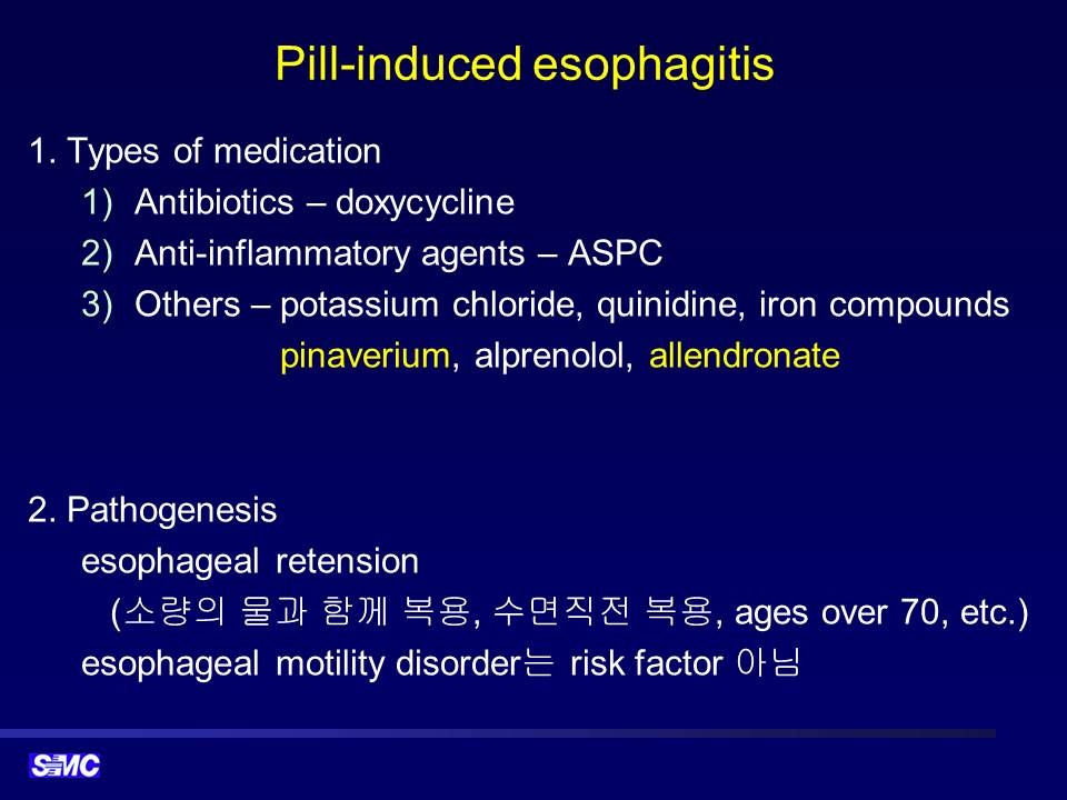
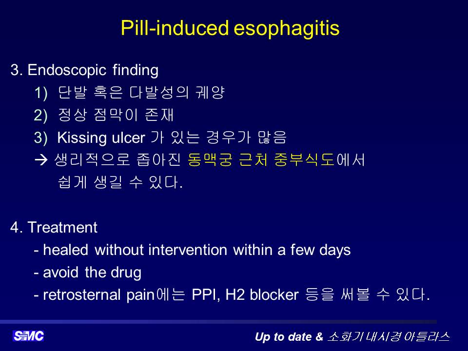
* 참고: EndoTODAY Pill-induced esophagitis
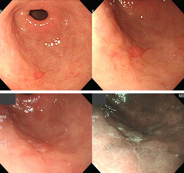
![]() 2. EGC IIc (Deformity after electrocauterization)
2. EGC IIc (Deformity after electrocauterization)
매우 이상하게 생긴 위암에 대하여 장시간 논의를 하였습니다. "중앙의 작은 함몰부만 보지 말고 조금 더 넓게 범위를 잡아야 합니다. Pale하면서 살짝 함몰된 부위, fold가 끊기면서 색조가 달라진 곳까지 암이라고 보는 것이 옳을 것 같습니다." 정도로 논의를 정리한 후 병리결과를 보았는데 예상보다 작았고 깊지 않았습니다.
Stomach, subtotal gastrectomy:
Early gastric carcinoma
1. Location : middle third, Center at lower body and anterior wall
2. Gross type : EGC type IIb+IIc
3. Histologic type : tubular adenocarcinoma, poorly differentiated
4. Histologic type by Lauren : diffuse
5. Size : 1.4x1.1 cm
6. Depth of invasion : invades mucosa (muscularis mucosa) (pT1a)
7. Resection margin: free from carcinoma, safety margin: proximal 7.8 cm, distal 8 cm
8. Lymph node metastasis : no metastasis in 53 regional lymph nodes (pN0) (0/53: "1", 0/10; "3", 0/21; "4", 0/5; "4ss", 0/0; "5", 0/0; "6", 0/1; "8a", 0/0; "7", 0/2; "9", 0/4; "11p", 0/3; "12a", 0/7)
9. Lymphatic invasion : not identified
10. Venous invasion : not identified
11. Perineural invasion : not identified
12. Peritoneal cytology : negative
하도 이상하여 집담회가 끝난 후 환자의 의무기록을 검토하였습니다. 역시나 사연이 있었습니다. Hematochezia로 한 병원 응급실을 방문하여 위암에서 출혈하는 것이 발견되어 지혈술을 받은 후 전원되셨던 분이었습니다. 앞서 논의한 사진은 지혈술 후 PPI 드시고 있는 사태로 전원 후 재검한 내시경 사진이었습니다.
(1) exposed vessel이 잘 보임, (2) electrocautery 직후, (3) 지혈술 다음 날 내시경 소견, (4)-(6) 내시경 지혈술 18일 후 추적내시경 소견
첫 내시경 사진(A)을 보면 훨씬 전형적인 함몰형 조기위암임을 알 수 있습니다. 조직검사 후 위암의 모양이 변형될 수 있습니다. 지혈술 후에는 훨씬 심하게 변형될 수 있습니다. 이번 집담회에서 논의가 매끄럽지 못했던 것은 지혈술 후 변형된 소견이었기 때문입니다. 첫 내시경 사진을 찾아보는 습관을 가집시다.
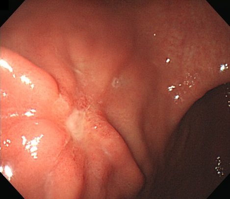
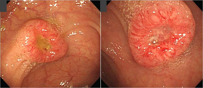
![]() 3. EGC
3. EGC
첫 조직검사에서 암으로 나오지 않아 제균치료 후 재검하여 암으로 나와 ESD를 시행하였습니다. 중앙의 붉은 부분만 병소라기보다는 좀 더 넓은 질병의 가능성을 고려해 할 것 같습니다.
Stomach, endoscopic submucosal dissection:
1. Location : antrum, greater curvature
2. Gross type : EGC type IIc
3. Histologic type : tubular adenocarcinoma, moderately differentiated
4. Histologic type by Lauren : intestinal
5. Size of carcinoma : (1) longest diameter, 16 mm (2) vertical diameter, 8 mm
6. Depth of invasion : invades submucosa, (depth of sm invasion : 200 ㎛) (pT1b)
7. Resection margin : free from carcinoma(N), safety margin : distal 6 mm, proximal 9 mm, anterior 10 mm, posterior 12 mm, deep 250 ㎛ (sm only)
8. Lymphatic invasion : not identified(N)
9. Venous invasion : not identified(N)
10. Perineural invasion : not identified(N)
11. Microscopic ulcer : absent
12. Histologic heterogeneity: absent
![]() 4. Colon cancer
4. Colon cancer
Anal verge 30 cm 입니다.
Sigmoid colon, anterior resection:
Adenocarcinoma, moderately differentiated
1. Location: sigmoid colon
2. Gross type: ulceroinfiltrative
3. Size: 1.6x1.5 cm
4. Depth of invasion: invades subserosa (pT3) (distance from nearest serosa: about >5.0 mm)
5. Resection margin: free from carcinoma, safety margin: proximal, 5.5 cm ; distal, 6 cm ; radial, >5.0 mm
6. Regional lymph node metastasis : no metastasis in all 12 regional lymph nodes(pN0) (0/12: pericolic, 0/12)
7. Lymphatic invasion: not identified
8. Venous invasion: not identified
9. Perineural invasion: not identified
10. Tumor budding : negative
11. Pathologic staging: pT3 N0 Mx
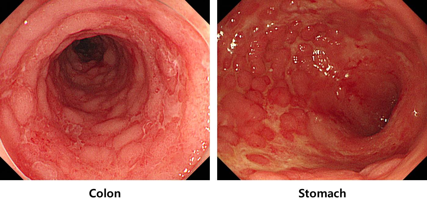
![]() 5. Intestinal tuberculosis
5. Intestinal tuberculosis
left: terminal ileum, right: ascending colon
* 참고: EndoTODAY 결핵성 장염
![]() 6. Systemic amyloidosis in Behcet's disease
6. Systemic amyloidosis in Behcet's disease
베체병 환자에서 발생한 systemic amyloidosis
* 참고 1: EndoTODAY 위 아밀로이드증
* 참고 2: EndoTODAY 대장 아밀로이드증
![]() [References]
[References]
1) SMC Endoscopy Unit 삼성서울병원 내시경실
2) SMC Monday GI conference 삼성서울병원 일원내시경교실 월요점심소화기집담회
3) SMC Thursday endoscopy conference 삼성서울병원 일원내시경교실 목요점심내시경집담회
© 일원내시경교실 바른내시경연구소 이준행. EndoTODAY Endoscopy Learning Center. Lee Jun Haeng.