EndoTODAY 내시경 교실
Beginner | ESA | Schedule | OPD
Seminars | Atlas | Recent | Links
![]() [Thursday Endoscopy Conference 20170420]
[Thursday Endoscopy Conference 20170420]
2017-4-20.
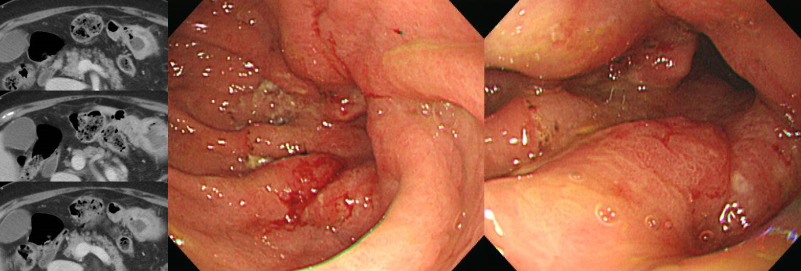
![]() 1. EGC (deep SM) with lymph node metastasis
1. EGC (deep SM) with lymph node metastasis
50대 여성의 epigastric pain으로 시행한 EGD였습니다. 집담회에서 voting을 했는데 위궤양, 위암, 위림프종 등 다양한 의견이 있었습니다. 저는 위궤양에 한 표 했는데.... 답은 암이었습니다. 창피는 계속됩니다.
Stomach, subtotal gastrectomy:
Early gastric carcinoma
1. Location : lower third, Center at antrum and greater curvature
2. Gross type : EGC type IIc
3. Histologic type : tubular adenocarcinoma, poorly differentiated
4. Histologic type by Lauren : diffuse
5. Size : 2.2x1.5 cm
6. Depth of invasion : invades submucosa (sm3) (pT1b)
7. Resection margin: free from carcinoma, safety margin: proximal 8.7 cm, distal 2 cm
8. Lymph node metastasis : metastasis to 1 out of 25 regional lymph nodes (pN1), (perinodal extension: absent) (1/25: LN3,5, 0/9; LN4,6, 0/4; "1", 0/0; "4ss", 0/0; "5", 0/0; "6", 1/4; "8a", 0/0; "7", 0/1; "9", 0/3; "11p", 0/2; "12a", 0/2)
9. Lymphatic invasion : present
10. Venous invasion : not identified
11. Perineural invasion : not identified
12. Peritoneal cytology : negative
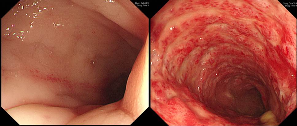
![]() 2. Ischemic colitis
2. Ischemic colitis
DM CKD로 혈액투석 중인 환자에서 급작스런 혈압 저하 및 복통 (혈변은 없었음)
![]() 3. Remnant gastric cancer
3. Remnant gastric cancer
40년 전 궤양 천공으로 subtotal gastrectomy 받은 분입니다.. 양 하지 부종으로 타원 방문하여 CT 검사를 하였고 위암 소견이 보여 내시경 검사를 받았고 진단되어 의뢰되었습니다. CT 판독은 "Gastrojejunostomy site 직상방에 약 4 cm segment에 걸쳐 enhancing wall thickening이 있음." 였는데... 다시 CT 사진을 보아도 진단이 쉽지 않았을 것 같습니다. 내시경 소견에서도 진단이 쉽지 않은 모양이었습니다. 뚜렷한 궤양은 없이 anastomosis site 직상방이 조금 부어 있는 정도가 대부분의 소견이었으므로.
Status post subtotal gastrectomy due to gastric ulcer perforation
Advanced gastric carcinoma
1. Location : lower third, Center at low body and greater curvature
2. Gross type : Borrmann type 3
3. Histologic type : tubular adenocarcinoma, moderately differentiated
4. Histologic type by Lauren : intestinal
5. Size : 4.5x4 cm
6. Depth of invasion : invades serosa (pT4a)
7. Resection margin: free from carcinoma, safety margin: proximal 4 cm, distal 1.5 cm
8. Lymph node metastasis : no metastasis in 20 regional lymph nodes (pN0) (0/20: "1", 0/1; "3", 0/1; "4", 0/1; "4sb", 0/0; "8a", 0/4; "7", 0/3; "9", 0/2; "11p", 0/5; "12a", 0/3)
9. Lymphatic invasion : present
10. Venous invasion : not identified
11. Perineural invasion : not identified
12. Peritoneal cytology : negative
13. AJCC stage by 7th edition: T4a N0
* 참고: EndoTODAY 잔위암
![]() 4. CMV gastritis
4. CMV gastritis
간이식 (DDLT, deceased donor liver transplantation) 한달 후 복통.
* 참고: EndoTODAY CMV 위염
![]() 5. Radiation proctitis with bleeding
5. Radiation proctitis with bleeding
전립선 암으로 방사선 치료 후 출혈
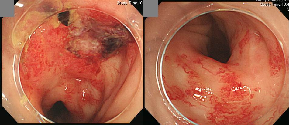

삼성서울병원 내시경 결과지 screenshot
* 참고: EndoTODAY 방사선 관련 위장관 손상
![]() [References]
[References]
1) SMC Endoscopy Unit 삼성서울병원 내시경실
2) SMC Monday GI conference 삼성서울병원 일원내시경교실 월요점심소화기집담회
3) SMC Thursday endoscopy conference 삼성서울병원 일원내시경교실 목요점심내시경집담회
© 일원내시경교실 바른내시경연구소 이준행. EndoTODAY Endoscopy Learning Center. Lee Jun Haeng.