 EndoTODAY 내시경 교실
Beginner | ESA | Schedule | OPD
Seminars | Atlas | Recent | Links
![]() [Gastric cancer 405]
[Gastric cancer 405]
001 | 101 | 201 | 301 | 401 | 501 | 601 | 701 | 801 | 901 | 1000
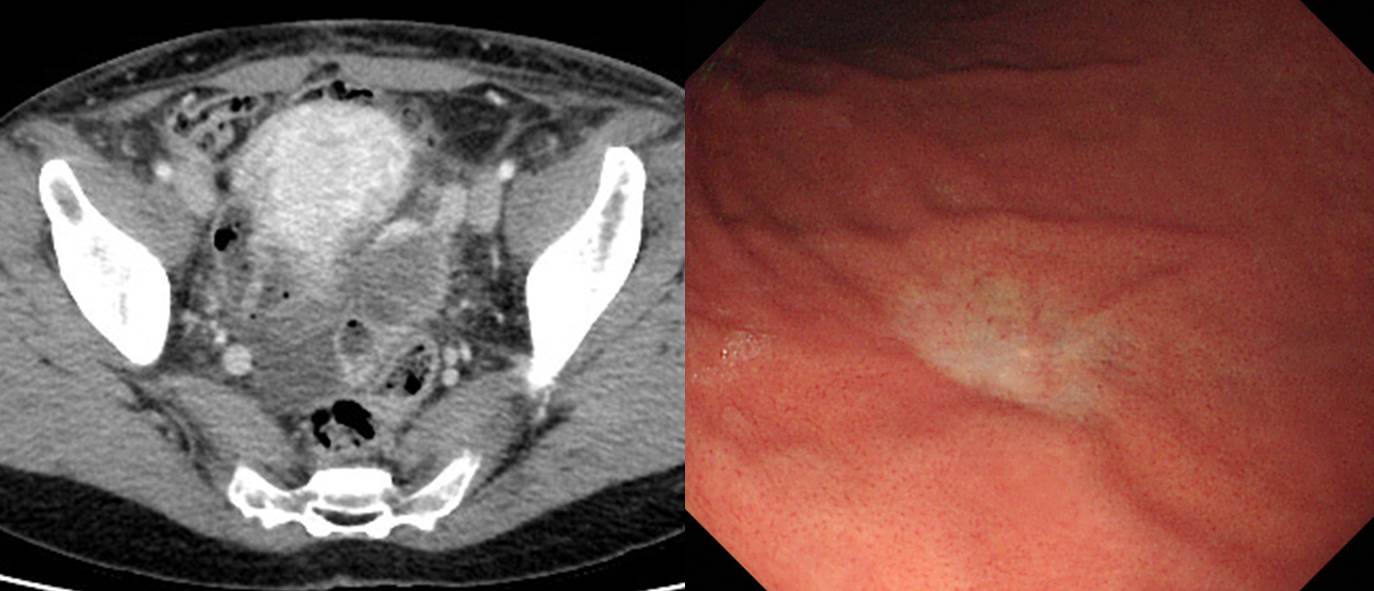
복수와 난소 종양으로 수술을 받은 환자입니다.
Ovary and salpinx, left oophorectomy, left salpingectomy, omentectomy, peritonectomy and total hysterectomy:
. Signet ring cell carcinoma, metastatic versus primary (see note) ;
1) Tumor size: 3.2x2.5x1.0 cm (left ovary)
2) Ovarian surface involvement: present
3) Involvement of left ovary and salpinx, uterus and "adhesion band"
4) No involvement of peritoneum and omentum
5) Lymphovascular invasion without D2-40 immunohistochemistry: Positive (frequent)
Omentum, omentectomy: no evidence of malignancy
Adhesion band, biopsy: tumor present
Peritoneum, peritonectomy: no evidence of malignancy
병리과 선생님은 특별히 다음과 같은 note를 남기셨습니다. "Note: Primary ovarian signet ring cell carcinoma는 매우 드물며 면역화학염색검사 결과상 종양세포들이 CK20(+), CDX-2(+), CK7(-) 소견을 보여 metastatic signet ring cell carcinoma from GI tract등의 가능성을 배제하기 어렵습니다. 전신적인 검사 및 전이암을 배제하기 위한 생검이 필요합니다."
위 내시경에서 작고 약간 함몰된 퇴색부위가 있었고 조직검사에서 signet ring cell carcinoma로 나왔습니다. 위의 작은 signet ring cell carcinoma가 난소로 전이되었던 경우로 판단하였습니다.
이 환자의 내시경에서 위암을 발견하였던 선생님께서 저에게 이런 말을 남겼습니다. "제 짧은 임상경험이지만 SRC 는 뭔가 주변 점막보다 pale 하고 whitish 한것이 특징인것 같습니다. 이 case 이 후에 focal 한 whitish discoloration 을 발견할때마다 조직검사를 시행해야할지 많이 고민하고 있습니다."
그렇습니다. SRC는 크기가 작을 때 발견하기 어렵습니다. SRC는 크기가 작아도 전이를 보이는 경우가 있습니다.
보만 4형 진행성 위암의 난소 전이에 대한 설명입니다.

© EndoTODAY Endoscopy Learning Center. Jun Haeng Lee. 일원내시경교실 바른내시경연구소 이준행