 EndoTODAY 내시경 교실
Beginner | ESA | Schedule | OPD
Seminars | Atlas | Recent | Links
 [Gastric cancer 487]
001 | 101 | 201 | 301 | 401 | 501 | 601 | 701 | 801 | 901 | 1000
[EGC after Ivor Lewis operation for esophageal cancer]
식도암이 발견되었습니다. 크기 및 깊이에 비하여 점막병소가 미약하였던 경우입니다. 이런 식도암은 놓치기 쉬운데, 검사자께서 참 잘 발견하신 경우입니다.

Esophagus and upper stomach, Ivor Lewis operation:
Invasive squamous cell carcinoma, moderately differentiated, distal esophagus:
1) tumor size: 4.5x3 cm
2) extension to perimuscular adventitia (pT3)
3) endolymphatic tumor emboli: not identified
4) perineural invasion: not identified
5) negative resection margins (proximal, 6.5 cm ; distal, 4 cm)
6) metastasis to 5 out of 52 regional lymph nodes (pN2) (5/52: "LC omentum", 0/0; "RRLN", 0/1; "LRLN", 0/6; "LD", 0/1; "5", 0/6; "7", 0/6; "8u", 0/2; "R9", 0/2; "L9", 0/1; "R10", 0/2; "L10", 0/4; "G1", 0/7; "G2", 0/2; "G3", 5/12)
식도암 치료는 잘 되었는데 6년 후 위암이 발견되었습니다. 매우 작았기 때문에 ESD로 치료하였습니다.
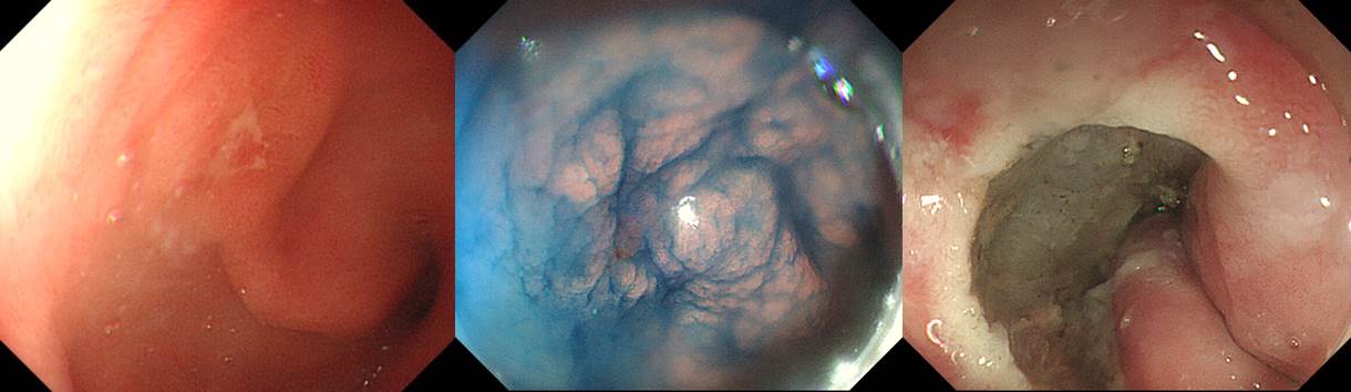
ESD. EGC
1. Location : antrum, anterior wall
2. Gross type : EGC type IIc
3. Histologic type : tubular adenocarcinoma, well differentiated
4. Histologic type by Lauren : intestinal
5. Size : (1) longest diameter, 6mm (2) vertical diameter, 6mm
6. Depth of invasion : invades mucosa (pT1a)
7. Resection margin: free from carcinoma (N), safety margin: proximal 10mm, distal 10mm, anterior 6mm, posterior 12mm
8. Lymphatic invasion : not identified
9. Venous invasion : not identified
10. Perineural invasion : not identified
11. Microscopic ulcer : absent
12. Histologic heterogeneity : absent
© 일원내시경교실 바른내시경연구소 이준행. EndoTODAY Endoscopy Learning Center. Lee Jun Haeng.