 EndoTODAY 내시경 교실
Beginner | ESA | Schedule | OPD
Seminars | Atlas | Recent | Links
![]() [Thursday Endoscopy Conference 20160630]
[Thursday Endoscopy Conference 20160630]
![]() 1. Early gastric cancer
1. Early gastric cancer
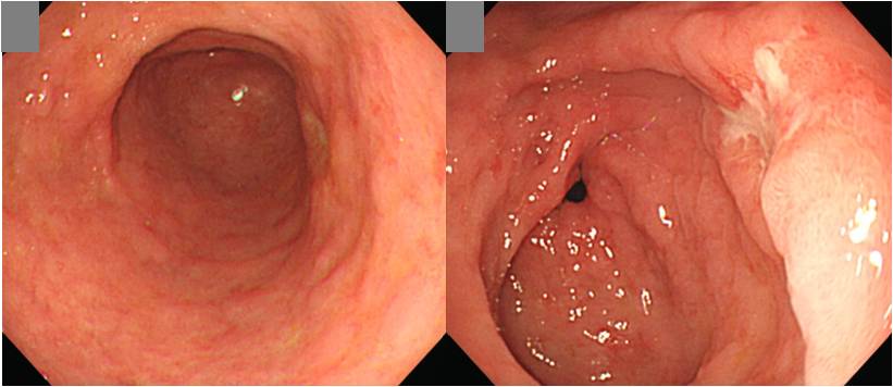
첫 조직검사에서 암이 나오지 않아 내시경 재검하여 암으로 확인되어 수술을 시행한 환자입니다.
Stomach, subtotal gastrectomy:
Early gastric carcinoma
1. Location : lower third Center at antrum and lesser curvature
2. Gross type : EGC type IIc
3. Histologic type : tubular adenocarcinoma, moderately differentiated
4. Histologic type by Lauren : intestinal
5. Size : 1.3x0.8 cm
6. Depth of invasion : invades submucosa (sm1) (pT1b)
7. Resection margin: free from carcinom, safety margin: proximal 9.6 cm, distal 2.3 cm
8. Lymph node metastasis : no metastasis in 32 regional lymph nodes (pN0)
9. Lymphatic invasion : not identified
10. Venous invasion : not identified
11. Perineural invasion : not identified
![]() 2. MALT lymphoma
2. MALT lymphoma
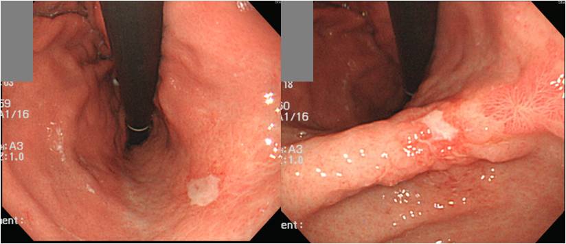
위체하부 소만의 shallow ulceration과 위각의 ulcer 및 scar처럼 보이는 병소가 있었습니다. Gastric MALToma 였습니다.
* 참고: EndoTODAY MALToma
![]() 3. Intestinal tuberculosis in patients on chemotherapy
3. Intestinal tuberculosis in patients on chemotherapy
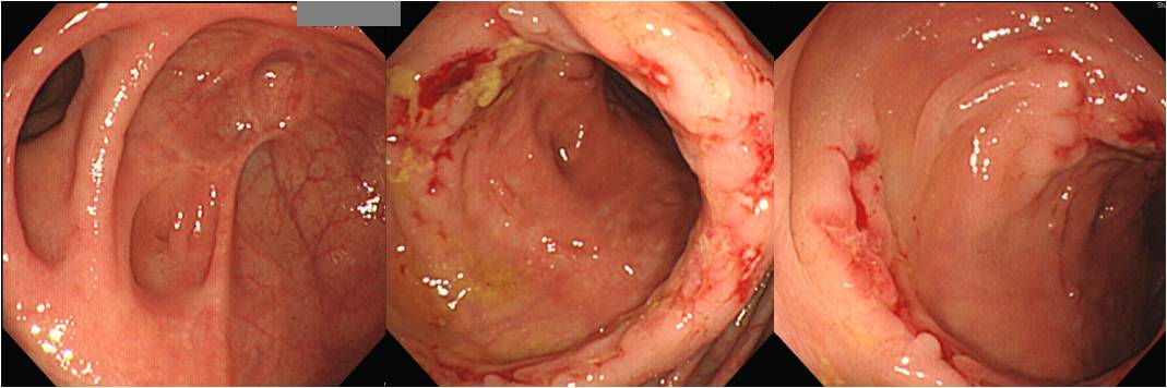
우측 conjunctiva의 malignant melanoma로 수술과 interferon을 포함한 항암화학요법을 받는 분인데 PET에서 상행결장의 uptake가 발견되었습니다. 장결핵이었습니다.
* 참고: EndoTODAY Gastrointestinal tuberculosis
![]() 4. Fundus EGC type I
4. Fundus EGC type I
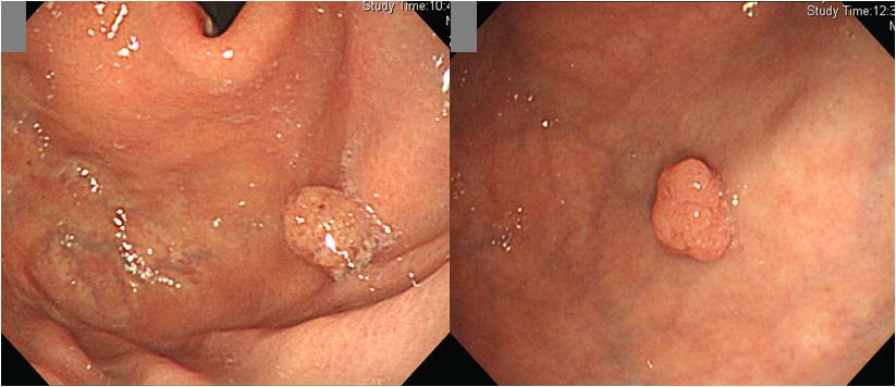
조직검사에서 과형성용종으로 나온 바 있던 환자의 추적내시경에서 용종이 커지면서 조직검사에서 adenoma with low grade dysplasia로 나와 의뢰된 환자입니다. EMR을 시행하였는데 의외의 결과가 나왔습니다.
Early gastric carcinoma
1. Location : fundus, anterior wall
2. Gross type : EGC type I
3. Histologic type : tubular adenocarcinoma, well differentiated (foveolar type), in adenoma (foveolar type) with high grade dysplasia
4. Histologic type by Lauren : intestinal
5. Size of carcinoma : (1) longest diameter, 8 mm (2) vertical diameter, 6 mm
6. Depth of invasion : invades mucosa (lamina propria) (pT1a)
7. Resection margin : free from carcinoma(N)
8. Lymphatic invasion : not identified(N)
9. Venous invasion : not identified(N)
10. Perineural invasion : not identified(N)
11. Microscopic ulcer : absent
12. Histologic heterogeneity: absent
![]() [References]
[References]
1) SMC Endoscopy Unit 삼성서울병원 내시경실
2) SMC Monday GI conference 삼성서울병원 일원내시경교실 월요점심소화기집담회
3) SMC Thursday endoscopy conference 삼성서울병원 일원내시경교실 목요점심내시경집담회
© EndoTODAY Endoscopy Learning Center. Jun Haeng Lee.