EndoTODAY 내시경 교실
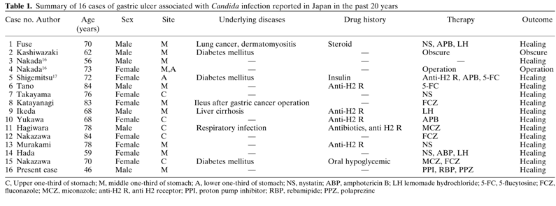
Beginner | ESA | Schedule | OPD
Seminars | Atlas | Recent | Links
![]() [Thursday Endoscopy Conference 20160929]
[Thursday Endoscopy Conference 20160929]
![]() 1. EGC
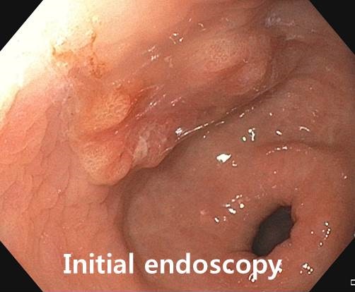
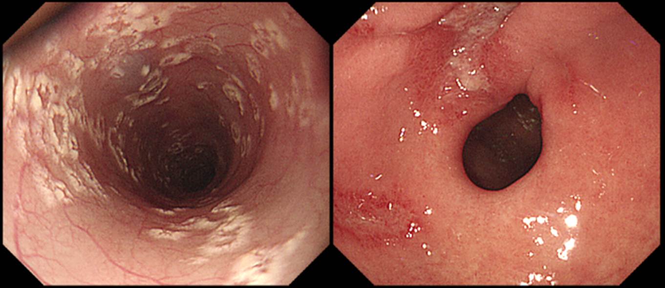
1. EGC
전정부의 ovoid한 IIa+IIc 병소였습니다. 장경 3 cm 정도였고 중앙부의 함몰이 있어 점막하암으로 추정하여 수술을 시행하였고 deep SM invasion이 나왔습니다.
Early gastric carcinoma
1. Location : middle third, Center at antrum and lesser curvature
2. Gross type : EGC type IIa
3. Histologic type: tubular adenocarcinoma, moderately differentiated
4. Histologic type by Lauren : intestinal
5. Size : 2.7x1.4 cm
6. Depth of invasion : invades submucosa (sm2) (pT1b)
7. Resection margin: free from carcinoma, safety margin: proximal 1.4 cm, distal 4 cm
8. Lymph node metastasis : no metastasis in 23 regional lymph nodes (pN0)
9. Lymphatic invasion : not identified
10. Venous invasion : not identified
11. Perineural invasion : not identified
12. AJCC stage by 7th edition: T1b N0
왜 ESD를 시도하지 않았는지 질문이 있었습니다. 사실 정확히 이야기하기는 어렵지만 저는 육안소견을 신봉하는 편입니다. EUS나 확대내시경을 하지 않더라도 경험 많은 내시경 의사의 섬세한 육안관찰 및 경험에 근거한 판단이 상당히 정확하다고 믿기 때문입니다. 이 환자의 경우는 첫 외부 내시경에서 점막하침윤이 의심되었습니다. 공기를 조금 빼고 찍은 사진에서는 깊어보였기 때문입니다. 조기위암에서 공기를 너무 많이 넣고 검사하면 병소가 납작해져서 심달도가 낮아 보입니다. 공기를 넣었다 뺐다 하면서 잘 관찰해야 하는 이유입니다.
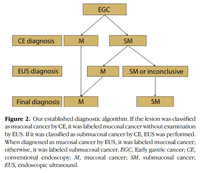
바로 수술을 결정하기보다는 EUS를 통하여 심달도 평가를 해 보는 것이 좋겠다는 한 교수님의 comment가 있었습니다. 의미있는 지적이라고 생각되었습니다. 관련된 오사카 대학의 논문을 소개합니다 (Tsujii Y. GIE. 2015). 내시경 육안소견이 점막암이면 EUS 없이 ESD를 시행하고, 내시경 육안소견이 점막하암으로 추정되면 EUS를 시행하여 위내시경에 의한 overstaging을 줄일 수 있다는 주장이었습니다. "CE (conventional endoscopy) accurately revealed mucosal cancer, and EUS efficiently salvaged the lesions that were over-diagnosed by CE."
점막암이 의심되면 바로 ESD를 하고, 점막하암이 의심되면 EUS를 해본다...... 이론적인 가능성이 있기는 합니다. 향후 이에 대한 평가는 필요해 보입니다.
* 참고: EndoTODAY EUS before ESD
![]() 2. Metastasis to the stomach from the lung cancer
2. Metastasis to the stomach from the lung cancer
비슷한 증례 몇 개 소개합니다. 모두 폐암 위전이입니다.
폐암 (large cell neuroendocrine carcinoma) 위전이
폐암 (adenocarcinoma) 위전이와 십이지장 전이
폐암 위전이
폐암 위전이
* 참고: EndoTODAY 위전이
![]() 3. 식도 칸디다 + CMV 위염
3. 식도 칸디다 + CMV 위염
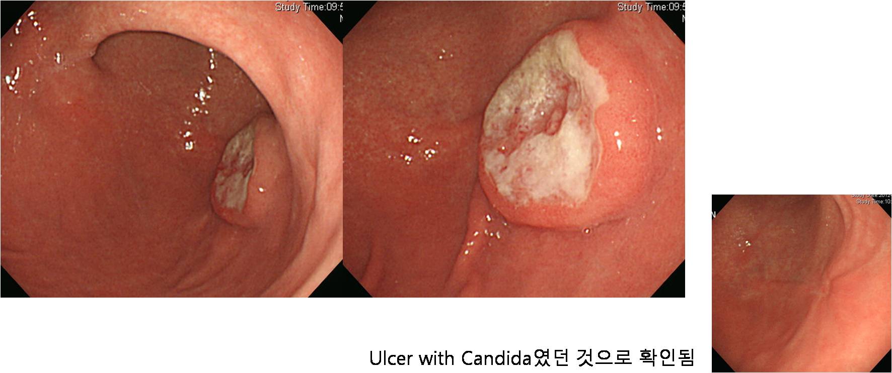
식도는 비교적 전형적인 칸디다 식도염입니다. 이 환자에서 위에 약간 이상한 모양의 궤양이 함께 발생하였는데 어떤 분이 칸디다 위궤양의 가능성을 언급하셨습니다. 최종 결론은 CMV gastritis 였습니다. 여하튼 제가 보아 온 칸디다 위궤양은 이와는 전혀 다른 모양이었습니다. 아래에 소개합니다.
"위궤양 + 캔디다"를 위암으로 오인하는 경우가 있습니다.
2011년 11월 7일 EndoTODAY와 위 조직검사 부분에서 다룬 바 있어서 약간 다듬어 옮깁니다.
위궤양 혹은 십이지장 궤양에 fungal colonization이 동반되는 경우가 있습니다. 캔디다 감염은 궤양 치유 속도에 영향을 미치지 않습니다. 치료도 달라지지 않습니다. Antifungal agent를 투여할 필요도 없습니다. 그냥 PPI만 주면 그만입니다.
칸디다 관련 궤양의 전형적인 예입니다. 캔디다가 먼저인지 궤양이 먼저인지 알 수 없지만 저는 궤양이 먼저라고 생각하고 있습니다.
통상의 위궤양 치료 후 경과관찰인데 완전히 호전되었습니다.
Candida 감염이 동반된 궤양을 설명한 어떤 저널 (Hirasaki S. J Gastroenterol 1999;34:688-693)의 일부를 옮깁니다. "Candida-infected gastric ulcer should be suspected in patients with a gastric submucosal tumor-like lesion with a thick, yellowish-white coated ulcer of unclear border on its summit, and this lesion should be distinguished from malignant diseases." 우리 증례를 그대로 설명해 주는 것 같습니다.
2011년 1월 6일 EndoTODAY의 증례는 위암과 캔디다가 모두 나온 경우였습니다. 큰 궤양형 위암에 캔디다가 자리잡은 경우라고 할 수 있습니다.
조직검사 poorly differentiated carcinoma, Candida (+)
![]() 4. Duodenal adenocarcinoma arising from duodenal adenoma
4. Duodenal adenocarcinoma arising from duodenal adenoma
조직검사에서 십이지장 선종으로 의뢰되었습니다. EMR을 하였는데 의외로 암이 나왔다고 합니다. 암은 1 mm도 안 되고 대부분 선종이라고 하니 다행입니다.
EMR for duodenal adenoma was done.
Duodenum, endoscopic submucosal dissection:
Adenocarcinoma, well differentiated (focal: < 10 %) arising from mainly adenoma (90 %)
1. Location : duodenum second portion
2. Size of carcinoma : < 1 mm
3. Size of adenoma : (1) longest diameter, 7 mm (2) vertical diameter, 4 mm
4. Depth of invasion : invades mucosa (lamina propria) (pT1a)
5. Resection margin : free from carcinoma(N)
6. Lymphatic invasion : not identified(N)
7. Venous invasion : not identified(N)
* 참고: EndoTODAY 십이지장암
![]() [References]
[References]
1) SMC Endoscopy Unit 삼성서울병원 내시경실
2) SMC Monday GI conference 삼성서울병원 일원내시경교실 월요점심소화기집담회
3) SMC Thursday endoscopy conference 삼성서울병원 일원내시경교실 목요점심내시경집담회
© 일원내시경교실 바른내시경연구소 이준행. EndoTODAY Endoscopy Learning Center. Lee Jun Haeng.