EndoTODAY 내시경 교실
Beginner | ESA | Schedule | OPD
Seminars | Atlas | Recent | Links
![]() [Thursday Endoscopy Conference 20161006]
[Thursday Endoscopy Conference 20161006]
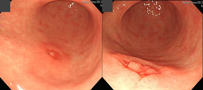
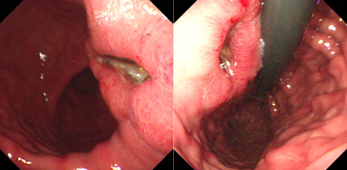
![]() 1. EGC IIa + IIc
1. EGC IIa + IIc
ESD가 시행되었고 아쉽게 deep SM invasion과 lymphatic invasion도 동반되어 있어 수술을 하였고 no residual tumor가 나왔습니다.
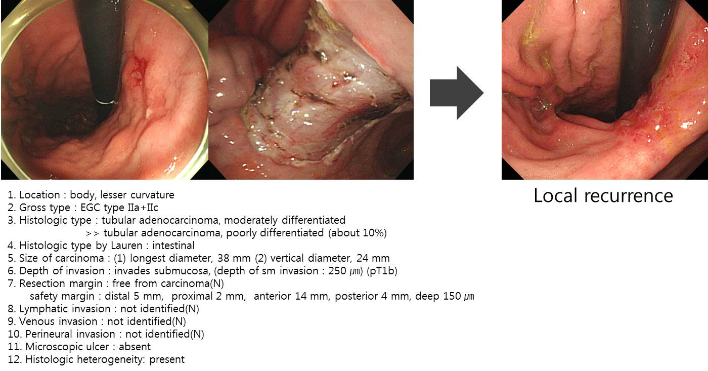
![]() 2. Local recurrence after ESD for EGC (EGC with SM invasion, mixed histology)
2. Local recurrence after ESD for EGC (EGC with SM invasion, mixed histology)
1) ESD 후 추적검사에서 육안소견이 정상 scar인 경우는 재발인 예가 거의 없습니다. 그러나 표면의 불규칙성이나 발적 등의 소견이 보이면 국소재발을 고려하여 조심스럽게 조직검사를 할 필요가 있습니다.
2) 미분화 혼재암인 경우 병소의 경계를 정하기 어렵습니다. ESD 후 resection margin involvement가 있거나 절재 여분이 충분하지 않을 수 있습니다 (EndoTODAY lateral margin involvement). 그 결과 local recur의 확률이 높을 것으로 생각합니다 (관련 연구 자료는 없습니다만...)
* 참고: EndoTODAY ESD 후 국소 재발
![]() 3. Local recurrence after esophageal cancer surgery
3. Local recurrence after esophageal cancer surgery
70대 여성입니다.
 일견 심해보지지 않는 식도암이 발견되었습니다. 그러나 EUS에서는 proper muscle invasion이 의심되었습니다.
 5주 후 재검입니다. 비교적 빨리 자라는 식도암으로 판단되었습니다. 림프절 전이도 의심되었습니다.
 수술 (3 hole operation) 후 병리결과는 생각보다 심했습니다. Adjuvant FOLFOX#6 시행하였습니다.
Esophagus and upper stomach, Ivor Lewis operation:
Invasive squamous cell carcinoma, well differentiated :
1) tumor size: 6.1x4.3 cm
2) extension to periesophageal soft tissue (adventitia)
3) endolymphatic tumor emboli: not identified
4) perineural invasion: not identified
5) resection margins: free from carcinoma, safety margin: proximal, 3.5 cm ; distal, 5 cm; circumferential (adventitial) margin(deep), < 50 ㎛
6) metastasis to 1 out of 41 regional lymph nodes (right recurrent laryngeal nerve LN, 1/3)
7) treatment effect: no prior treatment
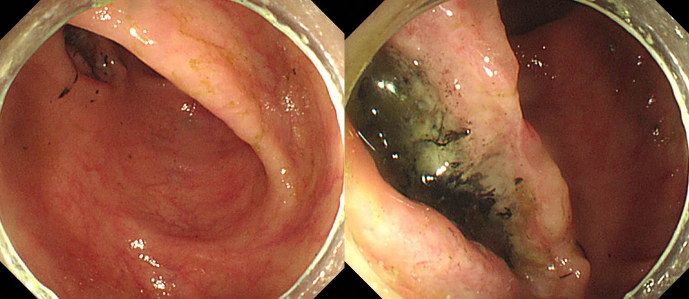
 3년 후 gastric tube에서 SMT-like 병소가 발견되었습니다. 겸자 조직검사는 특이소견이 없었습니다.
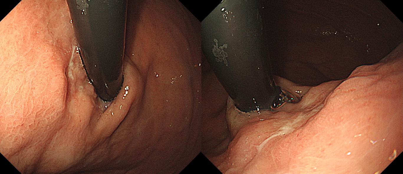
 4개월 후 재검에서 SMT-like 병소에 깊은 궤양이 형성되었습니다. 겸자 조직검사에서 squamous cell carcinoma로 확인되었습니다. 국소재발로 CCRT를 시행하였습니다.
* 김재준 교수님 comment: 전 내시경에서는 없었던 SMT 유사 병소가 intrathoracic stomach에서 발견되었기 때문에 통상의 SMT가 아니라 식도암의 국소 재발을 고려해야 합니다. 내시경 시행 의사가 내시경 결과지에 EUS-guided biopsy를 추천했더라면 더 좋았을 것 같습니다.
* 참고: EsoTODAY 식도암 국소 재발
![]() 4. DLBCL after liver transplantation
4. DLBCL after liver transplantation
간경변으로 간이식 시행받은 환자의 복통.
Ascending colon, cecum, appendix, and terminal ileum, extended right hemicolectomy:
Post-transplant lymphoproliferative disorder, diffuse large B cell lymphoma
1. Location: ascending colon
2. Gross type: ulceroinfiltrative
3. Size: 5x2.5 cm
4. Depth of invasion: invades subserosa or pericolic/perirectal adipose tissue (pT3)
5. Resection margin: free from carcinoma, safety margin: proximal, 11 cm ; distal, 30 cm
6. Regional lymph node metastasis : Involvement in 2 out of 27 regional lymph nodes(pN1b)
7. Pathologic staging: pT3 N1b
![]() 5. Cardia cancer
5. Cardia cancer
Cardia cancer는 늘 생각보다 심합니다.
Stomach, total gastrectomy:
Advanced gastric carcinoma
1. Location : upper third, Center at cardia
2. Gross type : Borrmann type (unclassifiable) (mimicking EGC type IIc)
3. Histologic type : tubular adenocarcinoma, poorly (poorly cohesive) differentiated
4. Histologic type by Lauren : diffuse
5. Size : 2.5x2.4 cm
6. Depth of invasion : invades muscularis propria (pT2)
7. Resection margin: free from carcinoma, safety margin: proximal 1 cm, distal 18 cm
8. Lymph node metastasis : metastasis to 1 out of 33 regional lymph nodes (pN1) ("9", 1/4)
9. Lymphatic invasion : present
10. Venous invasion : not identified
11. Perineural invasion : present
12. AJCC stage by 7th edition: T2 N1
PPT PDF 4.6 M
![]() [References]
[References]
1) SMC Endoscopy Unit 삼성서울병원 내시경실
2) SMC Monday GI conference 삼성서울병원 일원내시경교실 월요점심소화기집담회
3) SMC Thursday endoscopy conference 삼성서울병원 일원내시경교실 목요점심내시경집담회
© 일원내시경교실 바른내시경연구소 이준행. EndoTODAY Endoscopy Learning Center. Lee Jun Haeng.